
Article Figures & Data
Figures
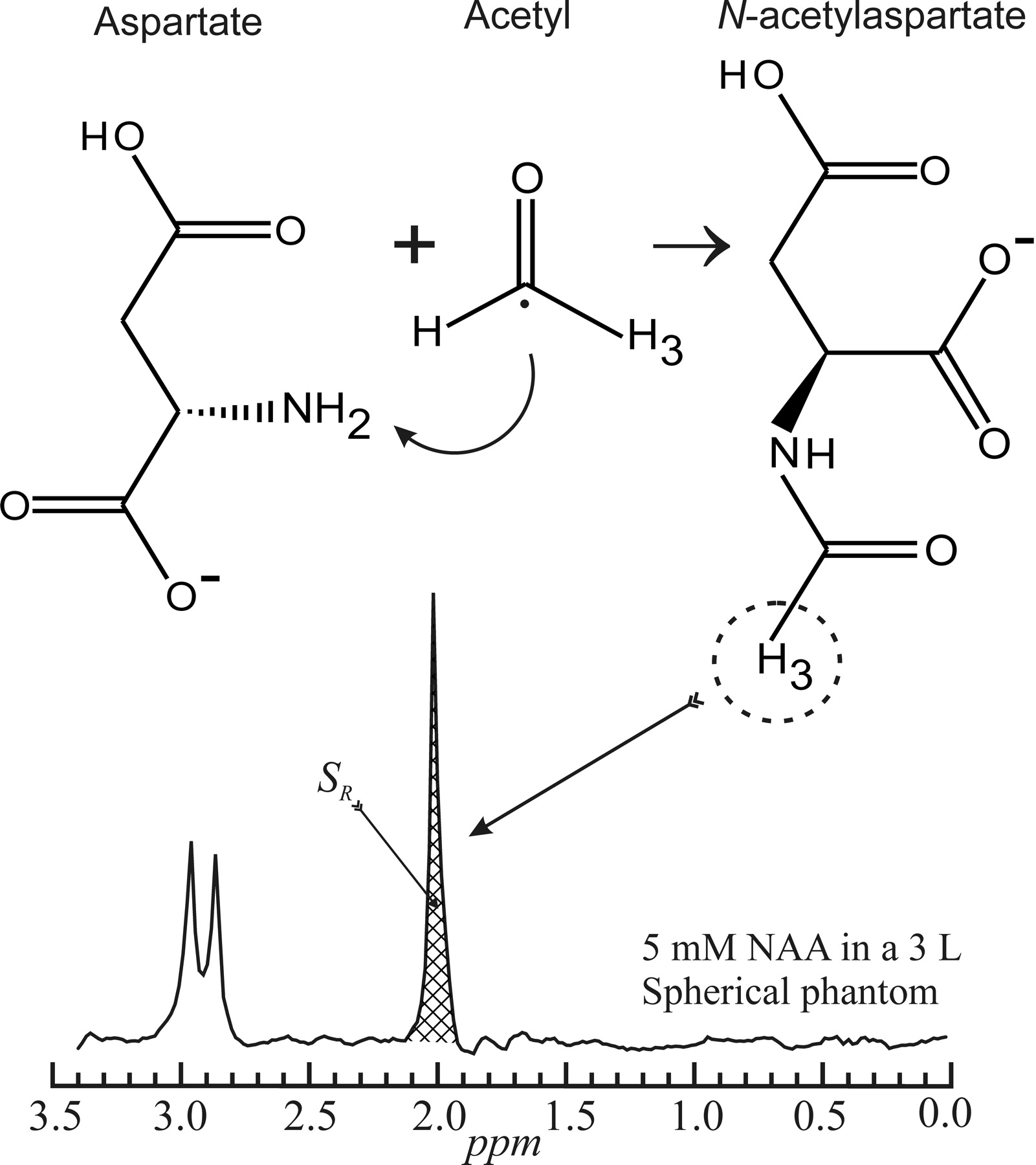
- Fig 1.
Schematic description of the structures and acetylization of aspartate into NAA in the mitochondria (top). The 3 protons of the methyl group of NAA provide the most prominent peak on the neurometabolite proton spectrum (bottom).
- Fig 2.
Top: Schematic comparison of relative VOI size and coverage between single-voxel, 2D or 3D multivoxel, and WBNAA in the human brain. Note that the single- and multivoxel VOIs cover only small fractions of the brain volume and must be kept away from the skull, missing most of the cortex, whereas WBNAA accounts for the entire brain. Bottom: The consequence of a 1-mm (1 guiding-image pixel) placemat error in each direction, x, y, and z, on a 1-cm voxel in a serial study. This 10% error leads to only 70% of the VOI common to both measurements. Note that though this error is technically unavoidable in localized spectroscopy, it is not an issue for WBNAA, as shown in the top portion of the lower panel.
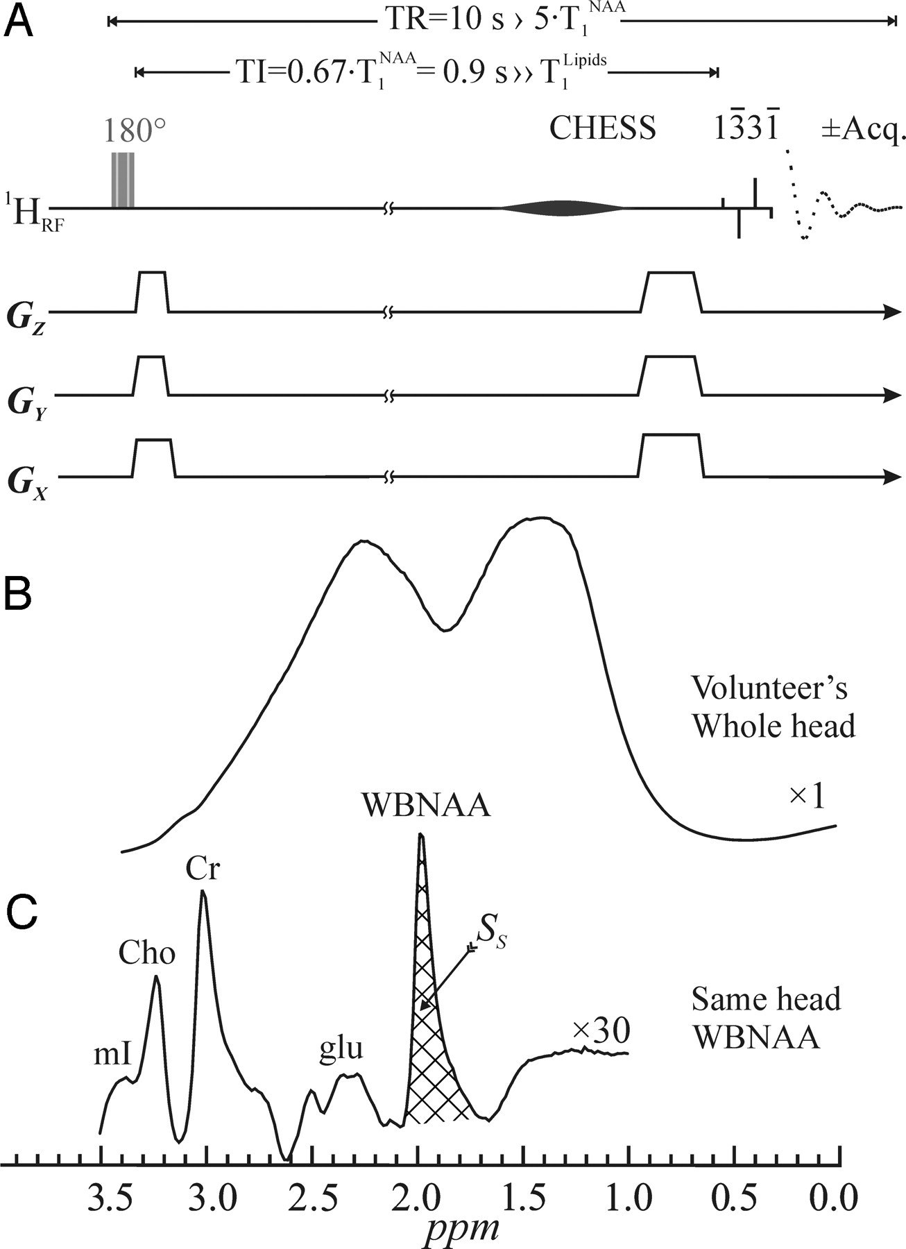
- Fig 3.
Top: A, Schematic representation of the 2-step WBNAA sequence. It comprises an alternating inversion pulse (applied every odd acquisition) followed by a TI designed to null the NAA signal. It ends with a chemical-shift-selective (CHESS) and a 13̄31̄ water-suppression pulse (4-ms interpulse delay at 1.5T). The latter also serves as the 90° readout pulse. Acquisition commences immediately (TE = 0), and every even acquisition is subtracted from every odd one. Because the TR is long, 10 seconds, no T1- or T2-weighting is incurred. Note that all the radio-frequency (RF) pulses are (spatially) nonselective and the gradient “blips” are just crushers. Center: B, The result of a 90° 13̄31̄ on a human head, demonstrating the problem of the immense metabolite-obscuring lipid signal from the bone marrow and adipose tissue. Bottom: C, The resulting spectrum from the WBNAA sequence on the same head as in B, demonstrating almost complete lipid suppression. Note that the NAA is implicitly localized to the brain, whereas with the other metabolites whose peaks are distinct and nonlocalized, it is impossible to ascertain where their signals come from. Also note the excellent SNR from this 2.5-minute acquisition.
- Fig 4.
Box plot showing the 25%, 50% (median), and 75% quartiles (box) and ±95% (whiskers) of the distributions of the subjects’ WBNAA concentrations in each of the 5 institutions, 6 scanners, 3 field strengths, and 2 manufacturers used in this study. Note that the differences among the distributions, 12.2 ± 1.2 mmol/L (means and SDs), for more than 120 healthy individuals are statistically insignificant, independent of these subjects’ ages or sex, indicating that the methodology is robust to instrumentation differences. HSR indicates Hospital San Rafael (Italy); FCCC, Fox Chase Cancer Center (USA); NYU, New York University (USA); Basel, Basel (Switzerland); U.PENN, University of Pennsylvania (USA).
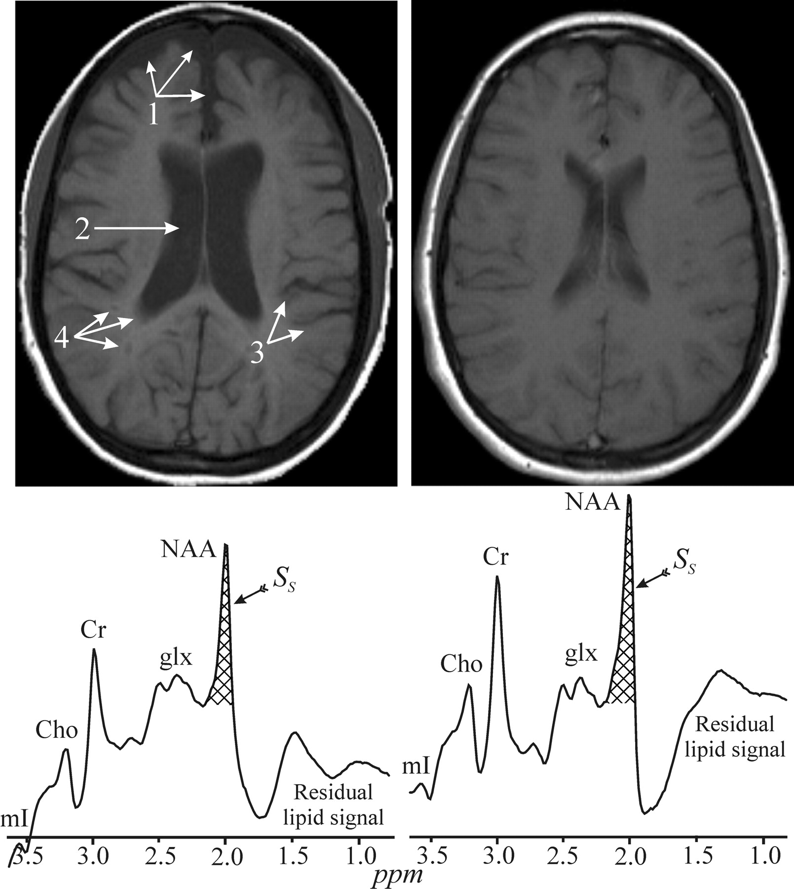
- Fig 5.
Top: Side-by-side axial T1 MR image from two 29-year-old-female patients with MS, both with a disease duration of 6 years and a very mild disability score (EDSS) of 1.5. Note that one exhibits noticeably more brain atrophy, reflected by enlarged subarachnoid spaces (1), ventricles (2), sulci (3) as well as T1-lesions (“black holes”) (4). These 2 images demonstrate the great heterogeneity in the MS course and pathogenesis. Bottom: The WBNAA spectrum from each of these women, normalized for brain tissue volume. Note that the one on the left is significantly lower than the one on the right, demonstrating that not only does she lose tissue faster but that the quality of the remaining tissue is worse (ie, neuronal dysfunction and damage precedes atrophy). mI indicates myo-Inositol; Cho, choline; Cr, creatine; glx, glutamine/glutamate; acq., acquisition.


In this issue


Jump to section
Related Articles
Cited By...
- Longitudinal Evaluation of Magnetic Resonance Spectroscopy Metabolites as Biomarkers in Huntingtons Disease
- Neuroimaging of Sports Concussions
- Whole-Brain N-Acetylaspartate Concentration Is Preserved during Mild Hypercapnia Challenge
- Alzheimer Disease: Focus on Computed Tomography
- Metabolic Changes in Patients with Aneurysmal Subarachnoid Hemorrhage Apart from Perfusion Deficits: Neuronal Mitochondrial Injury?
- Two-year serial whole-brain N-acetyl-L-aspartate in patients with relapsing-remitting multiple sclerosis
- Longitudinal Whole-Brain N-Acetylaspartate Concentration in Healthy Adults
- Diffuse White Matter Damage Is Absent in Neuromyelitis Optica
- Vitamin A Deficiency in Rats Induces Anatomic and Metabolic Changes Comparable with Those of Neurodegenerative Disorders
- Changes in NAA and lactate following ischemic stroke: A serial MR spectroscopic imaging study