Advertisement

Main menu

  • Home
  • Content
    • Current Issue
    • Accepted Manuscripts
    • Article Preview
    • Past Issue Archive
    • Video Articles
    • AJNR Case Collection
    • Case of the Week Archive
    • Case of the Month Archive
    • Classic Case Archive
  • Special Collections
    • AJNR Awards
    • Low-Field MRI
    • Alzheimer Disease
    • ASNR Foundation Special Collection
    • Photon-Counting CT
    • View All
  • Multimedia
    • AJNR Podcasts
    • AJNR SCANtastic
    • Trainee Corner
    • MRI Safety Corner
    • Imaging Protocols
  • For Authors
    • Submit a Manuscript
    • Submit a Video Article
    • Submit an eLetter to the Editor/Response
    • Manuscript Submission Guidelines
    • Statistical Tips
    • Fast Publishing of Accepted Manuscripts
    • Graphical Abstract Preparation
    • Imaging Protocol Submission
    • Author Policies
  • About Us
    • About AJNR
    • Editorial Board
    • Editorial Board Alumni
  • More
    • Become a Reviewer/Academy of Reviewers
    • Subscribers
    • Permissions
    • Alerts
    • Feedback
    • Advertisers
    • ASNR Home

User menu

  • Alerts
  • Log in

Search

  • Advanced search
American Journal of Neuroradiology

American Journal of Neuroradiology

ASHNR American Society of Functional Neuroradiology ASHNR American Society of Pediatric Neuroradiology ASSR
  • Alerts
  • Log in

Advanced Search

  • Home
  • Content
    • Current Issue
    • Accepted Manuscripts
    • Article Preview
    • Past Issue Archive
    • Video Articles
    • AJNR Case Collection
    • Case of the Week Archive
    • Case of the Month Archive
    • Classic Case Archive
  • Special Collections
    • AJNR Awards
    • Low-Field MRI
    • Alzheimer Disease
    • ASNR Foundation Special Collection
    • Photon-Counting CT
    • View All
  • Multimedia
    • AJNR Podcasts
    • AJNR SCANtastic
    • Trainee Corner
    • MRI Safety Corner
    • Imaging Protocols
  • For Authors
    • Submit a Manuscript
    • Submit a Video Article
    • Submit an eLetter to the Editor/Response
    • Manuscript Submission Guidelines
    • Statistical Tips
    • Fast Publishing of Accepted Manuscripts
    • Graphical Abstract Preparation
    • Imaging Protocol Submission
    • Author Policies
  • About Us
    • About AJNR
    • Editorial Board
    • Editorial Board Alumni
  • More
    • Become a Reviewer/Academy of Reviewers
    • Subscribers
    • Permissions
    • Alerts
    • Feedback
    • Advertisers
    • ASNR Home
  • Follow AJNR on Twitter
  • Visit AJNR on Facebook
  • Follow AJNR on Instagram
  • Join AJNR on LinkedIn
  • RSS Feeds

AJNR Awards, New Junior Editors, and more. Read the latest AJNR updates

Research ArticleBrain

Intracranial Dural Arteriovenous Fistulas with Retrograde Cortical Venous Drainage: Assessment with Cerebral Blood Volume by Dynamic Susceptibility Contrast Magnetic Resonance Imaging

K. Noguchi, M. Kubo, N. Kuwayama, Y. Kamisaki, G. Tomizawa, K. Kameda, H. Kawabe, S. Ogawa, N. Watanabe, S. Endo and H. Seto
American Journal of Neuroradiology June 2006, 27 (6) 1252-1256;
K. Noguchi
  • Find this author on Google Scholar
  • Find this author on PubMed
  • Search for this author on this site
M. Kubo
  • Find this author on Google Scholar
  • Find this author on PubMed
  • Search for this author on this site
N. Kuwayama
  • Find this author on Google Scholar
  • Find this author on PubMed
  • Search for this author on this site
Y. Kamisaki
  • Find this author on Google Scholar
  • Find this author on PubMed
  • Search for this author on this site
G. Tomizawa
  • Find this author on Google Scholar
  • Find this author on PubMed
  • Search for this author on this site
K. Kameda
  • Find this author on Google Scholar
  • Find this author on PubMed
  • Search for this author on this site
H. Kawabe
  • Find this author on Google Scholar
  • Find this author on PubMed
  • Search for this author on this site
S. Ogawa
  • Find this author on Google Scholar
  • Find this author on PubMed
  • Search for this author on this site
N. Watanabe
  • Find this author on Google Scholar
  • Find this author on PubMed
  • Search for this author on this site
S. Endo
  • Find this author on Google Scholar
  • Find this author on PubMed
  • Search for this author on this site
H. Seto
  • Find this author on Google Scholar
  • Find this author on PubMed
  • Search for this author on this site
  • Article
  • Figures & Data
  • Info & Metrics
  • Responses
  • References
  • PDF
Loading

Article Figures & Data

Figures

  • Tables
  • Fig 1.
    • Download figure
    • Open in new tab
    • Download powerpoint
    Fig 1.

    Schematic shows the region of interests for rCBV measurement on the target section.
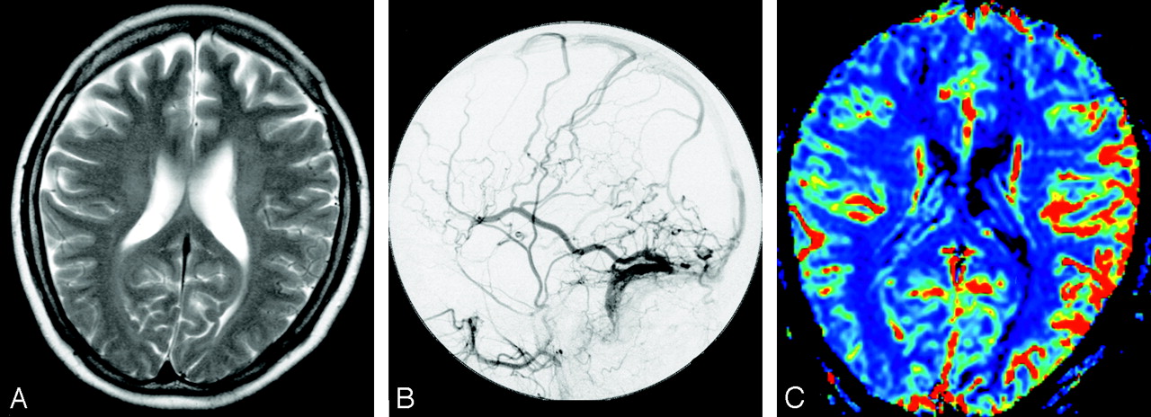
  • Fig 2.
    • Download figure
    • Open in new tab
    • Download powerpoint
    Fig 2.

    Dural arteriovenous fistula at left transverse-sigmoid sinus in a 52-year-old woman (case 7).

    A, T2-weighted image shows no prominent flow voids on the cortical sulci.

    B, Left external carotid angiogram depicts reflux in the vein of Labbe and retrograde cortical venous drainage with occlusion of left transverse sigmoid sinus.

    C, Color overlay of relative cerebral blood volume (rCBV) map shows marked increase of CBV in the left hemisphere (mean rCBV ratio, 1.32).
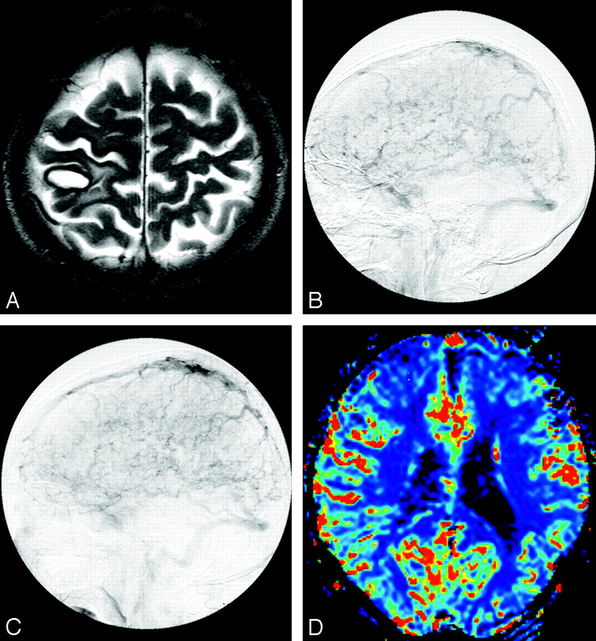
  • Fig 3.
    • Download figure
    • Open in new tab
    • Download powerpoint
    Fig 3.

    Dural arteriovenous fistula at superior sagittal sinus in a 70-year-old man (case 9).

    A, T2-weighted image shows late subacute subcortical hematoma in the parietal lobe.

    B, Venous phase of right common carotid angiogram depicts retrograde cortical venous drainage (RCVD) with occlusion of posterior part of superior sagittal sinus.

    C, Venous phase of left common carotid angiogram also depicts RCVD with occlusion of posterior part of superior sagittal sinus.

    D, Color overlay of relative cerebral blood volume (rCBV) map shows marked increase of CBV in the hemispheres bilaterally. The extent of increase of CBV is larger in the right hemisphere than in the left hemisphere (mean rCBV ratio, 1.37; however, the rCBV ratio may be underestimated because of the bilateral affected hemispheres).

  • Fig 4.
    • Download figure
    • Open in new tab
    • Download powerpoint
    Fig 4.

    Box and whisker plot show mean relative cerebral blood volume (rCBV) ratio in control subjects and in patients with dural arteriovenous fistulas (DAVF) with retrograde cortical venous drainage (RCVD). The mean rCBV ratio in patients with DAVF with RCVD is significantly higher than that in control subjects (P = .0002).

  • Fig 5.
    • Download figure
    • Open in new tab
    • Download powerpoint
    Fig 5.

    Dural arteriovenous fistula in left transverse-sigmoid sinus in a 71-year-old man (case 2).

    A, Color overlay of relative cerebral blood volume (rCBV) map shows marked increase of CBV in the left hemisphere (mean rCBV ratio, 1.94).

    B, Color overlay of rCBV map obtained after partial embolization shows improvement of increase of CBV in the left hemisphere (mean rCBV ratio, 1.74).

    C, Color overlay of rCBV map obtained after surgical operation (direct packing of the venous sinus) shows more improvement of increase of CBV in the left hemisphere (mean rCBV ratio, 1.23).

Tables

  • Figures
    • View popup
    Table 1:

    Summary of patient data

    Patient No.Main Clinical FindingSite of DAVFSite of RCVDAbnormal Flow VoidrCBV ratio
    1PapilledemaLeft TSSLeftYes1.42
    2Subarachnoid hemorrhageLeft TSSLeftYes1.94
    3Pulsatile tinnitisRight TSSRightNo1.14
    4ConvulsionLeft TSSLeftNo1.15
    5PapilledemaTSBothYes1.46
    6PapilledemaLeft TSSLeftNo1.24
    7Pulsatile tinnitisLeft TSSLeftNo1.32
    8PapilledemaTS, right TSSBothYes1.19
    9Subcortical HemorrhageSSSBothYes1.37
    10Conjunctival injectionRight CSLeftNo1.32
    Ref. 1Pulsatile tinnitisLeft TSSNoNo1.02
    Ref. 2Pulsatile tinnitisRight TSSNoNo1.03
    • Note:—DAVF indicates dural arteriovenous fistula; RCVD, retrograde cortical venous drainage; rCBV, relative cerebral blood volume; TSS, transverse-sigmoid sinus; TS, torcular herophili; SSS, superior sagittal sinus; CS, cavernous sinus; NA, not applicable.

PreviousNext
Back to top

In this issue

American Journal of Neuroradiology: 27 (6)
American Journal of Neuroradiology
Vol. 27, Issue 6
June 2006
  • Table of Contents
  • Index by author
Advertisement
Print
Download PDF
Email Article

Thank you for your interest in spreading the word on American Journal of Neuroradiology.

NOTE: We only request your email address so that the person you are recommending the page to knows that you wanted them to see it, and that it is not junk mail. We do not capture any email address.

Enter multiple addresses on separate lines or separate them with commas.
Intracranial Dural Arteriovenous Fistulas with Retrograde Cortical Venous Drainage: Assessment with Cerebral Blood Volume by Dynamic Susceptibility Contrast Magnetic Resonance Imaging
(Your Name) has sent you a message from American Journal of Neuroradiology
(Your Name) thought you would like to see the American Journal of Neuroradiology web site.
CAPTCHA
This question is for testing whether or not you are a human visitor and to prevent automated spam submissions.
Cite this article
K. Noguchi, M. Kubo, N. Kuwayama, Y. Kamisaki, G. Tomizawa, K. Kameda, H. Kawabe, S. Ogawa, N. Watanabe, S. Endo, H. Seto
Intracranial Dural Arteriovenous Fistulas with Retrograde Cortical Venous Drainage: Assessment with Cerebral Blood Volume by Dynamic Susceptibility Contrast Magnetic Resonance Imaging
American Journal of Neuroradiology Jun 2006, 27 (6) 1252-1256;

Citation Manager Formats

  • BibTeX
  • Bookends
  • EasyBib
  • EndNote (tagged)
  • EndNote 8 (xml)
  • Medlars
  • Mendeley
  • Papers
  • RefWorks Tagged
  • Ref Manager
  • RIS
  • Zotero
0 Responses
Respond to this article
Share
Bookmark this article
Intracranial Dural Arteriovenous Fistulas with Retrograde Cortical Venous Drainage: Assessment with Cerebral Blood Volume by Dynamic Susceptibility Contrast Magnetic Resonance Imaging
K. Noguchi, M. Kubo, N. Kuwayama, Y. Kamisaki, G. Tomizawa, K. Kameda, H. Kawabe, S. Ogawa, N. Watanabe, S. Endo, H. Seto
American Journal of Neuroradiology Jun 2006, 27 (6) 1252-1256;
del.icio.us logo Twitter logo Facebook logo Mendeley logo
  • Tweet Widget
  • Facebook Like
  • Google Plus One

Jump to section

  • Article
    • Abstract
    • Materials and Methods
    • Results
    • Discussion
    • Conclusions
    • References
  • Figures & Data
  • Info & Metrics
  • Responses
  • References
  • PDF

Related Articles

  • No related articles found.
  • PubMed
  • Google Scholar

Cited By...

  • Intracranial Dural Arteriovenous Fistula with Retrograde Cortical Venous Drainage: Use of Susceptibility-Weighted Imaging in Combination with Dynamic Susceptibility Contrast Imaging
  • Crossref
  • Google Scholar

This article has not yet been cited by articles in journals that are participating in Crossref Cited-by Linking.

More in this TOC Section

  • Evaluating the Effects of White Matter Multiple Sclerosis Lesions on the Volume Estimation of 6 Brain Tissue Segmentation Methods
  • Quiet PROPELLER MRI Techniques Match the Quality of Conventional PROPELLER Brain Imaging Techniques
  • Predictors of Reperfusion in Patients with Acute Ischemic Stroke
Show more Brain

Similar Articles

Advertisement

Indexed Content

  • Current Issue
  • Accepted Manuscripts
  • Article Preview
  • Past Issues
  • Editorials
  • Editor's Choice
  • Fellows' Journal Club
  • Letters to the Editor
  • Video Articles

Cases

  • Case Collection
  • Archive - Case of the Week
  • Archive - Case of the Month
  • Archive - Classic Case

More from AJNR

  • Trainee Corner
  • Imaging Protocols
  • MRI Safety Corner
  • Book Reviews

Multimedia

  • AJNR Podcasts
  • AJNR Scantastics

Resources

  • Turnaround Time
  • Submit a Manuscript
  • Submit a Video Article
  • Submit an eLetter to the Editor/Response
  • Manuscript Submission Guidelines
  • Statistical Tips
  • Fast Publishing of Accepted Manuscripts
  • Graphical Abstract Preparation
  • Imaging Protocol Submission
  • Evidence-Based Medicine Level Guide
  • Publishing Checklists
  • Author Policies
  • Become a Reviewer/Academy of Reviewers
  • News and Updates

About Us

  • About AJNR
  • Editorial Board
  • Editorial Board Alumni
  • Alerts
  • Permissions
  • Not an AJNR Subscriber? Join Now
  • Advertise with Us
  • Librarian Resources
  • Feedback
  • Terms and Conditions
  • AJNR Editorial Board Alumni

American Society of Neuroradiology

  • Not an ASNR Member? Join Now

© 2025 by the American Society of Neuroradiology All rights, including for text and data mining, AI training, and similar technologies, are reserved.
Print ISSN: 0195-6108 Online ISSN: 1936-959X

Powered by HighWire