
Article Figures & Data
Figures
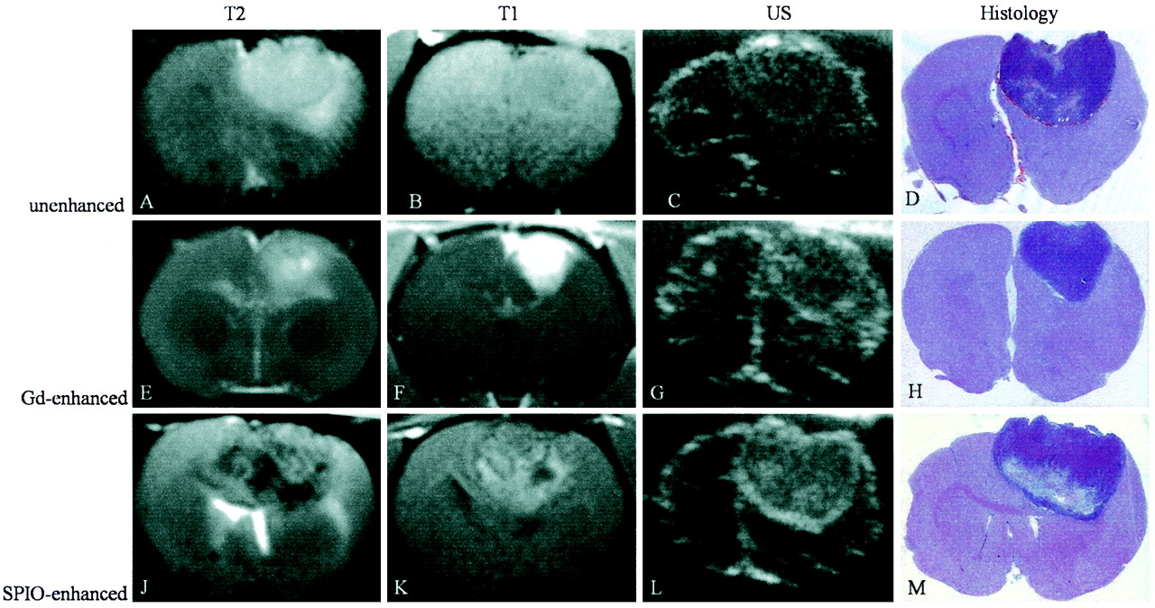
- Fig 1.
A–M, MR images (T2-weighted, 2500/96; T1-weighted, 600/14), sonograms (US), and photomicrographs of paraffin sections (hematoxylin-eosin stain; original magnification, X2) of C6 glioma in rat brain. Each row shows the different modalities in one animal. Images in the first row (A–D) were obtained without contrast media, the second row of images (E–H) was gadolinium-enhanced, and images in the third row (J–M) were SPIO-enhanced.
A–D, On the nonenhanced images, the C6 glioma is predominantly hyperintense on the T2-weighted image (A) and isointense on the T1-weighted image (B). On the sonogram (C), the tumor appears iso- to slightly hyperechoic to brain tissue. With hematoxylin-eosin staining (D), the corresponding area is clearly identifiable.
E–H, After injection of gadopentetate dimeglumine, the tumor is hyperintense on the T1-weighted image (F) and reveals similar characteristics on T2-weighted image (E), sonogram (G), and hematoxylin-eosin staining (H) to those of the tumor on nonenhanced images.
J–M, Twenty-four hours after injection of SPIO particles, the tumor is inhomogeneously hypo- to hyperintense on T2-weighted (J) and T1-weighted (K) images. On the sonogram (L), the tumor appears inhomogeneously hyperechoic to brain tissue with hyperechogic margins.
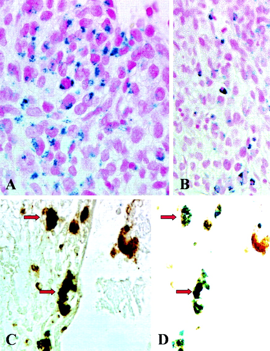
- Fig 2.
A–D, Histologic and immunohistologic localization of iron, tumor cells, and macrophages. Photomicrographs (original magnification, X500 [A, C, and D] and X330 [B]) of paraffin sections stained with Perl’s stain (A and B) for detection of SPIO particles show iron-laden tumor cells in the center of the tumor in A and mostly mononuclear cells in the periphery of the tumor in B. These cells represent iron-labeled macrophages as shown by double labeling with the antibody ED-1 (C). The black-appearing (due to overlap of blue and brown staining product) macrophages in C show discernible blue staining for iron and brown ED-1 immunoreactivity in D when the same section is examined by using a different optic filter of the microscope (identical cells are marked by arrows).


Tables
Imaging characteristics of the 36 experimental gliomas
Contrast Agent Hyperechogenicity on Sonogram* Size of Tumor on Sonogram (cm2)† Hypointensity on T2-Weighted MR Image* − + ++ − + ++ SPIO particles (n = 12) 1 3 8 0.11 ± 0.06 1 6 5 Gadopentetate dimeglumine (n = 12) 9 3 0 0.18 ± 0.1 12 0 0 None (n = 12) 9 2 1 0.15 ± 0.08 12 0 0 Note.—The − indicates absent; +, moderate; ++, extensive
* Data are number of tumors.
† Data are mean ± SD. Tumor size on sonogram was measured as maximal coronal area.
In this issue


Jump to section
Related Articles
Cited By...
- No citing articles found.