
Article Figures & Data
Figures
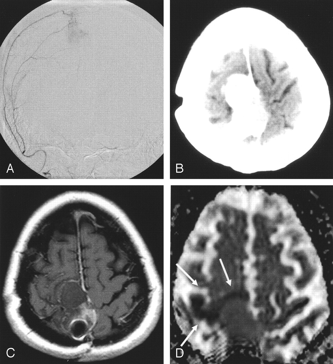
- Fig 1.
Patient 2. Peritumoral ischemia after embolization (Embospheres, 40–120 μm) of a recurrent frontal meningioma.
A, Predominant blood supply by the ipsilateral middle meningeal artery was embolized with spherical particles.
B, After the procedure, the patient had left-sided hemiparesis. CT shows attenuating pooling of contrast medium in the tumor.
C, Next day, T1-weighted spin-echo MR image shows no contrast enhancement, indicating complete devascularization of the tumor.
D, DC map shows a small, hypointense rim of brain parenchyma around the meningioma, indicating cytotoxic edema (arrows). This was interpreted as particles passing into the surrounding brain tissue via leptomeningeal collaterals.
- Fig 2.
Patient 11. Subarachnoid and intratumoral hemorrhage during embolization (Embospheres, 100–300 μm) of a right frontal-convexity meningioma.
A and B, Images show blood supply by the ipsilateral middle meningeal artery (A), which was subsequently devascularized with particles, and leptomeningeal branches of the middle cerebral artery (B).
C and D, At the end of the procedure, patient had sudden-onset headache. Angiograms show subarachnoid extravasation of contrast medium (arrows in C). Control run in the internal carotid artery (D) shows disappearance of the leptomeningeal supply, indicating complete tumor devascularization.
E, Postprocedural CT shows intratumoral and subarachnoid hemorrhage. At surgery, bleeding from intratumoral vessels were slight; the fresh intratumoral clot and tumor were easily removed. The patient recovered completely.
- Fig 3.
Patient 12. An 81-year-old woman with fatal subdural, subarachnoid, and intratumoral hemorrhages after embolization (Bead Block,100–300 μm).
A and B, Embolization of a large, right temporal meningioma with a predominant middle meningeal arterial supply.
C, Ipsilateral middle meningeal artery was superselectively probed and embolized with spherical particles.
D, Procedure was abandoned after the application of one vial because the patient had back pain. Control image reveals marked tumoral devascularization.
E and F, Afterward, the patient had no new neurologic symptoms, but 2 hours later, she was comatose with fixed, dilated pupils. CT shows extensive subdural (solid arrows), subarachnoid (dotted arrow) and intratumoral hemorrhage. Because of her age and clinical state, she did not undergo surgery and died the next day.
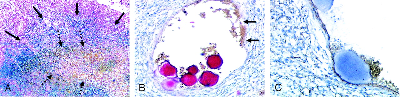
- Fig 4.
Representative histologic findings after periprocedural hemorrhage.
A, Patient 8. Section shows massive iron deposition (blue) indicating previous intratumoral hemorrhage (solid arrows) around acute hemorrhage (brown, dotted arrows) (hematoxylin-eosin and Prussian blue, original magnification ×8).
B and C, Patient 11. Pathologic vessels with variable wall thickness were seen in four of five patients (B, hematoxylin-eosin, original magnification ×20). In some areas, the wall is atypically thin relative to the lumen (arrows in B). These vessels were positive for actin, indicating arteries (C, original magnification ×50). Similar vessels, also filled with particles, were seen in other patients with hemorrhage.


Tables
Patient/Age (years)/Sex Localization Fluoroscopy Time (minutes) Embospheres (μm) Deficit Outcome 1/61/F Sphenoid wing 55 100–300 Hemiparesis, aphasia Resolved after 5 days 2/47/F Frontal convexity 31 40–120 Hemiparesis Persistent 3/49/M Frontal convexity 48 100–300 Hemiparesis, aphasia Residual hemiparesis, aphasia improved 4/71/F Frontal convexity 39 100–300 Hemiparesis Resolved after 48 hours 5/74/F Frontal convexity 31 100–300 Amaurosis Persistent 6/63/M Parietal convexity 69 100–300 Amaurosis Persistent Patient/Age (years)/Sex Localization Fluoroscopy Time (minutes) Particle (μm) Hemorrhage Deficity and Outcome Histologic Subtype Iron Deposition on Histology Pathologic Vessels 7/49/F Fossa posterior 25 Embospheres, 40–120 Intratumoral, subarachnoid Headache, resolved Rhabdoid, WHO III Yes Yes 8/60/F Frontal convexity 22 Embospheres, 100–300 intratumoral Headache, resolved Atypical, WHO II Yes Yes 9/66/F Frontal convexity 36 Embospheres, 100–300 Subdural, intratumoral Hemiparesis, resolved Fibromatous, WHO I No No 10/72/F Temporal convexity 41 Embospheres, 100–300 Subdural, intratumoral Hemiparesis, resolved Fibromatous, WHO I Yes Yes 11/75/F Frontal convexity 25 Embospheres, 100–300 Intratumoral, subarachnoid Headache, resolved Microcystic, WHO I No Yes 12/81/F Frontotemporal convexity 32 BeadBlock, 100–300 Intratumoral, subarachnoid, subdural Coma, death Unknown† Unknowna Unknowna a No autopsy.
In this issue


Jump to section
Related Articles
Cited By...
- Transophthalmic Artery Embolization of Anterior Skull Base Meningiomas: Technical Case Series
- Preoperative tumor embolization prolongs time to recurrence of meningiomas: a retrospective propensity-matched analysis
- Preoperative tumor embolization prolongs time to recurrence of meningiomas: a retrospective propensity-matched analysis
- Predictors of preoperative endovascular embolization of meningiomas: subanalysis of anatomic location and arterial supply
- Augmentation of N-butyl cyanoacrylate embolization of cranial, head, and neck tumors by simultaneous infusion of 5% dextrose solution
- Preoperative Embolization of Intracranial Meningiomas: Efficacy, Technical Considerations, and Complications
- Embolization of Meningiomas: Comparison of Safety between Calibrated Microspheres and Polyvinyl-Alcohol Particles as Embolic Agents
- Head, neck, and brain tumor embolization guidelines
- Preoperative Onyx Embolization of Meningiomas Fed by the Ophthalmic Artery: A Case Series
- Complications of Particle Embolization of Meningiomas: Frequency, Risk Factors, and Outcome