Advertisement

Main menu

  • Home
  • Content
    • Current Issue
    • Accepted Manuscripts
    • Article Preview
    • Past Issue Archive
    • Video Articles
    • AJNR Case Collection
    • Case of the Week Archive
    • Case of the Month Archive
    • Classic Case Archive
  • Special Collections
    • AJNR Awards
    • Low-Field MRI
    • Alzheimer Disease
    • ASNR Foundation Special Collection
    • Photon-Counting CT
    • View All
  • Multimedia
    • AJNR Podcasts
    • AJNR SCANtastic
    • Trainee Corner
    • MRI Safety Corner
    • Imaging Protocols
  • For Authors
    • Submit a Manuscript
    • Submit a Video Article
    • Submit an eLetter to the Editor/Response
    • Manuscript Submission Guidelines
    • Statistical Tips
    • Fast Publishing of Accepted Manuscripts
    • Graphical Abstract Preparation
    • Imaging Protocol Submission
    • Author Policies
  • About Us
    • About AJNR
    • Editorial Board
    • Editorial Board Alumni
  • More
    • Become a Reviewer/Academy of Reviewers
    • Subscribers
    • Permissions
    • Alerts
    • Feedback
    • Advertisers
    • ASNR Home

User menu

  • Alerts
  • Log in

Search

  • Advanced search
American Journal of Neuroradiology

American Journal of Neuroradiology

ASHNR American Society of Functional Neuroradiology ASHNR American Society of Pediatric Neuroradiology ASSR
  • Alerts
  • Log in

Advanced Search

  • Home
  • Content
    • Current Issue
    • Accepted Manuscripts
    • Article Preview
    • Past Issue Archive
    • Video Articles
    • AJNR Case Collection
    • Case of the Week Archive
    • Case of the Month Archive
    • Classic Case Archive
  • Special Collections
    • AJNR Awards
    • Low-Field MRI
    • Alzheimer Disease
    • ASNR Foundation Special Collection
    • Photon-Counting CT
    • View All
  • Multimedia
    • AJNR Podcasts
    • AJNR SCANtastic
    • Trainee Corner
    • MRI Safety Corner
    • Imaging Protocols
  • For Authors
    • Submit a Manuscript
    • Submit a Video Article
    • Submit an eLetter to the Editor/Response
    • Manuscript Submission Guidelines
    • Statistical Tips
    • Fast Publishing of Accepted Manuscripts
    • Graphical Abstract Preparation
    • Imaging Protocol Submission
    • Author Policies
  • About Us
    • About AJNR
    • Editorial Board
    • Editorial Board Alumni
  • More
    • Become a Reviewer/Academy of Reviewers
    • Subscribers
    • Permissions
    • Alerts
    • Feedback
    • Advertisers
    • ASNR Home
  • Follow AJNR on Twitter
  • Visit AJNR on Facebook
  • Follow AJNR on Instagram
  • Join AJNR on LinkedIn
  • RSS Feeds

AJNR Awards, New Junior Editors, and more. Read the latest AJNR updates

OtherBrain

MR Ventriculocisternography By Using 3D Balanced Steady-State Free Precession Imaging: Technical Note

Ming-Long Wu, Cheng-Wen Ko, Ting-Yi Chen, Ming-Ting Wu, Hsiao-Wen Chung, Teng-Yi Huang and Yi-Ru Lin
American Journal of Neuroradiology May 2005, 26 (5) 1170-1173;
Ming-Long Wu
  • Find this author on Google Scholar
  • Find this author on PubMed
  • Search for this author on this site
Cheng-Wen Ko
  • Find this author on Google Scholar
  • Find this author on PubMed
  • Search for this author on this site
Ting-Yi Chen
  • Find this author on Google Scholar
  • Find this author on PubMed
  • Search for this author on this site
Ming-Ting Wu
  • Find this author on Google Scholar
  • Find this author on PubMed
  • Search for this author on this site
Hsiao-Wen Chung
  • Find this author on Google Scholar
  • Find this author on PubMed
  • Search for this author on this site
Teng-Yi Huang
  • Find this author on Google Scholar
  • Find this author on PubMed
  • Search for this author on this site
Yi-Ru Lin
  • Find this author on Google Scholar
  • Find this author on PubMed
  • Search for this author on this site
  • Article
  • Figures & Data
  • Info & Metrics
  • Responses
  • References
  • PDF
Loading

Article Figures & Data

Figures

  • Fig 1.
    • Download figure
    • Open in new tab
    • Download powerpoint
    Fig 1.

    Steady-state signal intensities of CSF, gray matter (GM), and white matter (WM) as a function of flip angle. A flip angle of 70° is often chosen in MR ventriculocisternography to maximize CSF-parenchyma contrast.
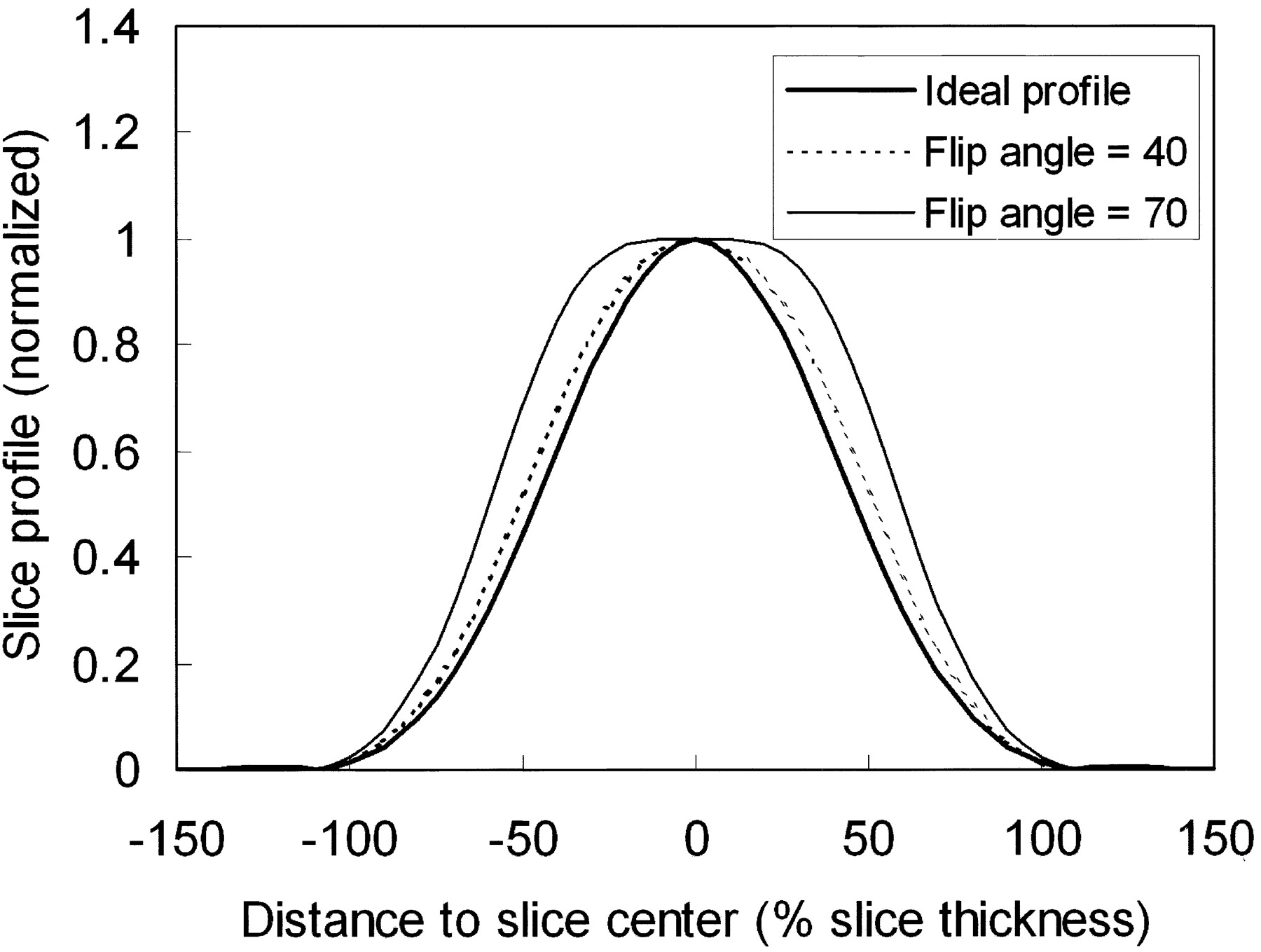
  • Fig 2.
    • Download figure
    • Open in new tab
    • Download powerpoint
    Fig 2.

    Steady-state signals across the imaging slab of a smoothed synch radio-frequency pulse lasting for 0.9 ms in time duration, plotted for two designated flip angles of 40° (dotted line) and 70° (thin solid line) along with the ideal flip-angle profile (thick solid line), all normalized to the value at the slab center. Note that, at larger designated flip angles, the actual slab profiles for the SSFP signals tend to become wider.

  • Fig 3.
    • Download figure
    • Open in new tab
    • Download powerpoint
    Fig 3.

    Percentage section aliasing, calculated as widening of the full-width-at-half-maximum of the steady-state signal intensity profile relative to the desired slab thickness, plotted as a function of the designated flip angle (dotted line). Experimental measurements (circles) from our subjects agreed fairly well with the theoretical deductions. Note that, at a 70° flip angle, the percentage section aliasing is nearly 30%, meaning that about one-third of the sections would be subject to interference from CSF outside the image slab. By lowering the flip angle to 30° or 40°, the percentage section aliasing decreases to <20%.
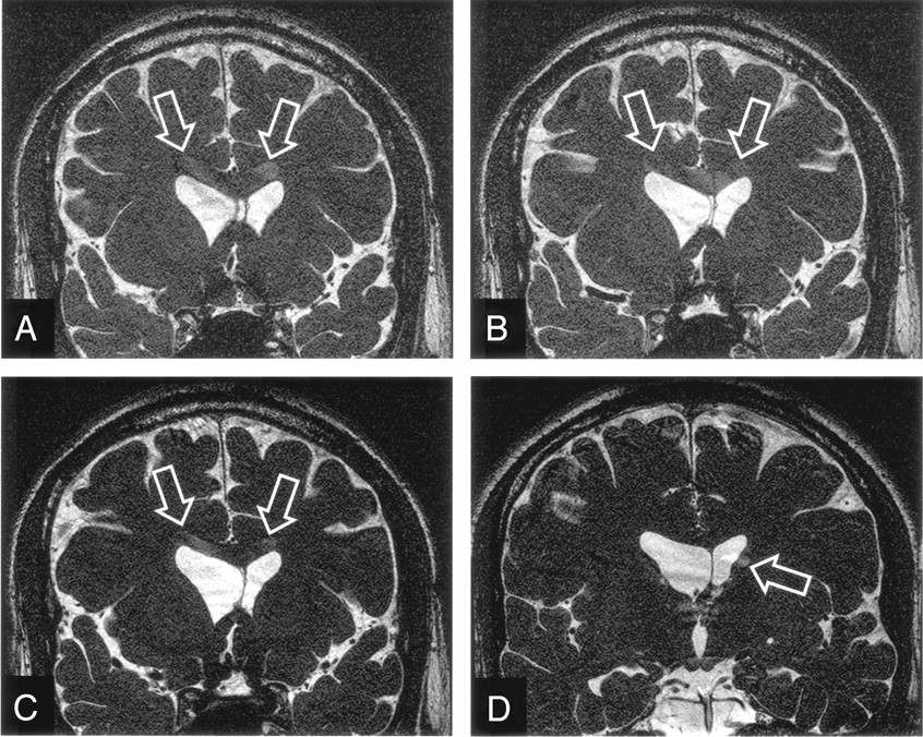
  • Fig 4.
    • Download figure
    • Open in new tab
    • Download powerpoint
    Fig 4.

    Increased section aliasing artifacts in 3D SSFP imaging on a 33-year male subject. Several inner sections, the 13th (A), 17th (B), and 21st (C) anterior sections in a 84-section volume acquired with a designated flip angle of 70°, are shown to exhibit prominent aliased CSF signals (A–C, arrows) from the lateral ventricles actually located at a posterior region (D). The posterior section also shows evidence of section aliasing from anterior region (D, arrow).

  • Fig 5.
    • Download figure
    • Open in new tab
    • Download powerpoint
    Fig 5.

    Contrast between CSF and white matter calculated as a function of the designated flip angle (dotted line), superimposed by experimental values (circles) measured from the images obtained from the healthy subjects recruited in our study. The measurements showed that the CSF-parenchyma contrast reduces as flip angle decreases, in good agreement with theoretical derivations.

  • Fig 6.
    • Download figure
    • Open in new tab
    • Download powerpoint
    Fig 6.

    Images acquired from a 23-year-old man healthy subject showing comparison of section aliasing artifacts in 3D SSFP imaging, as a function of the designated flip angle. The 8th anterior section in a 36-section volume acquired with a designated flip angle of 70° (A), exhibits prominent aliased signals from CSF (arrows). By lowering the flip angle to 50° (B) and 30° (C), the section aliasing decreases (arrows) and becomes invisible, respectively. Also note the continuous reduction in the contrast between CSF and brain parenchyma from A to C.

PreviousNext
Back to top

In this issue

American Journal of Neuroradiology: 26 (5)
American Journal of Neuroradiology
Vol. 26, Issue 5
1 May 2005
  • Table of Contents
  • Index by author
Advertisement
Print
Download PDF
Email Article

Thank you for your interest in spreading the word on American Journal of Neuroradiology.

NOTE: We only request your email address so that the person you are recommending the page to knows that you wanted them to see it, and that it is not junk mail. We do not capture any email address.

Enter multiple addresses on separate lines or separate them with commas.
MR Ventriculocisternography By Using 3D Balanced Steady-State Free Precession Imaging: Technical Note
(Your Name) has sent you a message from American Journal of Neuroradiology
(Your Name) thought you would like to see the American Journal of Neuroradiology web site.
CAPTCHA
This question is for testing whether or not you are a human visitor and to prevent automated spam submissions.
Cite this article
Ming-Long Wu, Cheng-Wen Ko, Ting-Yi Chen, Ming-Ting Wu, Hsiao-Wen Chung, Teng-Yi Huang, Yi-Ru Lin
MR Ventriculocisternography By Using 3D Balanced Steady-State Free Precession Imaging: Technical Note
American Journal of Neuroradiology May 2005, 26 (5) 1170-1173;

Citation Manager Formats

  • BibTeX
  • Bookends
  • EasyBib
  • EndNote (tagged)
  • EndNote 8 (xml)
  • Medlars
  • Mendeley
  • Papers
  • RefWorks Tagged
  • Ref Manager
  • RIS
  • Zotero
0 Responses
Respond to this article
Share
Bookmark this article
MR Ventriculocisternography By Using 3D Balanced Steady-State Free Precession Imaging: Technical Note
Ming-Long Wu, Cheng-Wen Ko, Ting-Yi Chen, Ming-Ting Wu, Hsiao-Wen Chung, Teng-Yi Huang, Yi-Ru Lin
American Journal of Neuroradiology May 2005, 26 (5) 1170-1173;
del.icio.us logo Twitter logo Facebook logo Mendeley logo
  • Tweet Widget
  • Facebook Like
  • Google Plus One

Jump to section

  • Article
    • Abstract
    • Description of Technique
    • Results
    • Discussion
    • Conclusion
    • Footnotes
    • References
  • Figures & Data
  • Info & Metrics
  • Responses
  • References
  • PDF

Related Articles

  • No related articles found.
  • PubMed
  • Google Scholar

Cited By...

  • Demonstration of Normal and Abnormal Fetal Brains Using 3D Printing from In Utero MR Imaging Data
  • 3D Double-Echo Steady-State with Water Excitation MR Imaging of the Intraparotid Facial Nerve at 1.5T: A Pilot Study
  • Is All "Communicating" Hydrocephalus Really Communicating? Prospective Study on the Value of 3D-Constructive Interference in Steady State Sequence at 3T
  • Crossref
  • Google Scholar

This article has not yet been cited by articles in journals that are participating in Crossref Cited-by Linking.

More in this TOC Section

  • Progression of Microstructural Damage in Spinocerebellar Ataxia Type 2: A Longitudinal DTI Study
  • SWI or T2*: Which MRI Sequence to Use in the Detection of Cerebral Microbleeds? The Karolinska Imaging Dementia Study
  • Statin Therapy Does Not Affect the Radiographic and Clinical Profile of Patients with TIA and Minor Stroke
Show more Brain

Similar Articles

Advertisement

Indexed Content

  • Current Issue
  • Accepted Manuscripts
  • Article Preview
  • Past Issues
  • Editorials
  • Editor's Choice
  • Fellows' Journal Club
  • Letters to the Editor
  • Video Articles

Cases

  • Case Collection
  • Archive - Case of the Week
  • Archive - Case of the Month
  • Archive - Classic Case

More from AJNR

  • Trainee Corner
  • Imaging Protocols
  • MRI Safety Corner
  • Book Reviews

Multimedia

  • AJNR Podcasts
  • AJNR Scantastics

Resources

  • Turnaround Time
  • Submit a Manuscript
  • Submit a Video Article
  • Submit an eLetter to the Editor/Response
  • Manuscript Submission Guidelines
  • Statistical Tips
  • Fast Publishing of Accepted Manuscripts
  • Graphical Abstract Preparation
  • Imaging Protocol Submission
  • Evidence-Based Medicine Level Guide
  • Publishing Checklists
  • Author Policies
  • Become a Reviewer/Academy of Reviewers
  • News and Updates

About Us

  • About AJNR
  • Editorial Board
  • Editorial Board Alumni
  • Alerts
  • Permissions
  • Not an AJNR Subscriber? Join Now
  • Advertise with Us
  • Librarian Resources
  • Feedback
  • Terms and Conditions
  • AJNR Editorial Board Alumni

American Society of Neuroradiology

  • Not an ASNR Member? Join Now

© 2025 by the American Society of Neuroradiology All rights, including for text and data mining, AI training, and similar technologies, are reserved.
Print ISSN: 0195-6108 Online ISSN: 1936-959X

Powered by HighWire