
Article Figures & Data
Figures
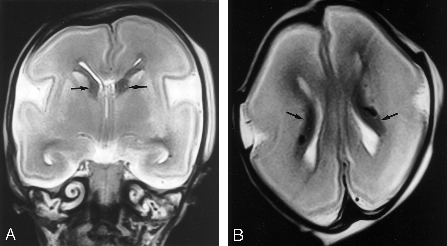
- Fig 1.
A, Coronal and B, axial brain MR images in a spontaneously aborted 22-week fetus. The image quality was assessed as excellent and findings reported as normal, which agreed with the autopsy report. The dark structures (arrows) in the immediate periventricular regions are the germinal matrices. Migrating neurons are shown in cerebral hemispheres as gray matter signal intensity against the high signal intensity of the white matter.
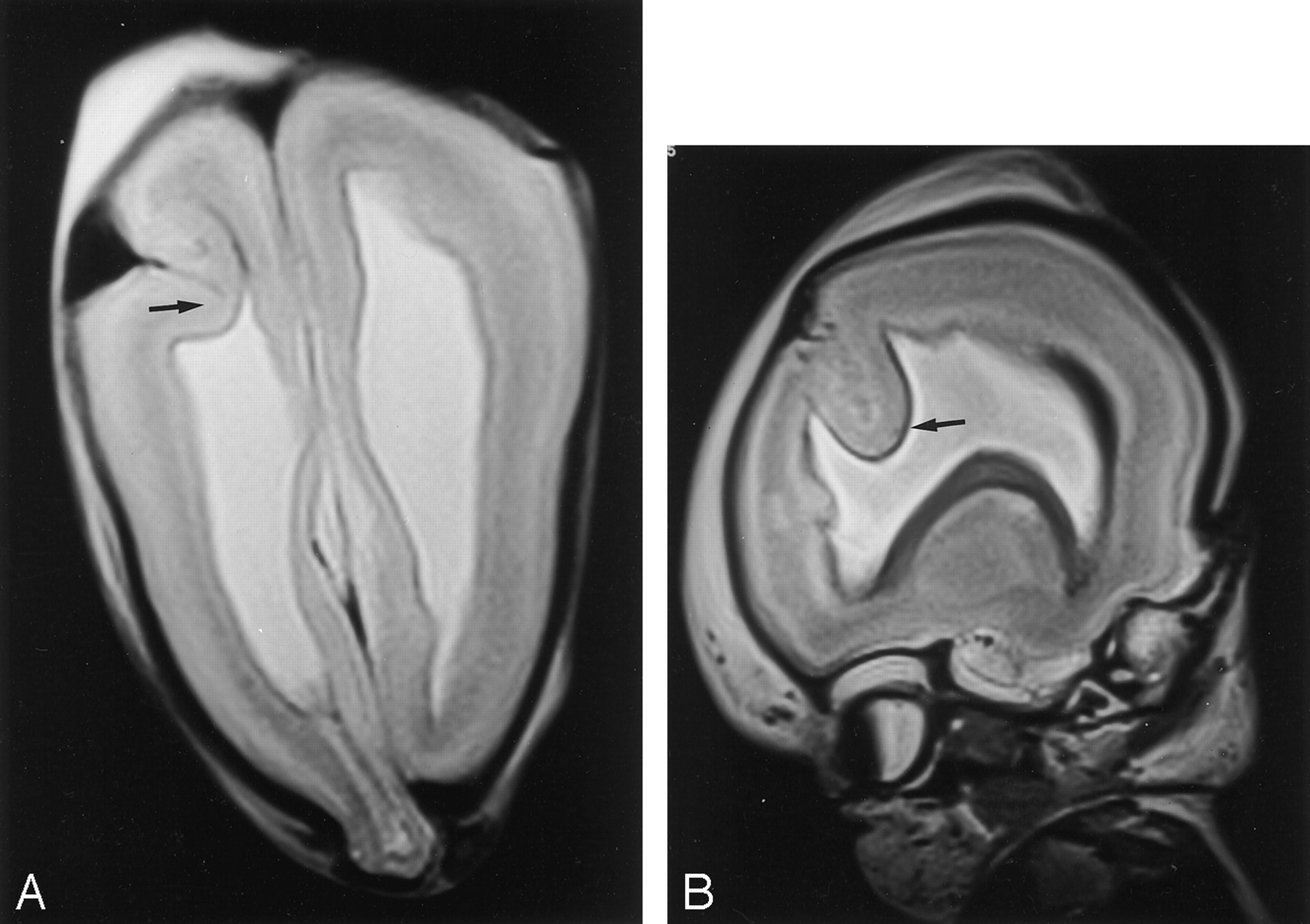
- Fig 2.
MR images of a fetus after therapeutic abortion at 18 weeks because of a sonographic diagnosis of alobar holoprosencephaly. Autopsy could not provide any information because of the poor state of the unfixed brain.
A, Axial and B, coronal brain MR images confirm the typical features of alobar holoprosencephaly: nonseparated cerebral hemispheres, holoventricle, and fused thalami. Note the single, poorly formed orbit (cyclopia) (arrow) and an azygous anterior cerebral artery (arrowheads).
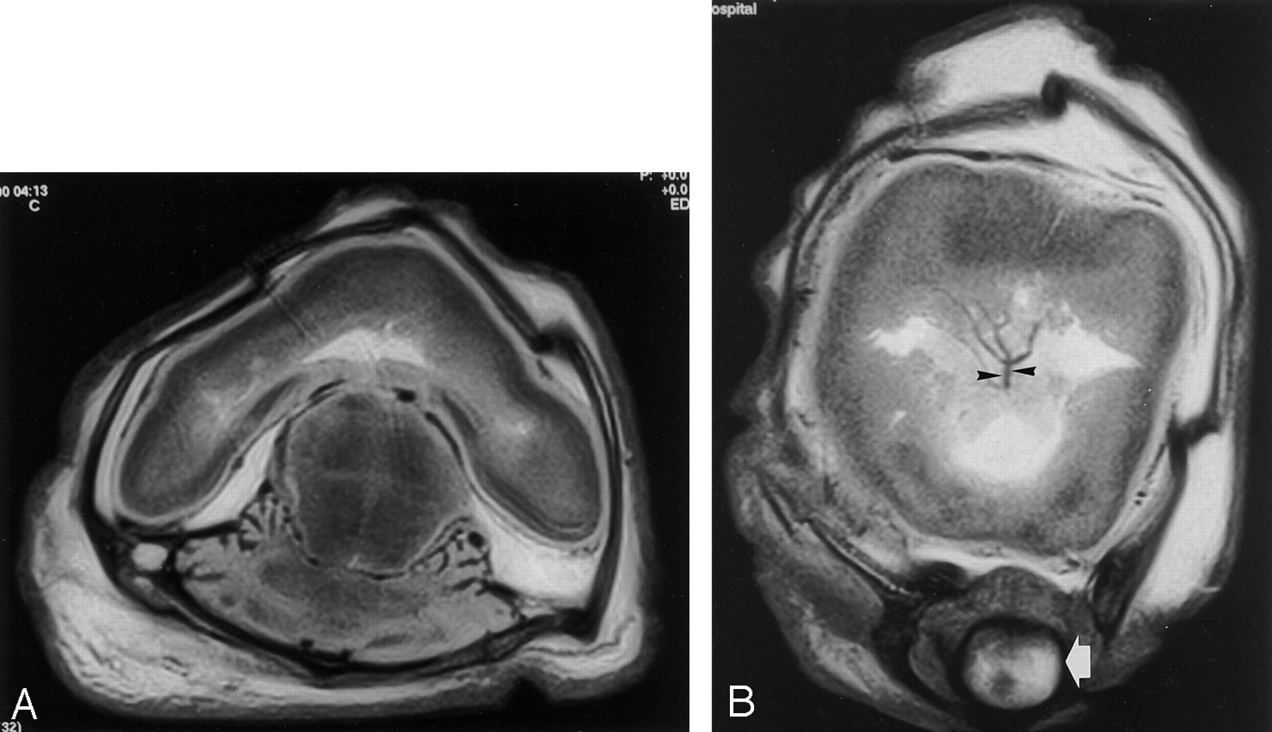
- Fig 3.
A, Sagittal spine and B, axial brain MR images in a 20-week fetus after a therapeutic abortion because of a sonographic diagnosis of myelomeningocele and Chiari II malformation. These findings were confirmed with postmortem MR imaging and autopsy. The spine MR image (A) shows a low thoracic myelomeningocele with an adjacent complicated fusion defect of the lower thoracic-upper lumbar vertebral bodies. There is also extensive syringohydromyelia. The cerebellar tonsils (arrow) are abnormally low (at C3), indicating a Chiari II abnormality, and images of the brain (not shown) confirmed the presence of a small posterior fossa. This brain MR image (B) shows ventriculomegaly and the “lemon-shaped” deformity recognized on sonograms in cases such as these.
- Fig 4.
A, Axial and B, right parasagittal MR images in a 21-week fetus after therapeutic abortion performed on the basis of a parietal meningocele on sonograms. This was reported as such at autopsy. MR images show that there is also brain and ventricle in the abnormality (therefore, technically a meningoencephalocystocele). In addition, the MR images show an area of cortical dysplasia (arrow) in the right frontal lobe that was not reported at sonography or autopsy. This was confirmed at consensus review. Note the generalized reduction in volume of migrating neurons in the right cerebral hemisphere.


Tables
- TABLE 1:
Results of Eight MR Examinations in Which No Structural Information Was Obtained from Autopsy Because of the Physical State of the Brain
Abnormality No. of Cases Normal 2 Agenesis of corpus callosum 2 Agenesis of corpus callosum and rhombencephalosynapsis 1 Holoprosencephaly (see Fig 2) 1 Germinal matrix hemorrhage 1 Hypoxic or ischemic damage 1 - TABLE 2:
Results of 17 Cases in Which MR Imaging and Autopsy Agreed on the Nature of Abnormal Findings on First Analysis
Abnormality No. of Cases Isolated ventriculomegaly 4 Myelomeningocele and Chiari II malformation (see Fig 3) 4 Dandy-Walker malformation 3 Holoprosencephaly 2 Germinal matrix hemorrhage 1 Hypoxic or ischemic damage 1 Ruptured arteriovenous malformation 1 Diastematomyelia 1 - TABLE 3:
Consensus Reports of Four Cases in Which MR and Autopsy Findings Disagreed on the Initial Assessment
MR Findings Autopsy Findings Consensus Report Parietal encephalocele, frontal cortical dysplasia (see Fig 4) Parietal meningocele Parietal encephalocele, frontal cortical dysplasia Ventriculomegaly due to aqueduct stenosis Dandy-Walker malformation Ventriculomegaly due to aqueduct stenosis Dandy-Walker malformation Normal Dandy-Walker malformation Colpocephaly Normal Normal
In this issue


Jump to section
Related Articles
Cited By...
- Are non-invasive or minimally invasive autopsy techniques for detecting cause of death in prenates, neonates and infants accurate? A systematic review of diagnostic test accuracy
- Long-term developmental outcome of children with a fetal diagnosis of isolated inferior vermian hypoplasia
- Antemortem cranial MRI compared with postmortem histopathologic examination of the brain in term infants with neonatal encephalopathy following perinatal asphyxia
- Less invasive autopsy: an evidenced based approach
- Corroboration of Normal and Abnormal Fetal Cerebral Lamination on Postmortem MR Imaging with Postmortem Examination
- A Prospective Study of Fetuses with Isolated Ventriculomegaly Investigated by Antenatal Sonography and In Utero MR Imaging
- A case of a Dandy-Walker variant: the importance of a multidisciplinary team approach using complementary techniques to obtain accurate diagnostic information
- Postmortem magnetic resonance imaging as an adjunct to perinatal autopsy for renal-tract abnormalities
- In utero magnetic resonance imaging for brain and spinal abnormalities in fetuses