Advertisement

Main menu

  • Home
  • Content
    • Current Issue
    • Accepted Manuscripts
    • Article Preview
    • Past Issue Archive
    • Video Articles
    • AJNR Case Collection
    • Case of the Week Archive
    • Case of the Month Archive
    • Classic Case Archive
  • Special Collections
    • AJNR Awards
    • Low-Field MRI
    • Alzheimer Disease
    • ASNR Foundation Special Collection
    • Photon-Counting CT
    • View All
  • Multimedia
    • AJNR Podcasts
    • AJNR SCANtastic
    • Trainee Corner
    • MRI Safety Corner
    • Imaging Protocols
  • For Authors
    • Submit a Manuscript
    • Submit a Video Article
    • Submit an eLetter to the Editor/Response
    • Manuscript Submission Guidelines
    • Statistical Tips
    • Fast Publishing of Accepted Manuscripts
    • Graphical Abstract Preparation
    • Imaging Protocol Submission
    • Author Policies
  • About Us
    • About AJNR
    • Editorial Board
    • Editorial Board Alumni
  • More
    • Become a Reviewer/Academy of Reviewers
    • Subscribers
    • Permissions
    • Alerts
    • Feedback
    • Advertisers
    • ASNR Home

User menu

  • Alerts
  • Log in

Search

  • Advanced search
American Journal of Neuroradiology

American Journal of Neuroradiology

ASHNR American Society of Functional Neuroradiology ASHNR American Society of Pediatric Neuroradiology ASSR
  • Alerts
  • Log in

Advanced Search

  • Home
  • Content
    • Current Issue
    • Accepted Manuscripts
    • Article Preview
    • Past Issue Archive
    • Video Articles
    • AJNR Case Collection
    • Case of the Week Archive
    • Case of the Month Archive
    • Classic Case Archive
  • Special Collections
    • AJNR Awards
    • Low-Field MRI
    • Alzheimer Disease
    • ASNR Foundation Special Collection
    • Photon-Counting CT
    • View All
  • Multimedia
    • AJNR Podcasts
    • AJNR SCANtastic
    • Trainee Corner
    • MRI Safety Corner
    • Imaging Protocols
  • For Authors
    • Submit a Manuscript
    • Submit a Video Article
    • Submit an eLetter to the Editor/Response
    • Manuscript Submission Guidelines
    • Statistical Tips
    • Fast Publishing of Accepted Manuscripts
    • Graphical Abstract Preparation
    • Imaging Protocol Submission
    • Author Policies
  • About Us
    • About AJNR
    • Editorial Board
    • Editorial Board Alumni
  • More
    • Become a Reviewer/Academy of Reviewers
    • Subscribers
    • Permissions
    • Alerts
    • Feedback
    • Advertisers
    • ASNR Home
  • Follow AJNR on Twitter
  • Visit AJNR on Facebook
  • Follow AJNR on Instagram
  • Join AJNR on LinkedIn
  • RSS Feeds

AJNR Awards, New Junior Editors, and more. Read the latest AJNR updates

OtherNeurointervention

Intraarterial Suction Thrombectomy in Acute Stroke

Helmi L. Lutsep, Wayne M. Clark, Gary M. Nesbit, Todd A. Kuether and Stan L. Barnwell
American Journal of Neuroradiology May 2002, 23 (5) 783-786;
Helmi L. Lutsep
  • Find this author on Google Scholar
  • Find this author on PubMed
  • Search for this author on this site
Wayne M. Clark
  • Find this author on Google Scholar
  • Find this author on PubMed
  • Search for this author on this site
Gary M. Nesbit
  • Find this author on Google Scholar
  • Find this author on PubMed
  • Search for this author on this site
Todd A. Kuether
  • Find this author on Google Scholar
  • Find this author on PubMed
  • Search for this author on this site
Stan L. Barnwell
  • Find this author on Google Scholar
  • Find this author on PubMed
  • Search for this author on this site
  • Article
  • Figures & Data
  • Info & Metrics
  • Responses
  • References
  • PDF
Loading

Article Figures & Data

Figures

  • Tables
  • Fig 1.
    • Download figure
    • Open in new tab
    • Download powerpoint
    Fig 1.

    Case 1. A 67-year-old man presented with resolving left hemiparesis.

    A, Baseline right lateral common carotid artery (CCA) angiogram shows near-occlusion of the right ICA with a string sign of slow flow into the proximal ICA.

    B, After predilation of the proximal ICA stenosis, this baseline right ICA angiogram reveals the presence of extensive thrombus.

    C, Baseline left CCA angiogram reveals some right ACA cross-filling but no right MCA filling.

    D, Postprocedural right lateral CCA angiogram shows less than 20% residual ICA stenosis. A Wallstent in the ICA is visible at the carotid bifurcation.

    E, Although the right MCA has a persistent mid-M1 occlusion, the postprocedural right CCA angiogram shows good distal filling of the MCA branches via pial collaterals from the ACA.

  • Fig 2.
    • Download figure
    • Open in new tab
    • Download powerpoint
    Fig 2.

    Case 2. A 72-year-old man had global aphasia and right hemiparesis.

    A, Baseline left lateral CCA angiogram shows complete occlusion of the cervical ICA. Flow through a 50% stenosis of the external carotid artery remains visible.

    B, Postprocedural left lateral CCA angiogram demonstrates essentially complete resolution of the ICA occlusion. The Wallstent placed in the left ICA is visible at the carotid bifurcation.
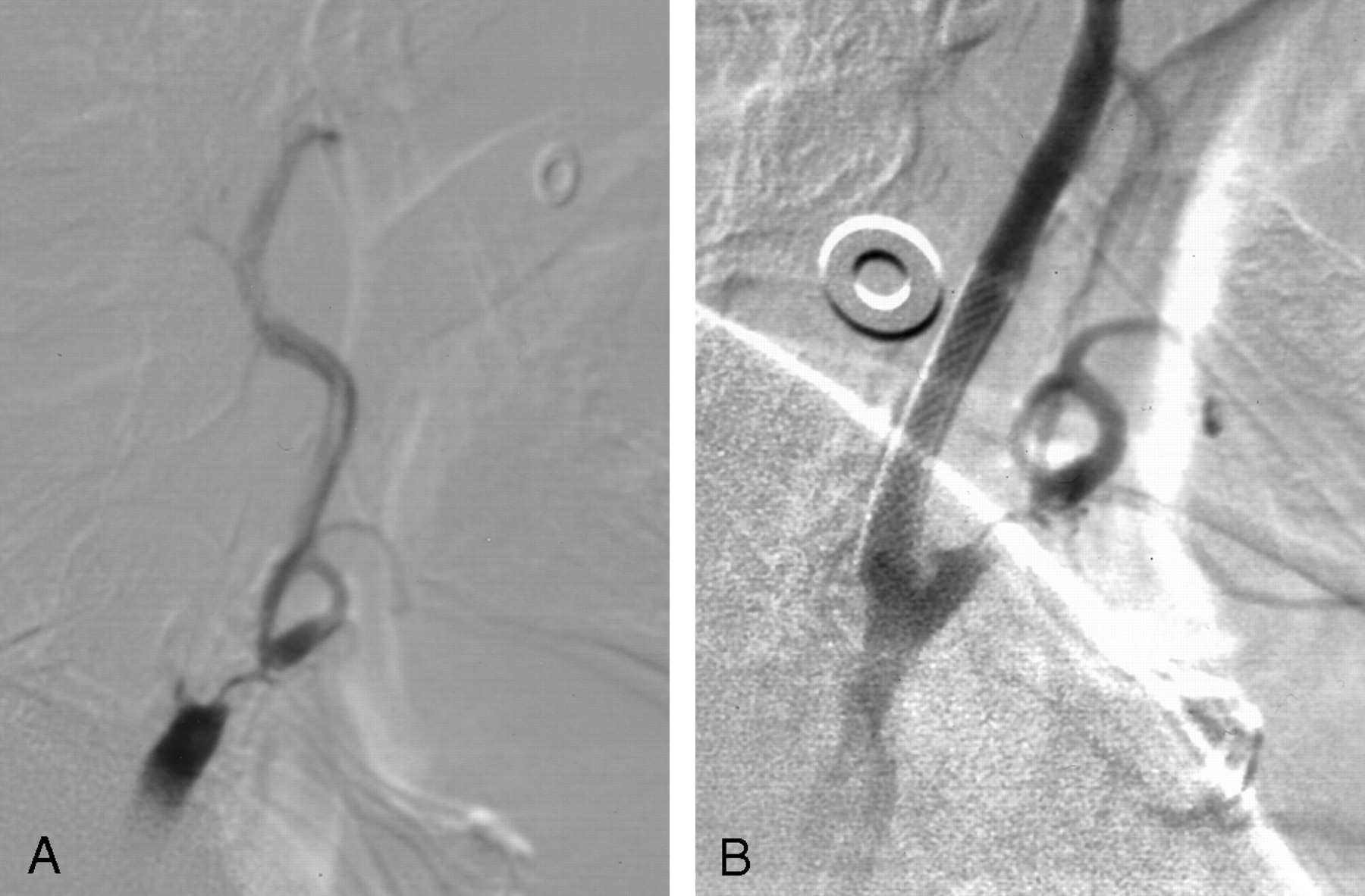
  • Fig 3.
    • Download figure
    • Open in new tab
    • Download powerpoint
    Fig 3.

    Case 3. A 49-year-old man with atrial fibrillation and a poor cardiac ejection fraction presented with left hemiparesis and right gaze preference

    A, Baseline right lateral CCA angiogram shows complete occlusion of the right ICA.

    B, Postprocedural right lateral CCA angiogram shows restored flow within the right ICA. A long segment of vessel narrowing is seen within the cervical ICA.

Tables

  • Figures
  • Clinical results with suction thrombectomy

    Case No./ Patient Age (y)Hours to TreatmentVesselNIHSS ScoreModified Rankin Score at 3 Months
    PreoperativeAt 3 Months
    1/573*Right ICA1221
    2/725Left ICA23223
    3/494.5Right ICA2243†
    • * Since worsening.

    • † Patient had a concurrent foot infection that limited his walking.

PreviousNext
Back to top

In this issue

American Journal of Neuroradiology: 23 (5)
American Journal of Neuroradiology
Vol. 23, Issue 5
1 May 2002
  • Table of Contents
  • Index by author
Advertisement
Print
Download PDF
Email Article

Thank you for your interest in spreading the word on American Journal of Neuroradiology.

NOTE: We only request your email address so that the person you are recommending the page to knows that you wanted them to see it, and that it is not junk mail. We do not capture any email address.

Enter multiple addresses on separate lines or separate them with commas.
Intraarterial Suction Thrombectomy in Acute Stroke
(Your Name) has sent you a message from American Journal of Neuroradiology
(Your Name) thought you would like to see the American Journal of Neuroradiology web site.
CAPTCHA
This question is for testing whether or not you are a human visitor and to prevent automated spam submissions.
Cite this article
Helmi L. Lutsep, Wayne M. Clark, Gary M. Nesbit, Todd A. Kuether, Stan L. Barnwell
Intraarterial Suction Thrombectomy in Acute Stroke
American Journal of Neuroradiology May 2002, 23 (5) 783-786;

Citation Manager Formats

  • BibTeX
  • Bookends
  • EasyBib
  • EndNote (tagged)
  • EndNote 8 (xml)
  • Medlars
  • Mendeley
  • Papers
  • RefWorks Tagged
  • Ref Manager
  • RIS
  • Zotero
0 Responses
Respond to this article
Share
Bookmark this article
Intraarterial Suction Thrombectomy in Acute Stroke
Helmi L. Lutsep, Wayne M. Clark, Gary M. Nesbit, Todd A. Kuether, Stan L. Barnwell
American Journal of Neuroradiology May 2002, 23 (5) 783-786;
del.icio.us logo Twitter logo Facebook logo Mendeley logo
  • Tweet Widget
  • Facebook Like
  • Google Plus One

Jump to section

  • Article
    • Abstract
    • Case Reports
    • Discussion
    • Footnotes
    • References
  • Figures & Data
  • Info & Metrics
  • Responses
  • References
  • PDF

Related Articles

  • No related articles found.
  • PubMed
  • Google Scholar

Cited By...

  • In vitro experiments of cerebral blood flow during aspiration thrombectomy: potential effects on cerebral perfusion pressure and collateral flow
  • Mechanical Thrombolysis and Stenting in Acute Ischemic Stroke
  • Treatment of acute middle cerebral artery occlusion with a Solitaire AB stent: preliminary experience
  • Endovascular treatment of basilar artery occlusion by manual aspiration thrombectomy
  • In Vivo Evaluation of the Phenox CRC Mechanical Thrombectomy Device in a Swine Model of Acute Vessel Occlusion
  • Endovascular Approaches to Acute Stroke, Part 1: Drugs, Devices, and Data
  • Mechanical Thromboembolectomy for Acute Ischemic Stroke: Comparison of the Catch Thromboectomy Device and the Merci Retriever In Vivo
  • Debunking 7 Myths That Hamper the Realization of Randomized Controlled Trials on Intra-Arterial Thrombolysis for Acute Ischemic Stroke
  • Mechanical Thrombectomy for Acute Ischemic Stroke: Thrombus-Device Interaction, Efficiency, and Complications In Vivo
  • Analysis of Thrombi Retrieved From Cerebral Arteries of Patients With Acute Ischemic Stroke
  • Mechanical Thrombolysis in Ischemic Stroke Attributable to Basilar Artery Occlusion as First-Line Treatment
  • Guidelines for the Early Management of Patients With Ischemic Stroke: 2005 Guidelines Update A Scientific Statement From the Stroke Council of the American Heart Association/American Stroke Association
  • Mechanical Thrombolysis in Acute Ischemic Stroke With Endovascular Photoacoustic Recanalization
  • Crossref
  • Google Scholar

This article has not yet been cited by articles in journals that are participating in Crossref Cited-by Linking.

More in this TOC Section

  • Rescue Reentry in Carotid Near-Occlusion
  • Contour Neurovascular System: Five Year Follow Up
  • Effect of SARS-CoV2 on Endovascular Thrombectomy
Show more Neurointervention

Similar Articles

Advertisement

Indexed Content

  • Current Issue
  • Accepted Manuscripts
  • Article Preview
  • Past Issues
  • Editorials
  • Editor's Choice
  • Fellows' Journal Club
  • Letters to the Editor
  • Video Articles

Cases

  • Case Collection
  • Archive - Case of the Week
  • Archive - Case of the Month
  • Archive - Classic Case

More from AJNR

  • Trainee Corner
  • Imaging Protocols
  • MRI Safety Corner
  • Book Reviews

Multimedia

  • AJNR Podcasts
  • AJNR Scantastics

Resources

  • Turnaround Time
  • Submit a Manuscript
  • Submit a Video Article
  • Submit an eLetter to the Editor/Response
  • Manuscript Submission Guidelines
  • Statistical Tips
  • Fast Publishing of Accepted Manuscripts
  • Graphical Abstract Preparation
  • Imaging Protocol Submission
  • Evidence-Based Medicine Level Guide
  • Publishing Checklists
  • Author Policies
  • Become a Reviewer/Academy of Reviewers
  • News and Updates

About Us

  • About AJNR
  • Editorial Board
  • Editorial Board Alumni
  • Alerts
  • Permissions
  • Not an AJNR Subscriber? Join Now
  • Advertise with Us
  • Librarian Resources
  • Feedback
  • Terms and Conditions
  • AJNR Editorial Board Alumni

American Society of Neuroradiology

  • Not an ASNR Member? Join Now

© 2025 by the American Society of Neuroradiology All rights, including for text and data mining, AI training, and similar technologies, are reserved.
Print ISSN: 0195-6108 Online ISSN: 1936-959X

Powered by HighWire