
Article Figures & Data
Figures
- fig 1.
Case 2: 50-year-old patient with severe SIHH.
A–F, MR images obtained in the acute phase (A and B), after 6 weeks (C and D), and after 6 months (E and F).
A and B, Sagittal T2-weighted FSE (A) and T1-weighted SE (B) images reveal a retrospinal fluid collection, isointense with CSF, at the C1–C2 level (large arrowheads). The collection extends far into the suboccipital region (small arrowheads, A). A substantial hygroma is located subtentorially (arrows, A).
C and D, Sagittal (C) and axial (D) 3D-CISS images reveal a similar retrospinal fluid collection at the C1–C2 level (arrowheads) and a ventral and dorsal spinal hygroma (arrows, C). The retrospinal fluid collection also extends bilaterally (arrows, D).
E and F, Sagittal (E) and axial (F) 3D-CISS images show the retrospinal fluid collection (arrowhead, E). An additional extradural fluid collection can be seen bilaterally (arrows, F). The superior segment of the ventral spinal hygroma decreased in size (closed arrows, E), and the subtentorial hygroma resolved completely (open arrow, E).
- fig 2.
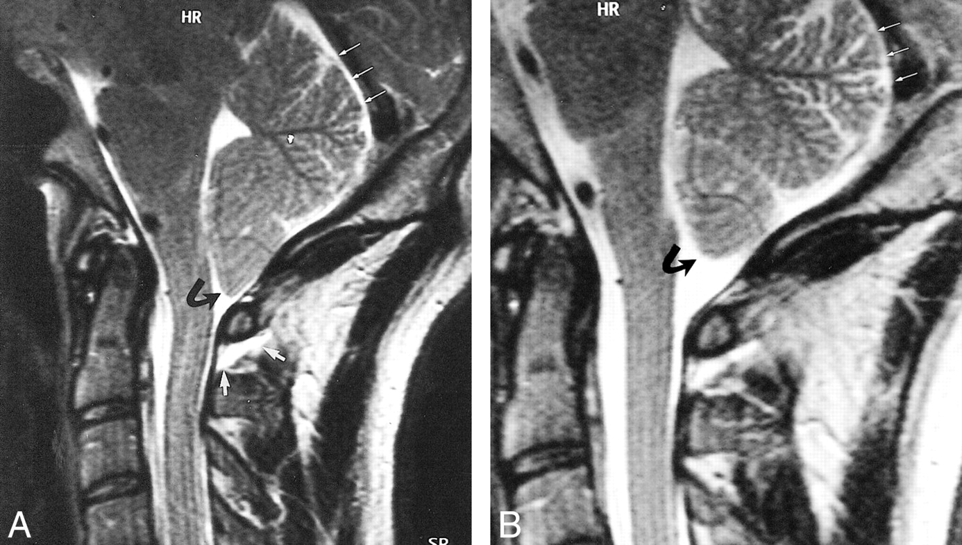
Case 3: 38-year-old patient with SIHH.
A, Sagittal T2-weighted FSE MR image obtained in the acute phase shows a retrospinal fluid collection that can be identified at the C1–C2 level (short white arrows), a descent of the tonsils (curved black arrow), and subtentorial hygroma (long white arrows).
B, Sagittal T2-weighted FSE image obtained after 4 weeks reveals complete regression of the tonsillar descent (curved black arrow) and of the subtentorial hygroma (straight white arrows).
- fig 3.
Case 9: 38-year old patient with SIHH. MR imaging was performed in the acute phase.
A–G, T2-weighted FSE images in the sagittal (A) and axial (D) planes reveal a retrospinal fluid collection (short thick arrow, A; arrowhead, D) and a subtentorial hygroma (thin double arrows, A). The anterior internal vertebral plexus is dilated, and large flow voids can be identified (long thick arrow, A; arrows, D). The flow voids of the dilated anterior internal vertebral plexus can be confirmed on corresponding axial slices obtained with a flow-sensitive 2D FLASH sequence (arrows, E). The dilated anterior internal vertebral plexus can also be identified on T1-weighted SE images in the sagittal (arrow, B) and axial (arrows, F) planes. The dilated anterior internal vertebral plexus is seen even better on contrast-enhanced T1-weighted SE images in the sagittal (arrow, C) and axial (arrows, G) planes.
- fig 4.
A, Posterior view of venous structures in suboccipital and upper cervical region. A, rectus capitis posterior major muscle; B, obliquus capitis superior muscle; C, obliquus capitis inferior muscle; D, semispinalis cervicis muscle; E, splenius cervicis muscle; F, atlantooccipital membrane; G, atlantoaxial membrane; H, dura;
SOVP, suboccipital venous plexus (note: slightly pulled up to visualize upper suboccipital triangle);
sPEVVP, superficial posterior external vertebral venous plexus;
DCV, deep cervical vein; PIVVP, posterior internal vertebral venous plexus; VA, vertebral artery; VV, vertebral veins; dPEVVP, deep posterior external vertebral venous plexus; VAVP, vertebral artery venous plexus; SOCS, suboccipital cavernous sinus; CEV, condylar emissary vein; MEV, mastoid emissary vein; I, atlas (posterior arch); II, axis (spinous process in A, body in B); asterisk, anastomotic vein.
B, Transverse section at the C1–C2 level. G, atlantoaxial membrane; H, dura; AIVVP, anterior internal vertebral venous plexus; IVV, intervertebral vein; PIVVP, posterior internal vertebral venous plexus; DCV, deep cervical vein; sPEVVP, superficial posterior external vertebral venous plexus; NL, nuchal ligament; dPEVVP, deep posterior external vertebral venous plexus; VA, vertebral artery; VAVP, vertebral artery venous plexus; LAAJ, lateral atlantoaxial joint; asterisk, anastomotic vein (between PIVVP and dPEVVP through G).


Tables
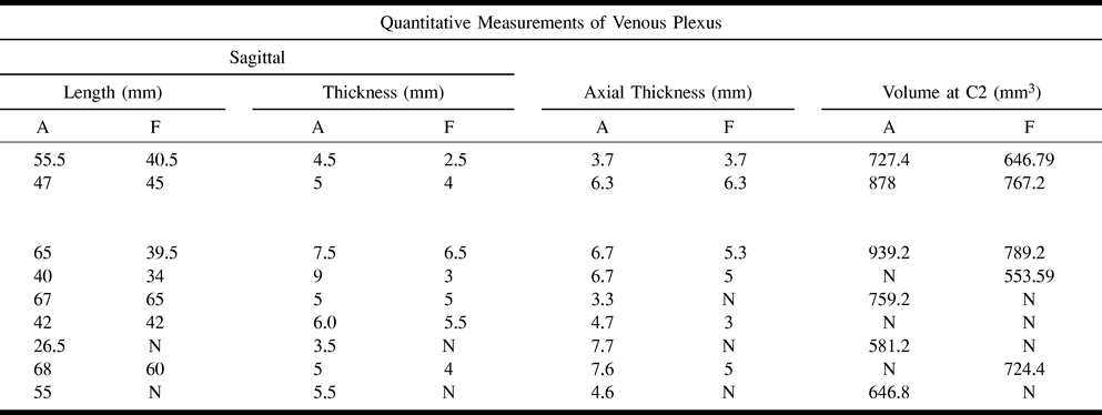
 TABLE 1A
MR imaging findings in patients with spontaneous intracranial hypotension headache
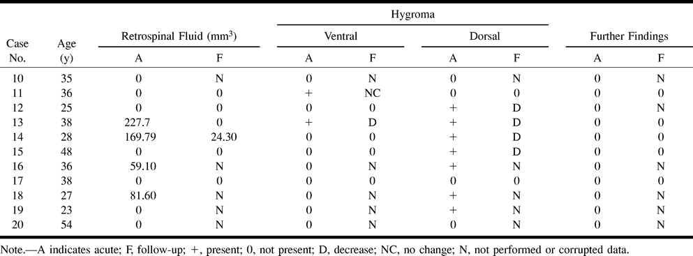
- TABLE 1B
MR imaging findings in patients with spontaneous intracranial hypotension headache
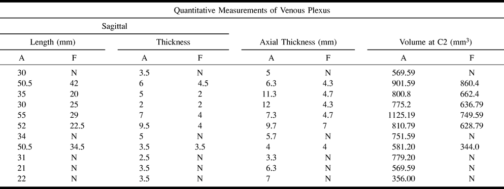
- TABLE 2A
MR imaging findings in patients with post–lumbar puncture headache
- TABLE 2B:
MR imaging findings in patients with post–lumbar puncture headache


In this issue


Jump to section
Related Articles
Cited By...
- Brain MRI features of postdural puncture headache
- Imaging Signs in Spontaneous Intracranial Hypotension: Prevalence and Relationship to CSF Pressure
- CT-Guided Epidural Blood Patching of Directly Observed or Potential Leak Sites for the Targeted Treatment of Spontaneous Intracranial Hypotension
- Teaching NeuroImages: False-positive magnetic resonance sign in spontaneous spinal CSF leak
- Diagnostic Value of Spinal MR Imaging in Spontaneous Intracranial Hypotension Syndrome
- Progressive cervical myelopathy secondary to chronic ventriculoperitoneal CSF overshunting
- Cranial Subdural Hygroma Complicating Thoracic Disc Surgery: A Case Report
- Cranial Subdural Hygroma Complicating Thoracic Disc Surgery: A Case Report
- Intracranial hypotension after chiropractic manipulation of the cervical spine
- CSF hypovolemia vs intracranial hypotension in "spontaneous intracranial hypotension syndrome"