Advertisement

Main menu

  • Home
  • Content
    • Current Issue
    • Accepted Manuscripts
    • Article Preview
    • Past Issue Archive
    • Video Articles
    • AJNR Case Collection
    • Case of the Week Archive
    • Case of the Month Archive
    • Classic Case Archive
  • Special Collections
    • AJNR Awards
    • Low-Field MRI
    • Alzheimer Disease
    • ASNR Foundation Special Collection
    • Photon-Counting CT
    • View All
  • Multimedia
    • AJNR Podcasts
    • AJNR SCANtastic
    • Trainee Corner
    • MRI Safety Corner
    • Imaging Protocols
  • For Authors
    • Submit a Manuscript
    • Submit a Video Article
    • Submit an eLetter to the Editor/Response
    • Manuscript Submission Guidelines
    • Statistical Tips
    • Fast Publishing of Accepted Manuscripts
    • Graphical Abstract Preparation
    • Imaging Protocol Submission
    • Author Policies
  • About Us
    • About AJNR
    • Editorial Board
    • Editorial Board Alumni
  • More
    • Become a Reviewer/Academy of Reviewers
    • Subscribers
    • Permissions
    • Alerts
    • Feedback
    • Advertisers
    • ASNR Home

User menu

  • Alerts
  • Log in

Search

  • Advanced search
American Journal of Neuroradiology

American Journal of Neuroradiology

ASHNR American Society of Functional Neuroradiology ASHNR American Society of Pediatric Neuroradiology ASSR
  • Alerts
  • Log in

Advanced Search

  • Home
  • Content
    • Current Issue
    • Accepted Manuscripts
    • Article Preview
    • Past Issue Archive
    • Video Articles
    • AJNR Case Collection
    • Case of the Week Archive
    • Case of the Month Archive
    • Classic Case Archive
  • Special Collections
    • AJNR Awards
    • Low-Field MRI
    • Alzheimer Disease
    • ASNR Foundation Special Collection
    • Photon-Counting CT
    • View All
  • Multimedia
    • AJNR Podcasts
    • AJNR SCANtastic
    • Trainee Corner
    • MRI Safety Corner
    • Imaging Protocols
  • For Authors
    • Submit a Manuscript
    • Submit a Video Article
    • Submit an eLetter to the Editor/Response
    • Manuscript Submission Guidelines
    • Statistical Tips
    • Fast Publishing of Accepted Manuscripts
    • Graphical Abstract Preparation
    • Imaging Protocol Submission
    • Author Policies
  • About Us
    • About AJNR
    • Editorial Board
    • Editorial Board Alumni
  • More
    • Become a Reviewer/Academy of Reviewers
    • Subscribers
    • Permissions
    • Alerts
    • Feedback
    • Advertisers
    • ASNR Home
  • Follow AJNR on Twitter
  • Visit AJNR on Facebook
  • Follow AJNR on Instagram
  • Join AJNR on LinkedIn
  • RSS Feeds

AJNR Awards, New Junior Editors, and more. Read the latest AJNR updates

Research ArticlePediatric Neuroimaging

Prenatal Diagnosis and Postnatal Follow-up of Pericallosal Lipoma: Report of Seven New Cases

Valérie Ickowitz, Danièle Eurin, Françoise Rypens, Pascale Sonigo, Isabelle Simon, Phillipe David, Francis Brunelle and Fred E. Avni
American Journal of Neuroradiology April 2001, 22 (4) 767-772;
Valérie Ickowitz
  • Find this author on Google Scholar
  • Find this author on PubMed
  • Search for this author on this site
Danièle Eurin
  • Find this author on Google Scholar
  • Find this author on PubMed
  • Search for this author on this site
Françoise Rypens
  • Find this author on Google Scholar
  • Find this author on PubMed
  • Search for this author on this site
Pascale Sonigo
  • Find this author on Google Scholar
  • Find this author on PubMed
  • Search for this author on this site
Isabelle Simon
  • Find this author on Google Scholar
  • Find this author on PubMed
  • Search for this author on this site
Phillipe David
  • Find this author on Google Scholar
  • Find this author on PubMed
  • Search for this author on this site
Francis Brunelle
  • Find this author on Google Scholar
  • Find this author on PubMed
  • Search for this author on this site
Fred E. Avni
  • Find this author on Google Scholar
  • Find this author on PubMed
  • Search for this author on this site
  • Article
  • Figures & Data
  • Info & Metrics
  • Responses
  • References
  • PDF
Loading

Article Figures & Data

Figures

  • Tables
  • fig 1.
    • Download figure
    • Open in new tab
    • Download powerpoint
    fig 1.

    Tubulo-nodular type of pericallosal lipoma. In utero and postnatal follow-up images (case 3).

    A, Sonogram obtained in utero at 35 weeks' gestation. A biparietal image is shown. A hyperechoic mass (M) can be seen within the anterior midline. The lipoma appears slightly less echogenic than the parietal bone; its margins are irregular. The mass is extending toward the frontal lobes (arrows).

    B, MR image obtained at birth. Sagittal view turbo spin-echo T1-weighted image (350/16/1) confirms the presence of the lipoma and the agenesis of the corpus callosum.

    C, MR image obtained at birth. Frontal view turbo spin-echo T1-weighted image (350/16/1) shows the lateral extend of the lipoma.

    D, MR image obtained at age 3 years. Mid-sagittal view turbo spin-echo T1-weighted sequence (450/15/1) shows the growth of the lipoma.

    E, MR image obtained at age 3 years. Similar findings are revealed by the frontal view turbo spin-echo T1-weighted sequence (450/15/1).

  • fig 2.
    • Download figure
    • Open in new tab
    • Download powerpoint
    fig 2.

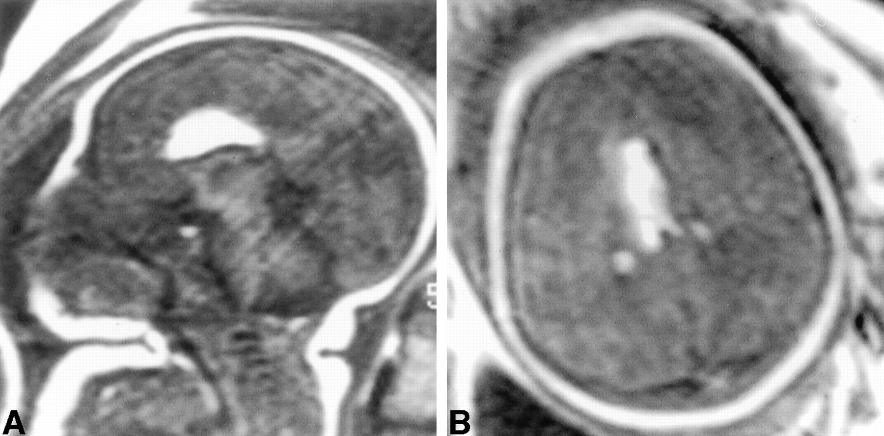
    Tubulo-nodular type. Fetal MR images (case 4).

    A, Mid-sagittal view turbo spin-echo T1-weighted sequence (400/17/1) shows typical lipoma and incomplete corpus callosum. Note that it was not possible to obtain this sagittal view image by using obstetric sonography.

    B, Transverse turbo spin-echo T1-weighted sequence (400/17/1) shows the lipoma and the extension toward the plexus choroids.

  • fig 3.
    • Download figure
    • Open in new tab
    • Download powerpoint
    fig 3.

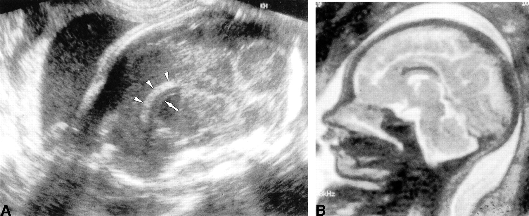
    Curvilinear pattern. Sonograms and fetal MR images (case 7).

    A, Obstetric sonograms obtained 26.5 weeks. Sagittal view image of the fetal head. The lipoma appears as a hyperechoic mass (arrowheads) with smooth margins parallel to the corpus callosum (arrow).

    B, Fetal MR image. Mid-sagittal fast spin-echo T2-weighted sequence (8000/122/2) shows a curvilinear hyposignal lipoma and a normal corpus callosum.

Tables

  • Figures
  • TABLE 1:
    • View popup
    • Download powerpoint
    TABLE 1:

    In utero and postpartum data of seven patients with pericallosal lipoma

  • TABLE 2:
    • View popup
    • Download powerpoint
    TABLE 2:

    Specific sonographic characteristics of the pericallosal lipomas

PreviousNext
Back to top

In this issue

American Journal of Neuroradiology
Vol. 22, Issue 4
1 Apr 2001
  • Table of Contents
  • Index by author
Advertisement
Print
Download PDF
Email Article

Thank you for your interest in spreading the word on American Journal of Neuroradiology.

NOTE: We only request your email address so that the person you are recommending the page to knows that you wanted them to see it, and that it is not junk mail. We do not capture any email address.

Enter multiple addresses on separate lines or separate them with commas.
Prenatal Diagnosis and Postnatal Follow-up of Pericallosal Lipoma: Report of Seven New Cases
(Your Name) has sent you a message from American Journal of Neuroradiology
(Your Name) thought you would like to see the American Journal of Neuroradiology web site.
CAPTCHA
This question is for testing whether or not you are a human visitor and to prevent automated spam submissions.
Cite this article
Valérie Ickowitz, Danièle Eurin, Françoise Rypens, Pascale Sonigo, Isabelle Simon, Phillipe David, Francis Brunelle, Fred E. Avni
Prenatal Diagnosis and Postnatal Follow-up of Pericallosal Lipoma: Report of Seven New Cases
American Journal of Neuroradiology Apr 2001, 22 (4) 767-772;

Citation Manager Formats

  • BibTeX
  • Bookends
  • EasyBib
  • EndNote (tagged)
  • EndNote 8 (xml)
  • Medlars
  • Mendeley
  • Papers
  • RefWorks Tagged
  • Ref Manager
  • RIS
  • Zotero
0 Responses
Respond to this article
Share
Bookmark this article
Prenatal Diagnosis and Postnatal Follow-up of Pericallosal Lipoma: Report of Seven New Cases
Valérie Ickowitz, Danièle Eurin, Françoise Rypens, Pascale Sonigo, Isabelle Simon, Phillipe David, Francis Brunelle, Fred E. Avni
American Journal of Neuroradiology Apr 2001, 22 (4) 767-772;
del.icio.us logo Twitter logo Facebook logo Mendeley logo
  • Tweet Widget
  • Facebook Like
  • Google Plus One

Jump to section

  • Article
    • Abstract
    • Methods
    • Results
    • Discussion
    • Conclusion
    • Footnotes
    • References
  • Figures & Data
  • Info & Metrics
  • Responses
  • References
  • PDF

Related Articles

  • No related articles found.
  • PubMed
  • Google Scholar

Cited By...

  • No citing articles found.
  • Crossref
  • Google Scholar

This article has not yet been cited by articles in journals that are participating in Crossref Cited-by Linking.

More in this TOC Section

  • Frontal Paraventricular Cysts
  • Sodium MRI in Pediatric Brain Tumors
  • FRACTURE MR in Congenital Vertebral Anomalies
Show more Pediatric Neuroimaging

Similar Articles

Advertisement

Indexed Content

  • Current Issue
  • Accepted Manuscripts
  • Article Preview
  • Past Issues
  • Editorials
  • Editor's Choice
  • Fellows' Journal Club
  • Letters to the Editor
  • Video Articles

Cases

  • Case Collection
  • Archive - Case of the Week
  • Archive - Case of the Month
  • Archive - Classic Case

More from AJNR

  • Trainee Corner
  • Imaging Protocols
  • MRI Safety Corner
  • Book Reviews

Multimedia

  • AJNR Podcasts
  • AJNR Scantastics

Resources

  • Turnaround Time
  • Submit a Manuscript
  • Submit a Video Article
  • Submit an eLetter to the Editor/Response
  • Manuscript Submission Guidelines
  • Statistical Tips
  • Fast Publishing of Accepted Manuscripts
  • Graphical Abstract Preparation
  • Imaging Protocol Submission
  • Evidence-Based Medicine Level Guide
  • Publishing Checklists
  • Author Policies
  • Become a Reviewer/Academy of Reviewers
  • News and Updates

About Us

  • About AJNR
  • Editorial Board
  • Editorial Board Alumni
  • Alerts
  • Permissions
  • Not an AJNR Subscriber? Join Now
  • Advertise with Us
  • Librarian Resources
  • Feedback
  • Terms and Conditions
  • AJNR Editorial Board Alumni

American Society of Neuroradiology

  • Not an ASNR Member? Join Now

© 2025 by the American Society of Neuroradiology All rights, including for text and data mining, AI training, and similar technologies, are reserved.
Print ISSN: 0195-6108 Online ISSN: 1936-959X

Powered by HighWire