AJNR Case Collection
Section Editors:
Anvita Pauranik, MD, University of British Columbia, Vancouver, British Columbia, Canada
Michael Travis Caton, MD, Mount Sinai South Nassau, New York
Simona Gaudino, MD, Università Cattolica del Sacro Cuore, Italy
Matthew S. Parsons, MD, Mallinckrodt Institute of Radiology, Missouri
Anat Yahav-Dovrat, MD, University of Toronto, Canada
Final Diagnosis: Posttransplant Lymphoproliferative Disease, Diffuse Large B-Cell Lymphoma


Imaging Findings:
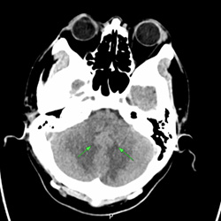
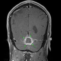
Initial head CT (A) demonstrates a faintly hyperdense mass in the cerebellar vermis (blue arrow), with extensive edema involving bilateral cerebellar hemispheres, resulting in effacement of the fourth ventricle. No hydrocephalus or brain herniation is observed. Subsequent MRI reveals a 2.6-cm ring-enhancing mass centered in the cerebellar vermis (B). Heterogeneous T1 and T2 signals are present within the lesion, with minimal intrinsic T1 hyperintensity. Intermediate diffusion restriction is demonstrated within the lesion, with an average ADC value of 918 x 10-6 x mm2/s (C). Perilesional susceptibility effect and adjacent fissural susceptibility effect are also noted (D). Similar to CT, the extensive edema involving bilateral cerebellar hemispheres and fourth ventricular effacement persists.


