Advertisement

Main menu

  • Home
  • Content
    • Current Issue
    • Accepted Manuscripts
    • Article Preview
    • Past Issue Archive
    • Video Articles
    • AJNR Case Collection
    • Case of the Week Archive
    • Case of the Month Archive
    • Classic Case Archive
  • Special Collections
    • AJNR Awards
    • Low-Field MRI
    • Alzheimer Disease
    • ASNR Foundation Special Collection
    • Photon-Counting CT
    • View All
  • Multimedia
    • AJNR Podcasts
    • AJNR SCANtastic
    • Trainee Corner
    • MRI Safety Corner
    • Imaging Protocols
  • For Authors
    • Submit a Manuscript
    • Submit a Video Article
    • Submit an eLetter to the Editor/Response
    • Manuscript Submission Guidelines
    • Statistical Tips
    • Fast Publishing of Accepted Manuscripts
    • Graphical Abstract Preparation
    • Imaging Protocol Submission
    • Author Policies
  • About Us
    • About AJNR
    • Editorial Board
    • Editorial Board Alumni
  • More
    • Become a Reviewer/Academy of Reviewers
    • Subscribers
    • Permissions
    • Alerts
    • Feedback
    • Advertisers
    • ASNR Home

User menu

  • Alerts
  • Log in

Search

  • Advanced search
American Journal of Neuroradiology

American Journal of Neuroradiology

ASHNR American Society of Functional Neuroradiology ASHNR American Society of Pediatric Neuroradiology ASSR
  • Alerts
  • Log in

Advanced Search

  • Home
  • Content
    • Current Issue
    • Accepted Manuscripts
    • Article Preview
    • Past Issue Archive
    • Video Articles
    • AJNR Case Collection
    • Case of the Week Archive
    • Case of the Month Archive
    • Classic Case Archive
  • Special Collections
    • AJNR Awards
    • Low-Field MRI
    • Alzheimer Disease
    • ASNR Foundation Special Collection
    • Photon-Counting CT
    • View All
  • Multimedia
    • AJNR Podcasts
    • AJNR SCANtastic
    • Trainee Corner
    • MRI Safety Corner
    • Imaging Protocols
  • For Authors
    • Submit a Manuscript
    • Submit a Video Article
    • Submit an eLetter to the Editor/Response
    • Manuscript Submission Guidelines
    • Statistical Tips
    • Fast Publishing of Accepted Manuscripts
    • Graphical Abstract Preparation
    • Imaging Protocol Submission
    • Author Policies
  • About Us
    • About AJNR
    • Editorial Board
    • Editorial Board Alumni
  • More
    • Become a Reviewer/Academy of Reviewers
    • Subscribers
    • Permissions
    • Alerts
    • Feedback
    • Advertisers
    • ASNR Home
  • Follow AJNR on Twitter
  • Visit AJNR on Facebook
  • Follow AJNR on Instagram
  • Join AJNR on LinkedIn
  • RSS Feeds

AJNR Awards, New Junior Editors, and more. Read the latest AJNR updates

Research ArticleNeurointervention

Optimizing Image Quality in Cerebral 3D Rotational Angiography: A Study on the Impact of Voxel Size

Seongsik Han, Byung Jun Kim, Pae Sun Suh, Hye Hyeon Moon, So-Yeon Kim, Jong-Tae Yoon, Boseong Kwon, Deok Hee Lee and Yunsun Song
American Journal of Neuroradiology May 2025, DOI: https://doi.org/10.3174/ajnr.A8672
Seongsik Han
aFrom the Department of Radiologic Technology (S.H.), Asan Medical Center, University of Ulsan College of Medicine, Seoul, Korea
  • Find this author on Google Scholar
  • Find this author on PubMed
  • Search for this author on this site
Byung Jun Kim
bAdvanced Therapies (B.J.K.), Siemens Healthineers Ltd., Seoul, Korea
  • Find this author on Google Scholar
  • Find this author on PubMed
  • Search for this author on this site
Pae Sun Suh
cDepartment of Radiology (P.S.S., H.H.M., S.-Y.K., B.K., D.H.L., Y.S.), Research Institute of Radiology, Asan Medical Center, University of Ulsan College of Medicine, Seoul, Korea
  • Find this author on Google Scholar
  • Find this author on PubMed
  • Search for this author on this site
  • ORCID record for Pae Sun Suh
Hye Hyeon Moon
cDepartment of Radiology (P.S.S., H.H.M., S.-Y.K., B.K., D.H.L., Y.S.), Research Institute of Radiology, Asan Medical Center, University of Ulsan College of Medicine, Seoul, Korea
  • Find this author on Google Scholar
  • Find this author on PubMed
  • Search for this author on this site
  • ORCID record for Hye Hyeon Moon
So-Yeon Kim
cDepartment of Radiology (P.S.S., H.H.M., S.-Y.K., B.K., D.H.L., Y.S.), Research Institute of Radiology, Asan Medical Center, University of Ulsan College of Medicine, Seoul, Korea
  • Find this author on Google Scholar
  • Find this author on PubMed
  • Search for this author on this site
Jong-Tae Yoon
dBiomedical Engineering Research Center (J.-T.Y.), Asan Institute for Life Sciences, Asan Medical Center, Seoul, Korea
  • Find this author on Google Scholar
  • Find this author on PubMed
  • Search for this author on this site
Boseong Kwon
cDepartment of Radiology (P.S.S., H.H.M., S.-Y.K., B.K., D.H.L., Y.S.), Research Institute of Radiology, Asan Medical Center, University of Ulsan College of Medicine, Seoul, Korea
  • Find this author on Google Scholar
  • Find this author on PubMed
  • Search for this author on this site
  • ORCID record for Boseong Kwon
Deok Hee Lee
cDepartment of Radiology (P.S.S., H.H.M., S.-Y.K., B.K., D.H.L., Y.S.), Research Institute of Radiology, Asan Medical Center, University of Ulsan College of Medicine, Seoul, Korea
  • Find this author on Google Scholar
  • Find this author on PubMed
  • Search for this author on this site
  • ORCID record for Deok Hee Lee
Yunsun Song
cDepartment of Radiology (P.S.S., H.H.M., S.-Y.K., B.K., D.H.L., Y.S.), Research Institute of Radiology, Asan Medical Center, University of Ulsan College of Medicine, Seoul, Korea
  • Find this author on Google Scholar
  • Find this author on PubMed
  • Search for this author on this site
  • ORCID record for Yunsun Song
  • Article
  • Figures & Data
  • Supplemental
  • Info & Metrics
  • Responses
  • References
  • PDF
Loading

Article Figures & Data

Figures

  • Figure
    • Download figure
    • Open in new tab
    • Download powerpoint
  • FIG 1.
    • Download figure
    • Open in new tab
    • Download powerpoint
    FIG 1.

    Secondary reconstruction with 2 voxel sizes (0.3 mm and 0.05 mm). A, FOV sizes for secondary reconstruction: 15.36 cm (green square, 0.3 mm × 512 matrix) and 2.56 cm (yellow square, 0.05 mm × 512 matrix). B and C, Fusion 3D images of the right (orange) and left (gray) internal carotid arteriograms (posterior view) are presented. The aneurysm at the anterior communicating artery origin of the right anterior cerebral artery is presented. A small subcallosal artery (arrow) arises from the anterior communicating artery. While the artery is depicted more clearly in the smaller voxel reconstruction (C), more noise is noted in the overall vessels. The window setting (W: window width, C: window center) was maintained across both reconstructed images to ensure a fair and unbiased comparative analysis.

  • FIG 2.
    • Download figure
    • Open in new tab
    • Download powerpoint
    FIG 2.

    Quantitative measurement of vessel intensity. A, Multiplanar reconstruction images of a vessel by using voxel sizes of 0.30 mm (right) and 0.05 mm (left). The line intensity profile was derived from a crossing line (indicated by the red line) perpendicular to the vessel. Regions of interest (circled) were designated to determine the maximum intensity and background noise. B. Line intensity profiles corresponding to each voxel size. C, Intensity profile with Gaussian curve fitting (black line). The slope of the curve was determined between the 20% and 80% intensity thresholds.
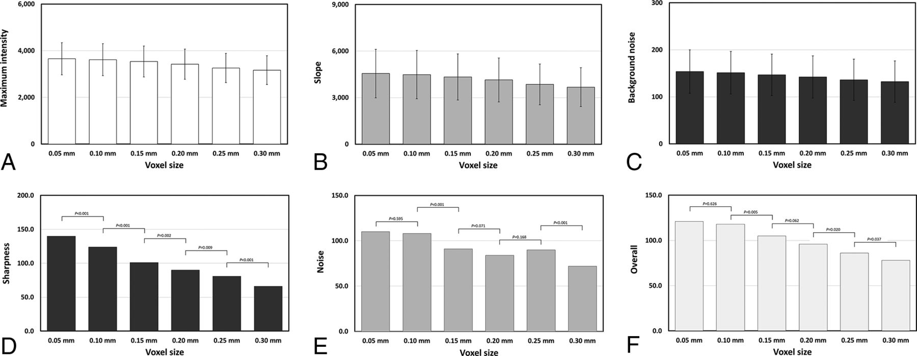
  • FIG 3.
    • Download figure
    • Open in new tab
    • Download powerpoint
    FIG 3.

    Quantitative and qualitative analyses of image quality across voxel sizes. Upper row, Quantitative metrics for image quality at varying voxel sizes (0.05 mm to 0.30 mm): A, Maximum intensity; B, Slope of grayscale intensity at the vessel margin; and C, Background noise levels. Lower row: Qualitative assessments of image quality by using a 5-point Likert scale, capturing cumulative scores from 3 raters: D, Image sharpness; E, Noise; and F, Overall image quality. Significant differences between adjacent voxel sizes are denoted with corresponding P values.
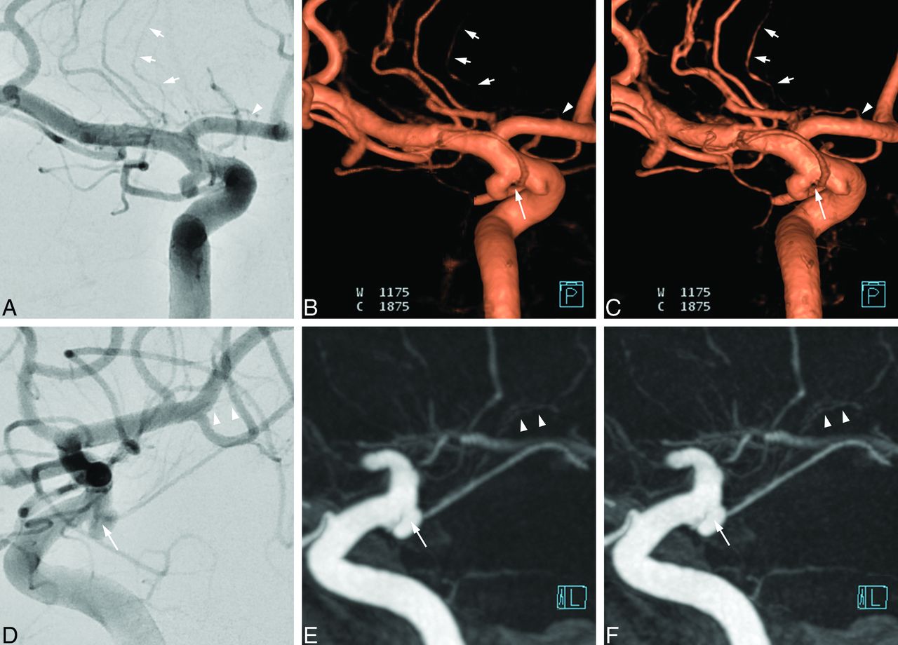
  • FIG 4.
    • Download figure
    • Open in new tab
    • Download powerpoint
    FIG 4.

    Impact of voxel size on 3DRA image quality: comparison with 2D DSA. Left internal carotid arteriography for a small aneurysm at the anterior choroidal artery (AChA) origin. The upper row shows posterior views, while the lower row presents left lateral projections. Images A and D are 2D DSA working views during the coiling procedure (A is flipped left-to-right for easier comparison). 3DRA images are reconstructed as volume-rendering images (upper row: B and C) and MIP images (lower row: E and F) by using 2 voxel sizes: 0.3 mm (B and E) and 0.05 mm (C and F). The 0.05 mm voxel size images (C and F) provide enhanced visualization of the AChA origin (long arrows), the lenticulostriate artery origin from the anterior cerebral artery (arrowheads, upper row), the lenticulostriate artery from the proximal middle cerebral artery (short arrows), and the thalamoperforators (arrowheads, lower row).

PreviousNext
Back to top
Advertisement
Print
Download PDF
Email Article

Thank you for your interest in spreading the word on American Journal of Neuroradiology.

NOTE: We only request your email address so that the person you are recommending the page to knows that you wanted them to see it, and that it is not junk mail. We do not capture any email address.

Enter multiple addresses on separate lines or separate them with commas.
Optimizing Image Quality in Cerebral 3D Rotational Angiography: A Study on the Impact of Voxel Size
(Your Name) has sent you a message from American Journal of Neuroradiology
(Your Name) thought you would like to see the American Journal of Neuroradiology web site.
CAPTCHA
This question is for testing whether or not you are a human visitor and to prevent automated spam submissions.
Cite this article
Seongsik Han, Byung Jun Kim, Pae Sun Suh, Hye Hyeon Moon, So-Yeon Kim, Jong-Tae Yoon, Boseong Kwon, Deok Hee Lee, Yunsun Song
Optimizing Image Quality in Cerebral 3D Rotational Angiography: A Study on the Impact of Voxel Size
American Journal of Neuroradiology May 2025, DOI: 10.3174/ajnr.A8672

Citation Manager Formats

  • BibTeX
  • Bookends
  • EasyBib
  • EndNote (tagged)
  • EndNote 8 (xml)
  • Medlars
  • Mendeley
  • Papers
  • RefWorks Tagged
  • Ref Manager
  • RIS
  • Zotero
0 Responses
Respond to this article
Share
Bookmark this article
Optimizing Voxel Size in 3D Rotational Angiography
Seongsik Han, Byung Jun Kim, Pae Sun Suh, Hye Hyeon Moon, So-Yeon Kim, Jong-Tae Yoon, Boseong Kwon, Deok Hee Lee, Yunsun Song
American Journal of Neuroradiology May 2025, DOI: 10.3174/ajnr.A8672
del.icio.us logo Twitter logo Facebook logo Mendeley logo
  • Tweet Widget
  • Facebook Like
  • Google Plus One
Purchase

Jump to section

  • Article
    • Graphical Abstract
    • Abstract
    • ABBREVIATIONS:
    • MATERIALS AND METHODS
    • RESULTS
    • DISCUSSION
    • CONCLUSIONS
    • Footnotes
    • References
  • Figures & Data
  • Supplemental
  • Info & Metrics
  • Responses
  • References
  • PDF

Related Articles

  • PubMed
  • Google Scholar

Cited By...

  • No citing articles found.
  • Crossref
  • Google Scholar

This article has not yet been cited by articles in journals that are participating in Crossref Cited-by Linking.

More in this TOC Section

  • Rescue Reentry in Carotid Near-Occlusion
  • Contour Neurovascular System: Five Year Follow Up
  • Effect of SARS-CoV2 on Endovascular Thrombectomy
Show more Neurointervention

Similar Articles

Advertisement

Indexed Content

  • Current Issue
  • Accepted Manuscripts
  • Article Preview
  • Past Issues
  • Editorials
  • Editor's Choice
  • Fellows' Journal Club
  • Letters to the Editor
  • Video Articles

Cases

  • Case Collection
  • Archive - Case of the Week
  • Archive - Case of the Month
  • Archive - Classic Case

More from AJNR

  • Trainee Corner
  • Imaging Protocols
  • MRI Safety Corner
  • Book Reviews

Multimedia

  • AJNR Podcasts
  • AJNR Scantastics

Resources

  • Turnaround Time
  • Submit a Manuscript
  • Submit a Video Article
  • Submit an eLetter to the Editor/Response
  • Manuscript Submission Guidelines
  • Statistical Tips
  • Fast Publishing of Accepted Manuscripts
  • Graphical Abstract Preparation
  • Imaging Protocol Submission
  • Evidence-Based Medicine Level Guide
  • Publishing Checklists
  • Author Policies
  • Become a Reviewer/Academy of Reviewers
  • News and Updates

About Us

  • About AJNR
  • Editorial Board
  • Editorial Board Alumni
  • Alerts
  • Permissions
  • Not an AJNR Subscriber? Join Now
  • Advertise with Us
  • Librarian Resources
  • Feedback
  • Terms and Conditions
  • AJNR Editorial Board Alumni

American Society of Neuroradiology

  • Not an ASNR Member? Join Now

© 2025 by the American Society of Neuroradiology All rights, including for text and data mining, AI training, and similar technologies, are reserved.
Print ISSN: 0195-6108 Online ISSN: 1936-959X

Powered by HighWire