
Article Figures & Data
Figures
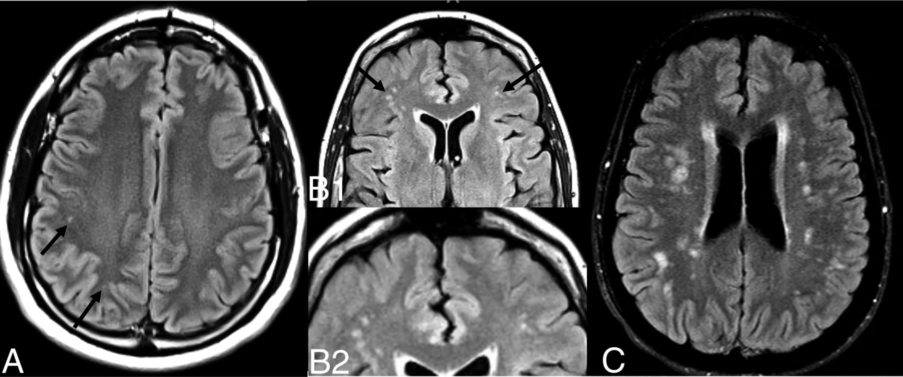
- FIG 1.
Illustrative examples of the classifications of FLAIR WMH. A, Patient with minimal punctate FLAIR hyperintensities without frontal predilection (arrows). B1, Patient with frontal-predominant migrainous-type WHM involving the subcortical and periventricular white matter (arrows; detailed inset B2), characteristically seen with migraines. C, Microangiopathic-type WMH with diffuse supratentorial lesions.
- FIG 2.
A, Bar graph illustrating the percentage of patients with patterns of WMH, which differed significantly between those with and without a myelographically-proved CVF (P = .005). B, Bar graph illustrating the frequency of myelographically-localized CVF in patients according to the pattern of WMH. There is an overall significant difference in CVF positivity across patterns of WMH (P = .006), with post hoc analyses demonstrating a significant difference between the minimal and migrainous WMH classes (*P = .002).
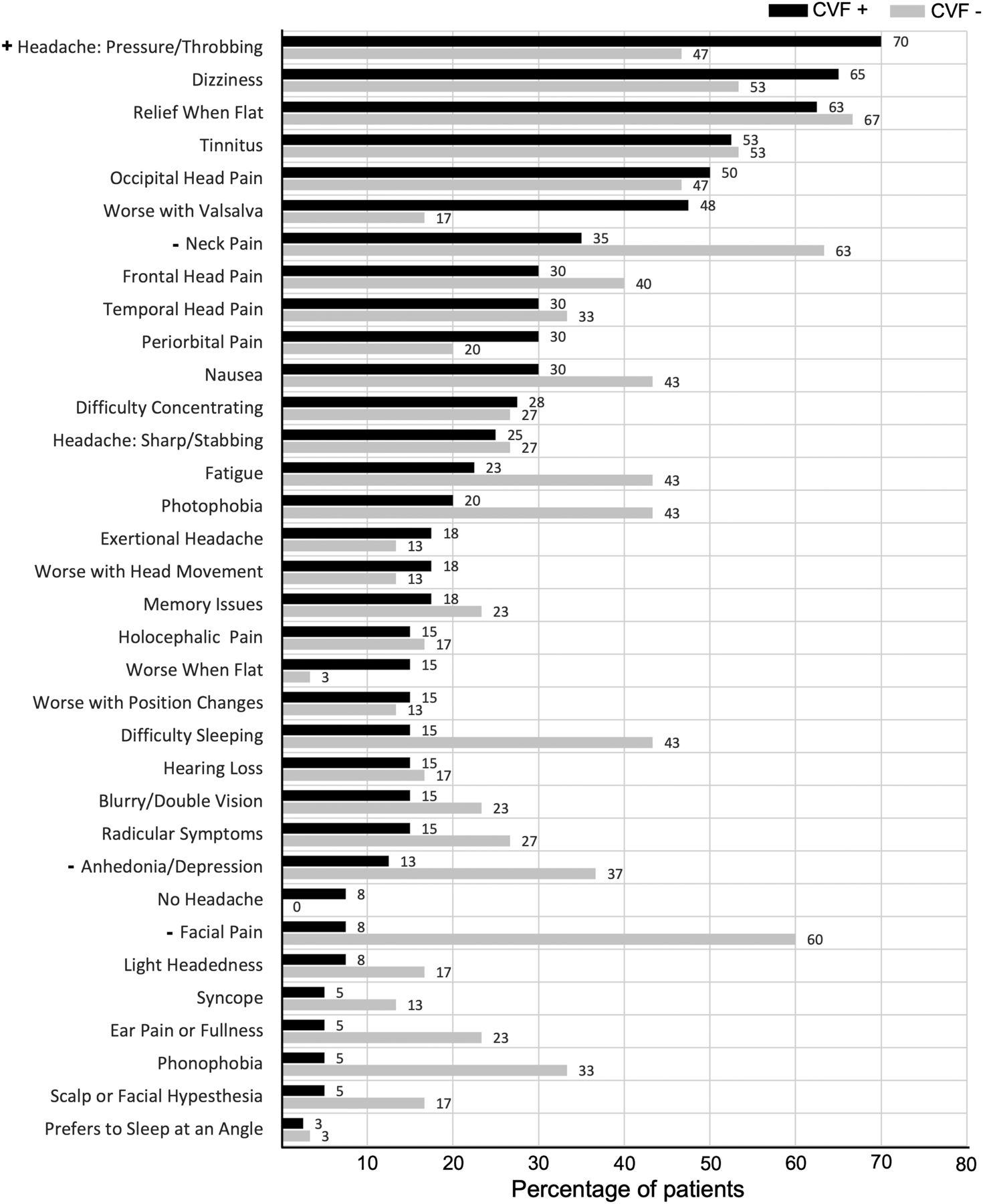
- FIG 3.
Bar graph illustrating the distribution of clinical symptoms and characteristics between patients with CVF+ and CVF– at presentation. Features identified via multivariate logistic regression to have a significant effect on the presence of CVF on dynamic CT myelography are denoted by positive (+) or negative (–) associations.


Tables
Characteristic CVF+ (n = 40) CVF– (n = 32) Mean age (range) (yr) 61.6 (SD, 9.2) (34–83) 58.2 (SD, 9.6) (22–71) Sex female (%)/male (%) 23 (58%)/17 (42%) 23 (72%)/9 (28%) Bern score Median (range; IQR) 6 (1–9; 3)a 1 (0–6; 2) Low probability 0–2 (%) 1 (2.5%) 28 (87.5%) Intermediate probability 3–4 (%) 10 (25%) 3 (9.4%) High probability 5–9 (%) 29 (72.5%) 1 (3.1%) WMH Mean total WMH (SD) 7.9 (8.2)a 20.7 (21.8) Mean frontal WMH (SD) 5.4 (6.5)a 14.6 (14.1) Median percentage frontal WMH (IQR) 53.3 (52.2) 70.6 (31.2) WMH pattern (No.) (%) Minimal 22 (55%) 10 (32.3%) Migrainous 5 (12.5%) 15 (46.9%) Microangiopathic 13 (32.5%) 7 (21.9%) Note:—IQR indicates interquartile range.
↵a Denotes P < .05 after Bonferroni adjustment. Patients with myelographically-proved CVF have higher Bern scores and fewer WMH. There is a strong correlation between the Bern score and CVF positivity (rpb = −0.82).
- Table 2:
The combined features of a low-probability Bern score and the presence of migrainous WMH resulted in the highest sensitivity, specificity, PPV, and NPV
Features Predictive of CVF− Sensitivity Specificity PPV NPV Bern score <5 0.725 0.969 0.967 0.738 Bern score <3 0.975 0.875 0.907 0.966 Presence of the pattern of migrainous WMH 0.900 0.469 0.679 0.789 Bern score <5 & presence of the pattern of migrainous WMH 0.65 1 1 0.696 Bern score <3 & presence of the pattern of migrainous WMH 0.975 0.938 0.951 0.968


Jump to section
Related Articles
Cited By...
- No citing articles found.