
Article Figures & Data
Figures
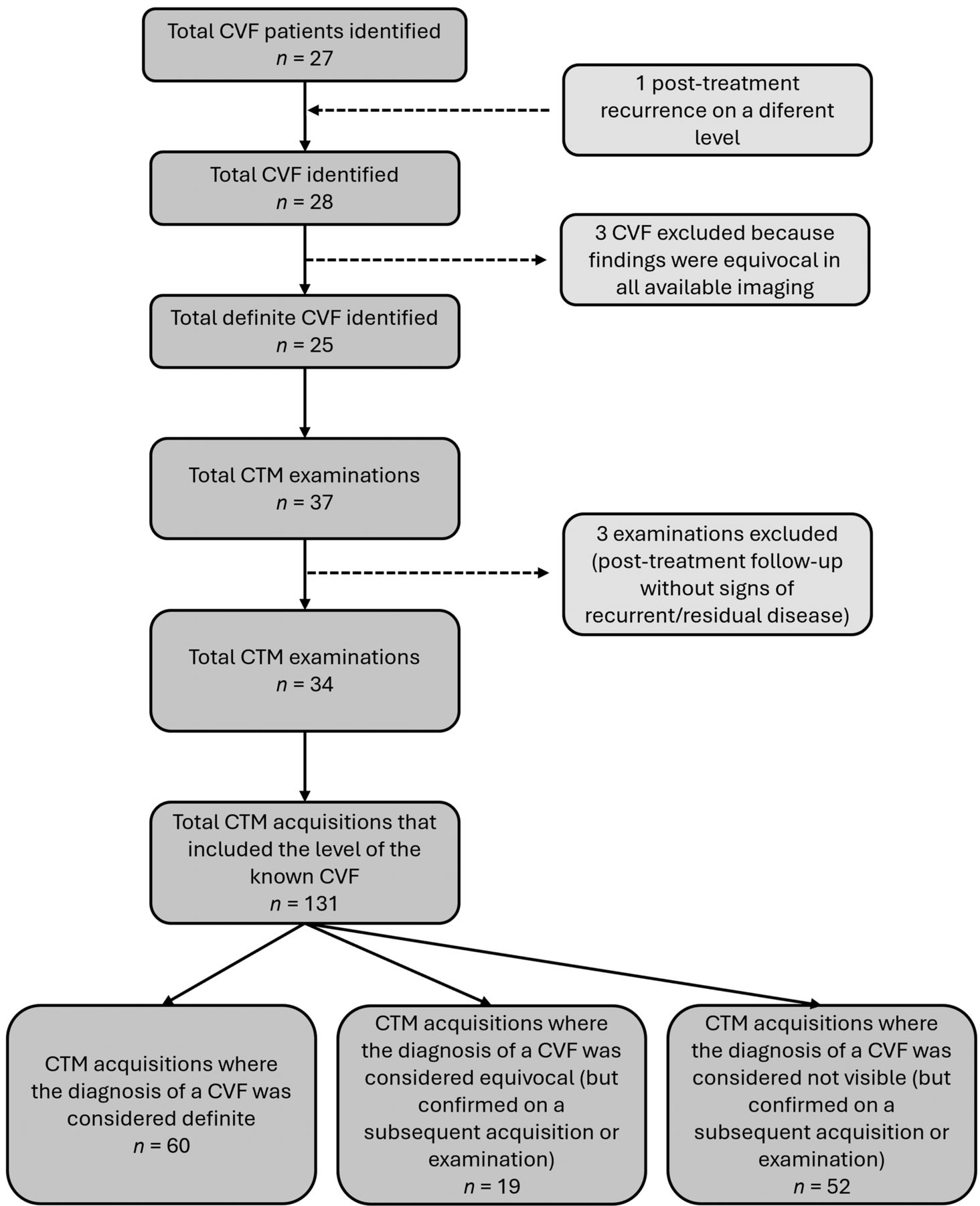
- FIG 1.
Flowchart of case selection.
- FIG 2.
Each row in A and B represents a single patient. Each dot represents a CTM acquisition. In A, the x-axis denotes the thecal sac attenuation on the level of a known CVF. In B, the x-axis denotes the time passed between contrast injection and the acquisition. Colors represent the subjective visibility of the CVF on that acquisition.
- FIG 3.
Boxplot graph showing the distribution of thecal sac contrast attenuation in each group of CVF visibility. The group with visible CVF showed a marked and statistically significant higher mean attenuation than the other 2 groups. Flowchart of case selection.
- FIG 4.
The y-axis in both images represents the attenuation of the draining vein of the known CVF. Each dot represents a CTM acquisition. Colors represent the subjective visibility of the CVF on that acquisition. In A, the x-axis denotes the thecal sac attenuation on the level of a known CVF, with a statistically significant relationship, even after multivariate regression analysis correcting for time and position. In B, the x-axis denotes the time passed between contrast injection and the acquisition, without a significant relationship to the attenuation of the vein, before and after multivariate regression correction.
- FIG 5.
Axial images from different acquisitions during 2 separate CTMs at the level of a confirmed CVF in a patient with spontaneous intracranial hypotension. A, Left static decubitus CTM with indefinite but suspicious finding for a CVF in the T10-T11 left epidural venous plexus (thecal sac attenuation: 1578 HU, vein attenuation: 180 HU, time passed: 21 minutes). As an isolated image, it could also represent the dural emergence of the dorsal nerve root. B, Left static decubitus CTM with a definitive CVF in the left T10-T11 foramen, represented by multiple epidural, foraminal, and extraforaminal vessels opacifying by contrast (thecal sac attenuation: 3070 HU, vein attenuation: 692 HU, time passed: 4 minutes). C, Prone acquisition after static decubitus CTM without any recognizable venous opacification (thecal sac attenuation: 1332HU, vein attenuation: 43 HU, time passed: 51 minutes).
- FIG 6.
Axial images from different acquisitions during a single CTM at the level of a confirmed CVF in a patient with spontaneous intracranial hypotension. A, Right static decubitus CTM with a definite right T11-T12 CVF draining to the paraspinal vein (thecal sac attenuation: 3020 HU, vein attenuation: 1871 HU, time passed: 4 minutes). B, Right static decubitus CTM with an equivocal finding for a CVF represented by a single faint contrast uptake apparently separate from the nerve root sleeve and diverticula (thecal sac attenuation: 685 HU, vein attenuation: 217 HU, time passed: 8 minutes). C, Prone acquisition after static decubitus CTM without any recognizable venous opacification (thecal sac attenuation: 988 HU, vein attenuation: 77 HU, time passed: 24 minutes).
- FIG 7.
Axial 5.7-mm maximum intensity projection images from 2 different acquisitions during a single CTM at the level of a confirmed CVF in a patient with spontaneous intracranial hypotension. A, Right static decubitus CTM at the T7-T8 level without any recognizable venous opacification (thecal sac attenuation: 996 HU, vein attenuation: 32 HU, time passed: 12 min). B, Right static decubitus CTM with a definite right T7-T8 CVF draining to the paraspinal vein (thecal sac attenuation: 1439 HU, vein attenuation: 321 HU, time passed: 21 minutes).


Tables
Scan Protocol Static Decubitus Ultrafast/Dynamic Conventional Total Examinations (n) 25 4 5 34 Acquisitions (n) 97 13 21 131 Sac attenuation (HU) 1531 1415 583 1368 CVF side down 2352 1736 739 2164 Other side down 542 344 547 534 Time after injection (min) 15.2 5 24.8 15.8 CVF C7-T1 1 T1-T2 2 T2-T3 1 T5-T6 3 T6-T7 4 T7-T8 3 T8-T9 3 T9-T10 1 T10-T11 7 T11-T12 1 Right-sided 16 (67%) CVF visibility Definite Equivocal Not visible Acquisitions (n) 60 19 52 Vein (HU) 639 198 5 Sac (HU) 2282 751 538 Definite Equivocal Not Visible 20 mL 50 17 34 10 mL 10 2 18


