
Article Figures & Data
Figures
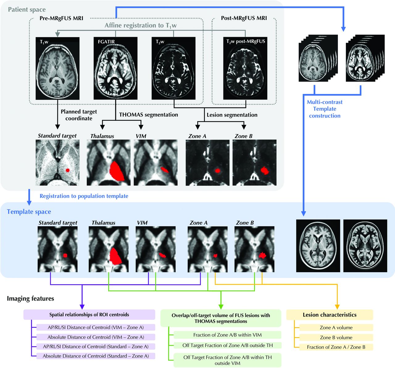
- FIG 1.
Imaging feature extraction pipeline. ROIs of standard target, FUS lesions and THOMAS segmentations were registered to the population-specific template generated from pre-operative T1-w and FGATIR images. Spatial relationships of ROI centroids, overlap of FUS lesions with segmentations and lesion volume characteristics were assessed. MRgFUS, MR-guided focused ultrasound; FGATIR, Fast Gray Matter Acquisition T1 Inversion Recovery; THOMAS, THalamus Optimized Multi Atlas Segmentation; VIM, ventral intermediate nucleus; TH, thalamus; AP, anterior-posterior; RL, right-left, SI, superior-inferior; ROI, region of interest.
- FIG 2.
Procedures to create standard target, FUS lesions and THOMAS segmentations of thalamus and VIM. A, Standard target ROIs were created from coordinates of the first sonication. B, FUS lesions were generated by subtracting the pre-operative T2-weighted image from the post-operative image and applying a thresholding method. C, The thalamus and VIM were segmented by applying THOMAS to pre-operative FGATIR images. The VIM here corresponds to the ventral part (inferior half) of the ventral lateral posterior nucleus in the Morel atlas. MRgFUS, MR-guided focused ultrasound; THOMAS, THalamus Optimized Multi Atlas Segmentation; VIM, ventral intermediate nucleus; FGATIR, Fast Gray Matter Acquisition T1 Inversion Recovery; ROI, region of interest.
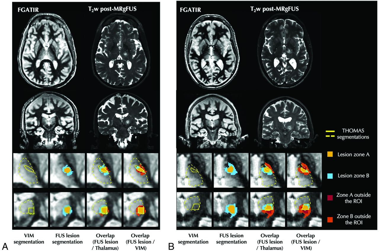
- FIG 3.
Example THOMAS segmentations (yellow line: thalamus; white line: VIM nucleus) and MRgFUS treated lesions (magenta: Zone A; cyan: Zone B) are shown in axial and coronal views. In the patient with side effects A, lesions extend farther out of the thalamus and segmented VIM, compared to the patient without side effects (B). FGATIR, Fast Gray Matter Acquisition T1 Inversion Recovery; MRgFUS, MR-guided focused ultrasound; THOMAS, Thalamus Optimized Multi Atlas Segmentation; VIM, ventral intermediate nucleus; ROI, region of interest.
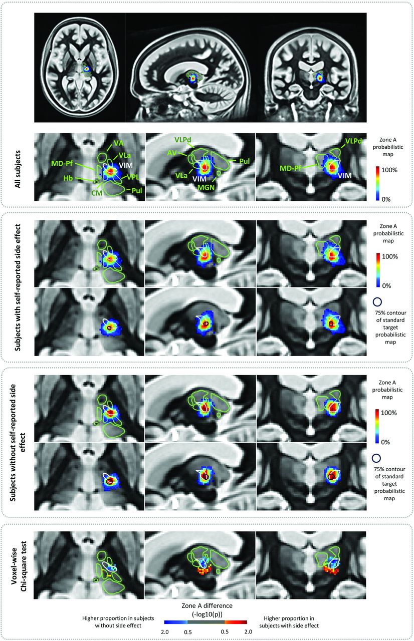
- FIG 4.
Probabilistic maps of Zone A in the population-specific template space. The spatial relationship between THOMAS-based thalamic nuclei segmentations, Zone A probabilistic maps, and standard targets are illustrated in different cohorts. The Zone A probabilistic map represents the voxel-wise percentage of FUS lesions across the subjects, and the standard target contour represents 75th percentile of standard coordinate ROI across the subjects. THOMAS, THalamus Optimized Multi Atlas Segmentation; FUS, focused ultrasound; VA, ventral anterior nucleus; VLa, ventral lateral anterior nucleus; VLP, ventral lateral posterior nucleus; VPL, ventral posterior lateral nucleus; VPLd, dorsal part of ventral posterior lateral nucleus; Pul, pulvinar nucleus; CM, centromedian nucleus; MD-Pf, mediodorsal-parafascicular nucleus; Hb, habenula; MGN, medial geniculate nucleus; ROI, region of interest.
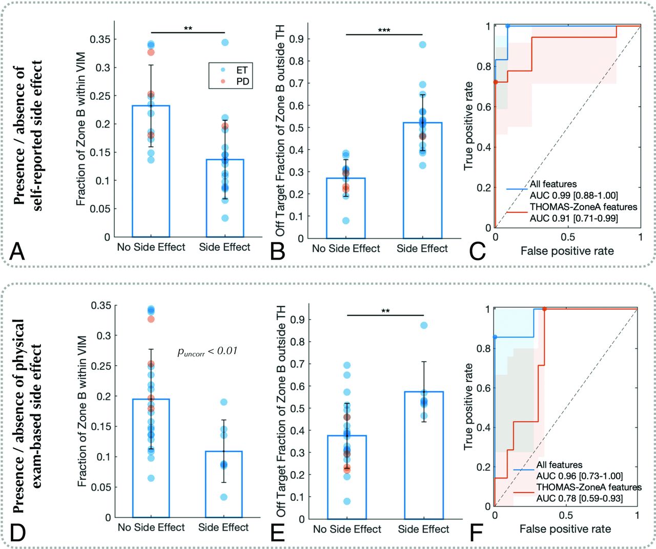
- FIG 5.
Results of imaging feature comparisons between patients with self-reported or physical exam-based gait/balance side effects and those without. In patients with side effects, Zone B shows less overlap with VIM segmentation and the off-target fraction of Zone B outside TH segmentation is greater (Graphs A–B and D–E). Graphs C and F show receiver operating characteristic curves of multivariate prediction models of side effects using imaging features. ET, essential tremor; PD, Parkinson’s disease; VIM, ventral intermediate nucleus; TH, thalamus; AUC, area under the curve.


Tables
Comparisons of imaging features between patients with and without side effect
MRI Features Self-Reported Side Effect Physical Exam-Based Side Effect Gait/Balance Gait/balance > 1 Month Gait/Balance p-Value p-Value p-Value FUS lesion volume Volume of Zone A 0.21 0.31 0.96 Volume of Zone B 0.01 0.15 0.14 Fraction of Zone A/Zone B 0.01 0.45 0.04 Overlap between FUS lesions and THOMAS segmentations Fraction of Zone A within VIMa 0.006 0.09 0.03 Fraction of Zone B within VIMa 0.001 0.01 0.01 Off Target Fraction of Zone A outside THb < 0.001 0.02 0.02 Off Target Fraction of Zone B outside THb < 0.001 0.008 0.003 Off Target Fraction of Zone A within THc 0.57 0.22 1 Off Target Fraction of Zone B within THc < 0.001 0.07 0.05 Distance between FUS core lesion and THOMAS segmentations AP Distance of Centroid (VIM - Zone A) 0.57 0.45 0.52 RL Distance of Centroid (VIM - Zone A) 0.49 0.52 0.34 SI Distance of Centroid (VIM - Zone A) < 0.001 0.27 0.1 Absolute Distance of Centroid (VIM - Zone A) < 0.001 0.19 0.04 Distance between FUS core lesion and standard coordinate AP Distance of Centroid (Standard - Zone A) 0.64 0.32 0.005 RL Distance of Centroid (Standard - Zone A) 0.17 0.94 0.74 SI Distance of Centroid (Standard - Zone A) 0.43 0.69 0.7 Absolute Distance of Centroid (Standard - Zone A) 0.23 0.65 0.7 ↵a Volume of overlap between Zone A (or B) and VIM divided by that of Zone A (or B).
↵b Volume of Zone A (or B) outside TH divided by that of Zone A (or B).
↵c Volume of Zone A within TH but outside VIM divided by that of Zone A (or B).
P-values in the table represent uncorrected p-values. The bolded items represent statistical tests with Benjamini–Hochberg corrected p-values < 0.05.
Abbreviations: FUS, focused ultrasound; THOMAS, THalamus Optimized Multi Atlas Segmentation; VIM, ventral intermediate nucleus; TH, thalamus; AP, anterior-posterior; RL, right-left, SI, superior-inferior.


Jump to section
Related Articles
Cited By...
- No citing articles found.