Advertisement

Main menu

  • Home
  • Content
    • Current Issue
    • Accepted Manuscripts
    • Article Preview
    • Past Issue Archive
    • Video Articles
    • AJNR Case Collection
    • Case of the Week Archive
    • Case of the Month Archive
    • Classic Case Archive
  • Special Collections
    • AJNR Awards
    • Low-Field MRI
    • Alzheimer Disease
    • ASNR Foundation Special Collection
    • Photon-Counting CT
    • View All
  • Multimedia
    • AJNR Podcasts
    • AJNR SCANtastic
    • Trainee Corner
    • MRI Safety Corner
    • Imaging Protocols
  • For Authors
    • Submit a Manuscript
    • Submit a Video Article
    • Submit an eLetter to the Editor/Response
    • Manuscript Submission Guidelines
    • Statistical Tips
    • Fast Publishing of Accepted Manuscripts
    • Graphical Abstract Preparation
    • Imaging Protocol Submission
    • Author Policies
  • About Us
    • About AJNR
    • Editorial Board
    • Editorial Board Alumni
  • More
    • Become a Reviewer/Academy of Reviewers
    • Subscribers
    • Permissions
    • Alerts
    • Feedback
    • Advertisers
    • ASNR Home

User menu

  • Alerts
  • Log in

Search

  • Advanced search
American Journal of Neuroradiology

American Journal of Neuroradiology

ASHNR American Society of Functional Neuroradiology ASHNR American Society of Pediatric Neuroradiology ASSR
  • Alerts
  • Log in

Advanced Search

  • Home
  • Content
    • Current Issue
    • Accepted Manuscripts
    • Article Preview
    • Past Issue Archive
    • Video Articles
    • AJNR Case Collection
    • Case of the Week Archive
    • Case of the Month Archive
    • Classic Case Archive
  • Special Collections
    • AJNR Awards
    • Low-Field MRI
    • Alzheimer Disease
    • ASNR Foundation Special Collection
    • Photon-Counting CT
    • View All
  • Multimedia
    • AJNR Podcasts
    • AJNR SCANtastic
    • Trainee Corner
    • MRI Safety Corner
    • Imaging Protocols
  • For Authors
    • Submit a Manuscript
    • Submit a Video Article
    • Submit an eLetter to the Editor/Response
    • Manuscript Submission Guidelines
    • Statistical Tips
    • Fast Publishing of Accepted Manuscripts
    • Graphical Abstract Preparation
    • Imaging Protocol Submission
    • Author Policies
  • About Us
    • About AJNR
    • Editorial Board
    • Editorial Board Alumni
  • More
    • Become a Reviewer/Academy of Reviewers
    • Subscribers
    • Permissions
    • Alerts
    • Feedback
    • Advertisers
    • ASNR Home
  • Follow AJNR on Twitter
  • Visit AJNR on Facebook
  • Follow AJNR on Instagram
  • Join AJNR on LinkedIn
  • RSS Feeds

AJNR Awards, New Junior Editors, and more. Read the latest AJNR updates

Review ArticleReview Articles

Cerebrovascular Anomalies in the Fetus

Camilo Jaimes, Suely Fazio Ferraciolli and Darren B Orbach
American Journal of Neuroradiology September 2024, DOI: https://doi.org/10.3174/ajnr.A8377
Camilo Jaimes
aFrom the Departments of Radiology (C.J., S.F.F.), Massachusetts General Hospital, Boston, Massachusetts
bPediatric Imaging Research Center (C.J., S.F.F.), MA General Hospital, Boston, Massachusetts
cHarvard Medical School (C.J., S.F.F., D.B.O.), Boston, Massachusetts
  • Find this author on Google Scholar
  • Find this author on PubMed
  • Search for this author on this site
  • ORCID record for Camilo Jaimes
Suely Fazio Ferraciolli
aFrom the Departments of Radiology (C.J., S.F.F.), Massachusetts General Hospital, Boston, Massachusetts
bPediatric Imaging Research Center (C.J., S.F.F.), MA General Hospital, Boston, Massachusetts
cHarvard Medical School (C.J., S.F.F., D.B.O.), Boston, Massachusetts
  • Find this author on Google Scholar
  • Find this author on PubMed
  • Search for this author on this site
  • ORCID record for Suely Fazio Ferraciolli
Darren B Orbach
cHarvard Medical School (C.J., S.F.F., D.B.O.), Boston, Massachusetts
dDepartment of Interventional Neuroradiology (D.B.O.), Boston Children’s Hospital, Boston, Massachusetts
  • Find this author on Google Scholar
  • Find this author on PubMed
  • Search for this author on this site
  • ORCID record for Darren B Orbach
  • Article
  • Figures & Data
  • Supplemental
  • Info & Metrics
  • Responses
  • References
  • PDF
Loading

Article Figures & Data

Figures

  • Tables
  • FIG 1.
    • Download figure
    • Open in new tab
    • Download powerpoint
    FIG 1.

    Vein of Galen malformation in a 35-week-old fetus. SSFSE in the (A) coronal, (B) axial, and (C) sagittal planes shows a varix in the quadrigeminal cistern corresponding to the dilated median prosencephalic vein. No parenchymal injury was identified.

  • FIG 2.
    • Download figure
    • Open in new tab
    • Download powerpoint
    FIG 2.

    Vein of Galen malformation with parenchymal injuries in a 35-week-old fetus. A, Axial T2 SSFSE shows a dilated median prosencephalic varix, falcine sinus, and torcular, as well as ventriculomegaly. B, Axial T2 SSFSE shows areas of cystic degeneration (black arrow) and T2 hyperintensity in the left frontal lobe (white arrow), representing venous ischemia. C, Neonatal coronal T2 image demonstrates progression of parenchymal involvement, with new areas of ischemic injury in the right frontal lobe and insula (white arrow) associated with moderate ventriculomegaly.

  • FIG 3.
    • Download figure
    • Open in new tab
    • Download powerpoint
    FIG 3.

    Parenchymal injuries in 2 fetuses with vein of Galen malformations. A, Axial DWI in a 31-week-old fetus shows restricted diffusion (white arrow) in the left temporal lobe. B, Coronal T2 SSFSE shows generalized volume loss, cystic change (black arrow), and T2 hyperintense signal (white arrows) related to encephalomalacia. C, Axial T2* echo-planar image in a different 24-week-old fetus shows hemorrhage in the left periventricular region.
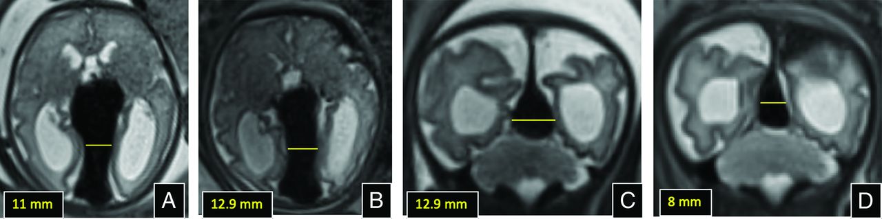
  • FIG 4.
    • Download figure
    • Open in new tab
    • Download powerpoint
    FIG 4.

    Measurement of the mediolateral width of the falcine sinus for consideration of fetal intervention and postintervention imaging. A, Axial T2 image at 32 weeks soon after the initial diagnosis of VOGM, with intact parenchyma and mild ventriculomegaly. B, Axial T2 image at 34 weeks, just before fetal embolization, shows interval growth in sinus width. C and D, Coronal T2 images prefetal embolization and 1-day post embolization respectively, showing diminution in width of the falcine sinus, indicative of diminution in lesional flow.

  • FIG 5.
    • Download figure
    • Open in new tab
    • Download powerpoint
    FIG 5.

    Pial arteriovenous fistula in a 32-week-old fetus. A, Sagittal T2 SSFSE shows a flow void in the frontal interhemispheric region (arrow) that drains into the deep venous system and eventually into the torcular. B, Sagittal T2 SSFSE through the right hemisphere shows the prominent pial vessels (white arrows), congestion of medullary veins, and developing periventricular cystic change. C, Coronal T2 SSFSE shows right greater than left parenchymal cystic changes (arrow), related to encephalomalacia and associated mild ventriculomegaly. D, Coronal T2* echo-planar image obtained 2 weeks later shows areas of blooming in the periventricular regions that correspond to areas of T1 shortening on (E) coronal T1 gradient-echo imaging (white arrows in D and E, respectively), related to venous congestion and subacute blood products.

  • FIG 6.
    • Download figure
    • Open in new tab
    • Download powerpoint
    FIG 6.

    Dural sinus malformation in a 33-week-old fetus. A, Sagittal T2 SSFSE shows marked dilation of the posterior aspect of the superior sagittal sinus and torcular. B, Sagittal DWI confirms the presence of clot within the dilated sinus (arrow). The brain parenchyma appears normal.
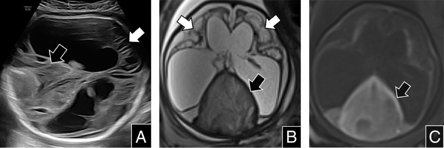
  • FIG 7.
    • Download figure
    • Open in new tab
    • Download powerpoint
    FIG 7.

    Dural sinus malformation in a 32-week-old fetus who was referred with a diagnosis of ventriculomegaly. A, Fetal sonography revealed a dilated dural sinus with internal echogenicity representing clot (black arrow). The echogenicity of the parenchyma was also abnormal (white arrow), with cystic changes and volume loss associated with severe ventriculomegaly. B, Axial T2 SSFSE shows severe parenchymal injury with cystic encephalomalacia (white arrows), severe ex vacuo ventriculomegaly, and intermediate signal from clot within the distended torcular (black arrow). C, Axial T1 gradient-echo shows blood products within the torcular that demonstrate T1 hyperintensity (black arrow).
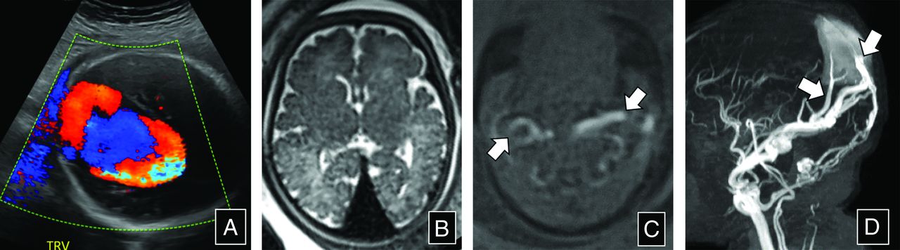
  • FIG 8.
    • Download figure
    • Open in new tab
    • Download powerpoint
    FIG 8.

    Dural sinus malformation in a 33-week-old fetus. A, Fetal sonography with color Doppler shows turbulent flow within a dilated torcular. B, Axial T2 SSFSE shows an enlarged flow void in the region of the torcular, without signs of clot. There were no sonographic changes in the brain parenchyma. C, Axial T1 gradient-echo shows flow-related signal within enlarged external carotid arteries bilaterally (arrows). D, 3D maximum intensity projection from the postnatal time-of-flight MRA shows arterialized flow within the dural sinus malformations and middle meningeal and occipital artery feeders (arrows).
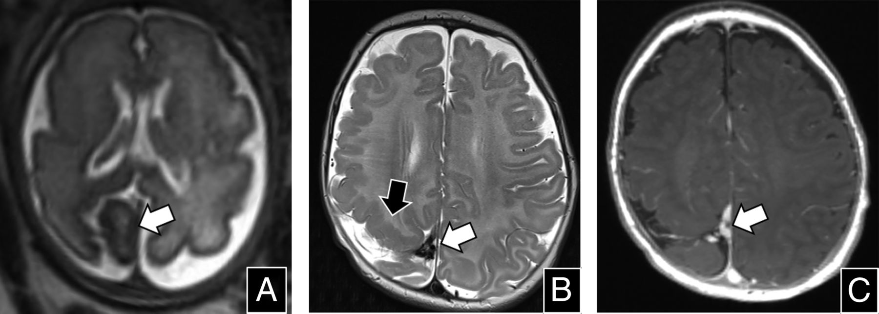
  • FIG 9.
    • Download figure
    • Open in new tab
    • Download powerpoint
    FIG 9.

    Intracranial VM in a 31-week-old fetus (white arrows in A to C). A, Axial T2 SSFSE shows a globoid soft tissue structure with internal signal heterogeneity located in the right posterior parafalcine region (arrow); the subjacent parietal parenchyma appears abnormally folded and hypointense. B, Axial T2 and (C) postcontrast axial MPRAGE demonstrate partial involution of the structure since the fetal examination (white arrows in B and C), with polymicrogyria adjacent to the lesion (black arrow in B). Persistent enhancement and a somewhat tubular and branching architecture are apparent on postcontrast imaging (C).

Tables

  • Figures
  • MRI protocol for evaluation of fetal cerebrovascular anomalies

    SequencePlaneTR/TE (msec)In-Plane Voxel Dimension (mm)Slice Thickness mm (Through-plane)Acceleration Factor (iPat)
    T2 SSFSE (maternal)aSag, Cor1400/1181×13–42
    T2 SSFSESag, Cor, Ax1400/991×13–42
    b-SSFPSag, Cor, Ax4.25 /1.811×13–42
    T1-weighted gradient-echoSag, Cor, Ax3.64/1.351×122
    Echo-planar T2* imagingSag, Cor, Ax6670/811×12–
    DWIbAx, Cor3000–4600/682×23–42
    • Note:—Parameters provided for a 3T Magnetom VIDA (Siemens) utilizing an 18-channel abdominal coil.

    • Ax indicates axial; Cor, coronal; Sag, sagittal.

    • aMaternal acquisition utilizes a large field of view of approximately 400 × 400 mm to provide full coverage of the uterus.

    • bDiffusion parameters include 6–12 directions, b = 0 sec/mm2 and b = 700 seconds/mm2.

PreviousNext
Back to top
Advertisement
Print
Download PDF
Email Article

Thank you for your interest in spreading the word on American Journal of Neuroradiology.

NOTE: We only request your email address so that the person you are recommending the page to knows that you wanted them to see it, and that it is not junk mail. We do not capture any email address.

Enter multiple addresses on separate lines or separate them with commas.
Cerebrovascular Anomalies in the Fetus
(Your Name) has sent you a message from American Journal of Neuroradiology
(Your Name) thought you would like to see the American Journal of Neuroradiology web site.
CAPTCHA
This question is for testing whether or not you are a human visitor and to prevent automated spam submissions.
Cite this article
Camilo Jaimes, Suely Fazio Ferraciolli, Darren B Orbach
Cerebrovascular Anomalies in the Fetus
American Journal of Neuroradiology Sep 2024, DOI: 10.3174/ajnr.A8377

Citation Manager Formats

  • BibTeX
  • Bookends
  • EasyBib
  • EndNote (tagged)
  • EndNote 8 (xml)
  • Medlars
  • Mendeley
  • Papers
  • RefWorks Tagged
  • Ref Manager
  • RIS
  • Zotero
0 Responses
Respond to this article
Share
Bookmark this article
Cerebrovascular Anomalies in the Fetus
Camilo Jaimes, Suely Fazio Ferraciolli, Darren B Orbach
American Journal of Neuroradiology Sep 2024, DOI: 10.3174/ajnr.A8377
del.icio.us logo Twitter logo Facebook logo Mendeley logo
  • Tweet Widget
  • Facebook Like
  • Google Plus One
Purchase

Jump to section

  • Article
    • SUMMARY:
    • ABBREVIATIONS:
    • IMAGING
    • FETAL CEREBROVASCULAR ANOMALIES
    • TAKE HOME POINTS
    • CONCLUSIONS
    • Footnotes
    • References
  • Figures & Data
  • Supplemental
  • Info & Metrics
  • Responses
  • References
  • PDF

Related Articles

  • PubMed
  • Google Scholar

Cited By...

  • No citing articles found.
  • Crossref
  • Google Scholar

This article has not yet been cited by articles in journals that are participating in Crossref Cited-by Linking.

More in this TOC Section

  • An Atlas of Neonatal Neurovascular Imaging Anatomy as Depicted with Microvascular Imaging: The Intracranial Arteries
  • An Atlas of Neonatal Neurovascular Imaging Anatomy as Depicted with Microvascular Imaging: The Intracranial Veins
  • Clinical Translation of Hyperpolarized 13C Metabolic Probes for Glioma Imaging
Show more Review Articles

Similar Articles

Advertisement

Indexed Content

  • Current Issue
  • Accepted Manuscripts
  • Article Preview
  • Past Issues
  • Editorials
  • Editor's Choice
  • Fellows' Journal Club
  • Letters to the Editor
  • Video Articles

Cases

  • Case Collection
  • Archive - Case of the Week
  • Archive - Case of the Month
  • Archive - Classic Case

More from AJNR

  • Trainee Corner
  • Imaging Protocols
  • MRI Safety Corner
  • Book Reviews

Multimedia

  • AJNR Podcasts
  • AJNR Scantastics

Resources

  • Turnaround Time
  • Submit a Manuscript
  • Submit a Video Article
  • Submit an eLetter to the Editor/Response
  • Manuscript Submission Guidelines
  • Statistical Tips
  • Fast Publishing of Accepted Manuscripts
  • Graphical Abstract Preparation
  • Imaging Protocol Submission
  • Evidence-Based Medicine Level Guide
  • Publishing Checklists
  • Author Policies
  • Become a Reviewer/Academy of Reviewers
  • News and Updates

About Us

  • About AJNR
  • Editorial Board
  • Editorial Board Alumni
  • Alerts
  • Permissions
  • Not an AJNR Subscriber? Join Now
  • Advertise with Us
  • Librarian Resources
  • Feedback
  • Terms and Conditions
  • AJNR Editorial Board Alumni

American Society of Neuroradiology

  • Not an ASNR Member? Join Now

© 2025 by the American Society of Neuroradiology All rights, including for text and data mining, AI training, and similar technologies, are reserved.
Print ISSN: 0195-6108 Online ISSN: 1936-959X

Powered by HighWire