
Article Figures & Data
Figures
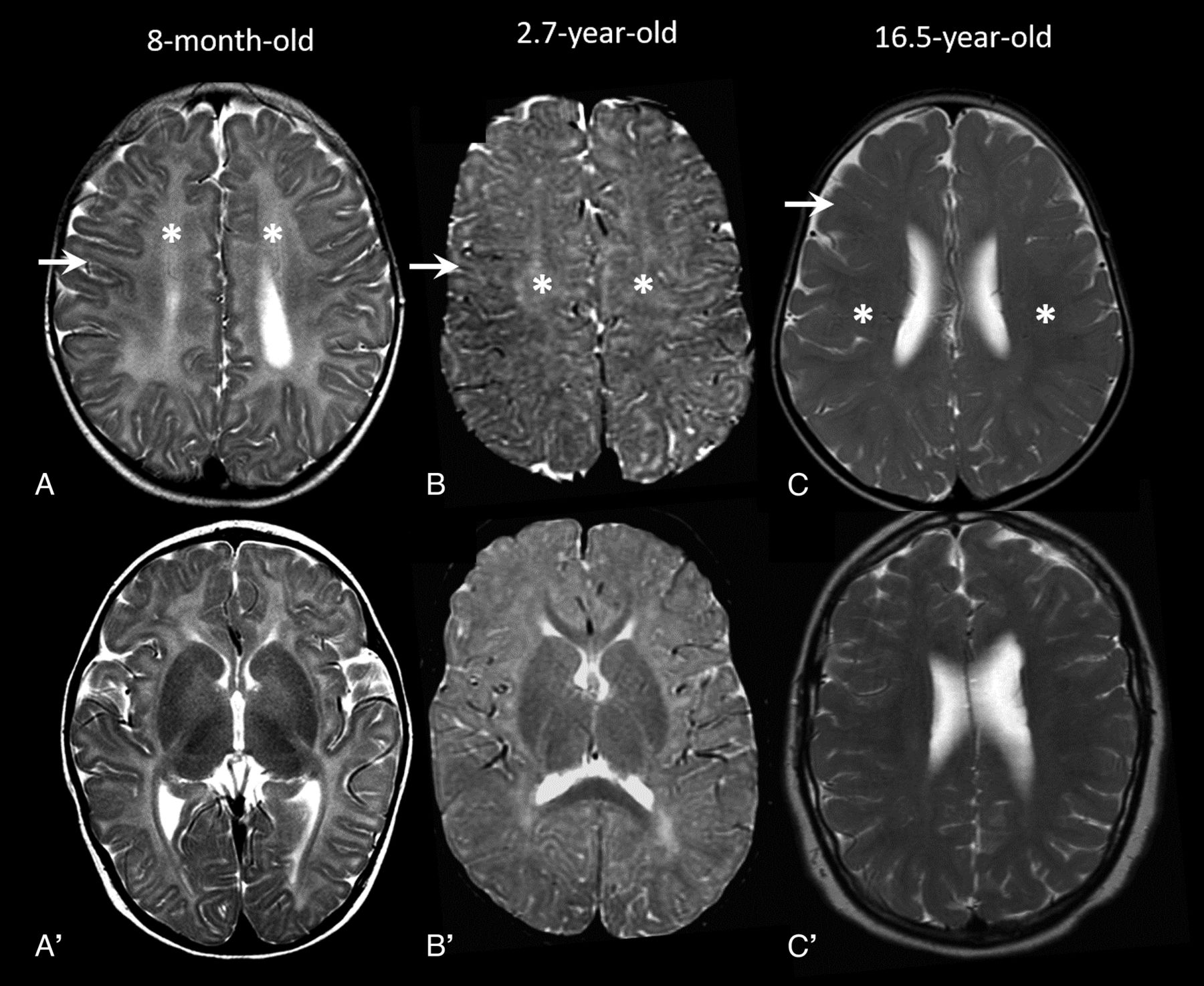
- FIG 1.
MR imaging patterns in 3 different cases. A and A', A PMD-like pattern with diffuse T2-hyperintensity of white matter (asterisk) and preserved GWMD (arrow). B and B', The “intermediate pattern” with mixed areas of preservation and loss of GWMD (arrow) with dirty inhomogeneous white matter appearance (asterisk). C and C'. The “washed-out pattern” with milder diffuse abnormal signal (asterisk) with diffusely poor GWMD (arrow).
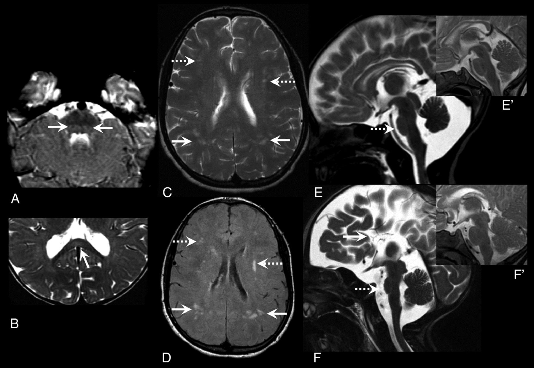
- FIG 2.
Focal signal changes and small pons. Axial T2 images show relatively better myelination and T2 hypointensity of the medial lemniscus (arrows, A) in the dorsal brainstem (closed-eye sign) and focal midsplenial T2 hyperintensity (arrow, B). Axial T2 (C) and FLAIR (D) images in an 18-year-old with ring chromosome 18 show multiple scattered focal hyperintensities in the frontal (dashed arrows) and parietal (solid arrows) white matter in the background of a washed-out MR imaging pattern. Midline sagittal sections in 2 trisomy 18 cases at 1 and 3 months (E and F, respectively) showing a reduced APD of pons (dashed arrow, E and F) and agenesis of CC in case 2 (arrow, F). Normal appearance of the pons in age-matched controls provided for reference in the inset images (E' and F').
- FIG 3.
Venn diagram and table detailing MR imaging core feature distribution across etiology subgroups (90% or greater: +++; 50%–70%: ++; <20%: +).


Tables
Clinical Characteristics (n = 36) Age at first imaging (months) 19.6 (4.3–59.3) Sex (F:M) 25:11 (2.2:1) Diagnosis 18q deletion (18q-) - 23 (64) Trisomy 18 - 7/tetrasomy 18 - 1 (Total 8 [22]) Ring chromosome 18 - 4 (11)18q inversion 1 (3) Clinical data (denominator indicates cases who underwent dedicated assessment for the specific feature and available documentation) Microcephalya 13/19 (68) Short staturea 13/14 (93) Visual impairment 11/15 (73) Hearing impairment 9/15 (60) Cleft palate 4/12 (33) Epilepsy 8/18 (44) Genitourinary 9/14 (64) Cardiac 11/16 (69) Imaging characteristics (n = 36, 50 MRIs) At least 1 abnormal MR imaging: 33/36 (92, n = 50 for the rows below) Abnormal white matter 35 (70) Abnormal CC 30 (60) Dysplastic/agenetic CC 8 (16) CC-APD <3rd percentilea 18/47 (38) Body and/or splenium thickness <3rd percentilea 25/47 (53) APD pons <3rd percentilea 10 (20) CCD vermis <3rd percentilea 14 (28) Other findings Periventricular nodular heterotopia 3 Ectopic posterior pituitary 3 Holoprosencephaly variant (thickened lamina rostralis and fused fornices, CNPAS with SMMCIb absent olfactory bulbs), aqueductal stenosis, polymicrogyria 1 each - Table 2:
Comparative analysis of the spectrum of imaging findings across the 18q chromosome abnormalities
18q- Group (n = 23) Ring chromosome 18 Group (n = 4) Trisomy 18 Group (Trisomy 18 n = 7, Tetrasomy 18 n = 1) P Value Number of MRIs (n = 49) 34 (69) 5 (10) 10 (21) - Age at MR imaging 51.8 (17.8–114.4) 18.1 (14.9–212.2) 6.8 (0.17–43.7) .06 Abnormal white matter 25 (74) 5 (100) 5 (50) .11 PMD-like pattern 4 (12) 1 (20) 2 (20) .37 Intermediate pattern 7 (21) 2 (40) 2 (20) Washed-out pattern 14 (41) 2 (40) 1 (10) Normal 9 (26) 0 5 (50) Myelination lag 17.2 (3.3–19.6) 27.0 (19.2–33.0) 9.0 (3.0–15.1) .005 (18 ring vs 18 trisomy) .03 (18 ring vs 18q-) Multifocal white matter hyperintensities 0 3 (60) 1 (10) .001 (18 ring vs others) APD pons (mm) 18.5 (16.9–19.4) 15.3 (15.2–19.5) 13.0 (10.4–17.3) .002 (18 trisomy vs 18q-) Pons APD <3rd percentile 3 (9) 0 (0) 7 (70) <.001 (18 trisomy vs others) Pons/FOD ratio 0.13 (0.12–0.14) 0.13 (0.12–0.14) 0.11 (0.10–0.12) .002 (18 trisomy vs 18q-) .02 (18 trisomy vs 18 ring) CCD vermis (mm) 41.8 (37.1–45.7) 38.8 (36.8–43.5) 27.6 (19.4–35.0) <.001 (18 trisomy vs 18q-) CCD vermis <3rd percentile 7 (21) 0 (0) 7 (70) .003 (18 trisomy vs others) CCD vermis/FOD ratio 0.30 (0.27–0.33) 0.31 (0.30–0.35) 0.23 (0.19–0.25) <.001 (18 trisomy vs 18q-) .002 (18 trisomy vs 18 ring) TCD (mm) 88.4 (81.7–94.3) 78.2 (72.2–87.9) 63.9 (47.6–88.3) .04 TCD/FOD ratio 0.65 (0.59–0.68) 0.63 (0.59–0.72) 0.56 (0.49–0.63) .06 Note:—Numbers in brackets indicate percentages and interquartile range as applicable.
- Table 3:
Venn diagram and table detailing MR imaging core feature distribution across etiology subgroups (90% or greater: +++; 50%–70%: ++; <20%: +)
Core Features Ring chromosome 18 18q Deletion (18q-) Trisomy/Tetrasomy 18 1. Delayed myelination +++ ++ ++ 2. Multifocal white matter changes ++ − + 3. Small pons (APD pons <3rd percentile) − + +++ 4. Small vermis (CCD vermis <3rd percentile) − + +++


Jump to section
Related Articles
Cited By...
- No citing articles found.