
Article Figures & Data
Figures
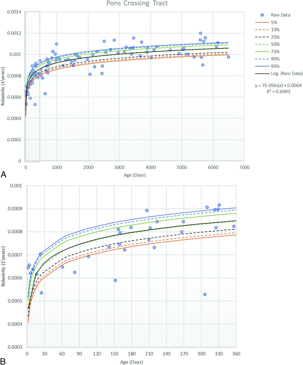
- FIG 1.
Scatterplot with trendline and percentile lines of the pons crossing tract. This includes the entire cohort (A) as well as a magnified view for the cohort in the first year of life (B).
- FIG 2.
Scatterplot with trendline and percentile lines of the middle cerebellar peduncle. This includes the entire cohort (A) as well as a magnified view for the cohort in the first year of life (B).
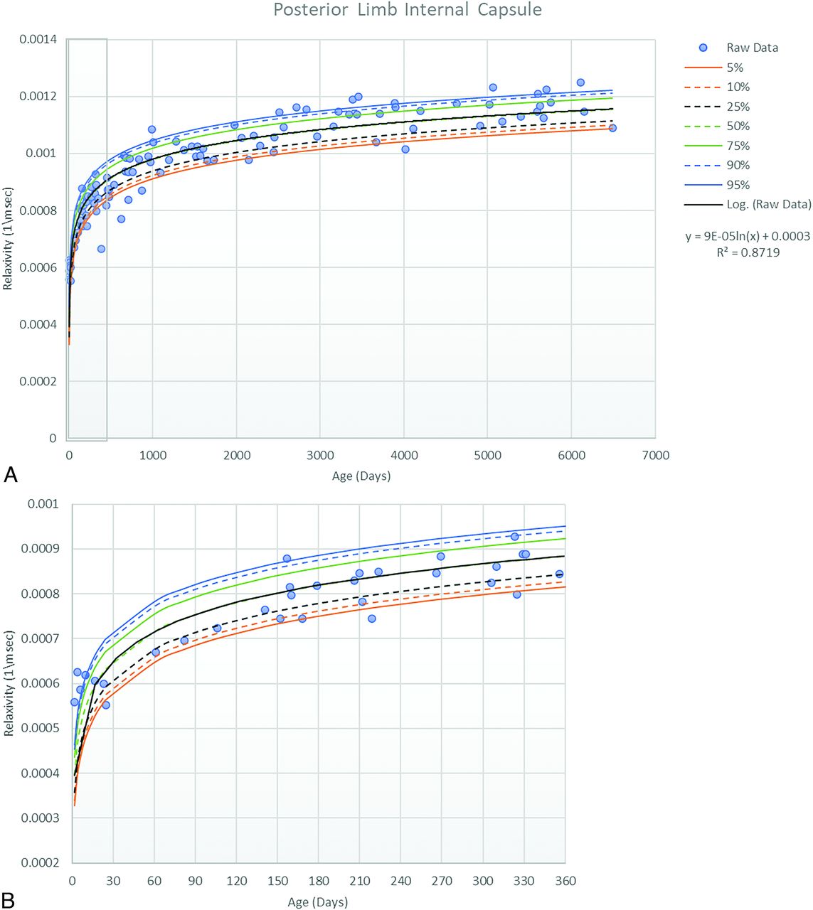
- FIG 3.
Scatterplot with trendline and percentile lines of the posterior limb of the internal capsule. This includes the entire cohort (A) as well as a magnified view for the cohort in the first year of life (B).
- FIG 4.
Scatterplot with trendline and percentile lines of the anterior limb of the internal capsule. This includes the entire cohort (A) as well as a magnified view for the cohort in the first year of life (B).
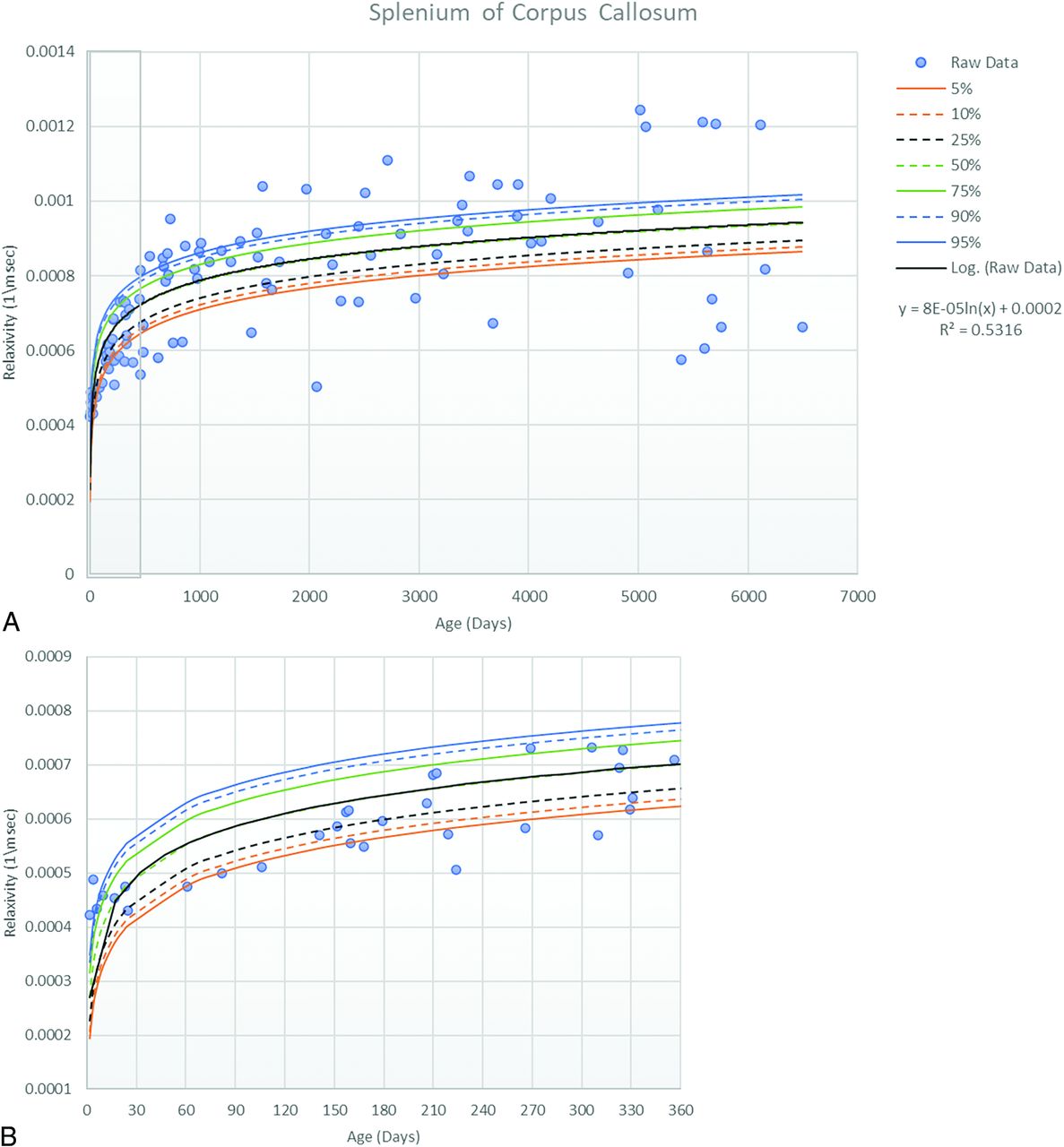
- FIG 5.
Scatterplot with trendline and percentile lines of the splenium of the corpus callosum. This includes the entire cohort (A) as well as a magnified view for the cohort in the first year of life (B).
- FIG 6.
Scatterplot with trendline and percentile lines of the genu of the corpus callosum. This includes the entire cohort (A) as well as a magnified view for the cohort in the first year of life (B).
- FIG 7.
Scatterplot with the trendline of select white matter atlas regions. This includes the entire cohort (A), and the magnified view of the cohort in the first year of life (B). ACR indicates anterior corona radiata; ALIC, anterior limb of internal capsule; BCC, body of corpus callosum; CP, cerebral peduncle; CST, corticospinal tract; EC, external capsule; GCC, genu of corpus callosum; ICP, inferior cerebellar peduncle; MCP, middle cerebellar peduncle; ML, medial lemniscus; PCR, posterior corona radiata; PCT, pontine crossing tract; PLIC, posterior limb of the internal capsule; PTR, posterior thalamic radiation; RLIC, retrolenticular internal capsule; SCC, splenium corpus callosum; SCP, superior cerebellar peduncle; SCR, superior corona radiata; UNC, uncinate fasiculus.
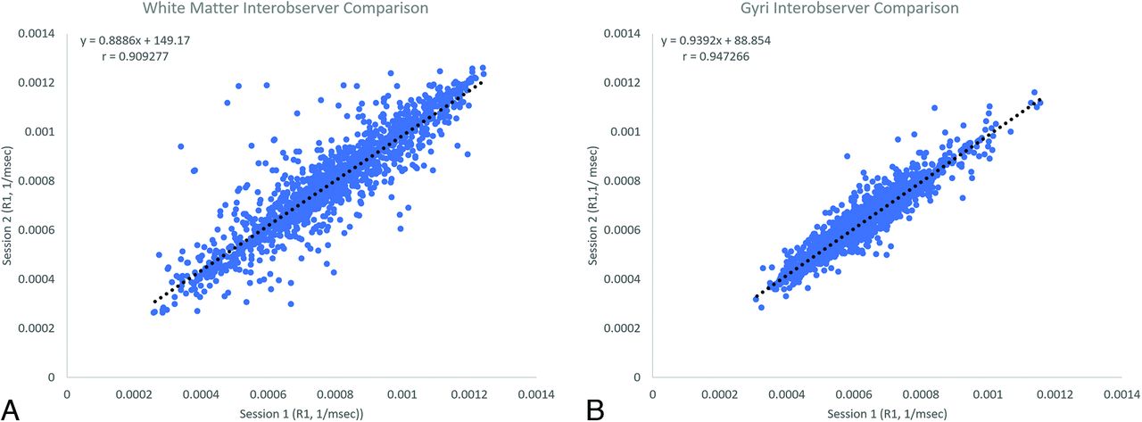
- FIG 8.
A, Pearson Product Moment Interobserver Comparison for white matter. B, Pearson Product Moment Interobserver Comparison for gyral anatomy.


Tables
Indications for MR imaging
Indications Seizures or spasms 26 Headaches 22 Developmental delay 16 Cutaneous or subcutaneous head and neck lesions 10 Abnormal eye movement 7 Attention deficit/hyperactivity disorder 4 Chiari I 4 Non-CNS malformations 3 Macrocephaly 2 Narcolepsy 2 Autism 1 Failure to thrive 1 Hearing loss 1 Hypertonia 1 Hypoxic-ischemic encephalopathy 1 Nonaccidental trauma work-up 1 Vomiting 1 Total 103


Jump to section
Related Articles
Cited By...
- No citing articles found.