Advertisement

Main menu

  • Home
  • Content
    • Current Issue
    • Accepted Manuscripts
    • Article Preview
    • Past Issue Archive
    • Video Articles
    • AJNR Case Collection
    • Case of the Week Archive
    • Case of the Month Archive
    • Classic Case Archive
  • Special Collections
    • AJNR Awards
    • Low-Field MRI
    • Alzheimer Disease
    • ASNR Foundation Special Collection
    • Photon-Counting CT
    • View All
  • Multimedia
    • AJNR Podcasts
    • AJNR SCANtastic
    • Trainee Corner
    • MRI Safety Corner
    • Imaging Protocols
  • For Authors
    • Submit a Manuscript
    • Submit a Video Article
    • Submit an eLetter to the Editor/Response
    • Manuscript Submission Guidelines
    • Statistical Tips
    • Fast Publishing of Accepted Manuscripts
    • Graphical Abstract Preparation
    • Imaging Protocol Submission
    • Author Policies
  • About Us
    • About AJNR
    • Editorial Board
    • Editorial Board Alumni
  • More
    • Become a Reviewer/Academy of Reviewers
    • Subscribers
    • Permissions
    • Alerts
    • Feedback
    • Advertisers
    • ASNR Home

User menu

  • Alerts
  • Log in

Search

  • Advanced search
American Journal of Neuroradiology

American Journal of Neuroradiology

ASHNR American Society of Functional Neuroradiology ASHNR American Society of Pediatric Neuroradiology ASSR
  • Alerts
  • Log in

Advanced Search

  • Home
  • Content
    • Current Issue
    • Accepted Manuscripts
    • Article Preview
    • Past Issue Archive
    • Video Articles
    • AJNR Case Collection
    • Case of the Week Archive
    • Case of the Month Archive
    • Classic Case Archive
  • Special Collections
    • AJNR Awards
    • Low-Field MRI
    • Alzheimer Disease
    • ASNR Foundation Special Collection
    • Photon-Counting CT
    • View All
  • Multimedia
    • AJNR Podcasts
    • AJNR SCANtastic
    • Trainee Corner
    • MRI Safety Corner
    • Imaging Protocols
  • For Authors
    • Submit a Manuscript
    • Submit a Video Article
    • Submit an eLetter to the Editor/Response
    • Manuscript Submission Guidelines
    • Statistical Tips
    • Fast Publishing of Accepted Manuscripts
    • Graphical Abstract Preparation
    • Imaging Protocol Submission
    • Author Policies
  • About Us
    • About AJNR
    • Editorial Board
    • Editorial Board Alumni
  • More
    • Become a Reviewer/Academy of Reviewers
    • Subscribers
    • Permissions
    • Alerts
    • Feedback
    • Advertisers
    • ASNR Home
  • Follow AJNR on Twitter
  • Visit AJNR on Facebook
  • Follow AJNR on Instagram
  • Join AJNR on LinkedIn
  • RSS Feeds

AJNR Awards, New Junior Editors, and more. Read the latest AJNR updates

Research ArticleULTRA-HIGH-FIELD MRI/IMAGING OF EPILEPSY/DEMYELINATING DISEASES/INFLAMMATION/INFECTION

Multiparametric Characterization and Spatial Distribution of Different MS Lesion Phenotypes

Francesco Tazza, Giacomo Boffa, Simona Schiavi, Caterina Lapucci, Gian Franco Piredda, Emilio Cipriano, Domenico Zacà, Luca Roccatagliata, Tom Hilbert, Tobias Kober, Matilde Inglese and Mauro Costagli
American Journal of Neuroradiology May 2024, DOI: https://doi.org/10.3174/ajnr.A8271
Francesco Tazza
aFrom the Departments of Neuroscience, Rehabilitation, Ophthalmology, Genetics, Maternal and Child Health (F.T., G.B., S.S., E.C., M.I., M.C.), University of Genoa, Genoa, Italy
  • Find this author on Google Scholar
  • Find this author on PubMed
  • Search for this author on this site
  • ORCID record for Francesco Tazza
Giacomo Boffa
aFrom the Departments of Neuroscience, Rehabilitation, Ophthalmology, Genetics, Maternal and Child Health (F.T., G.B., S.S., E.C., M.I., M.C.), University of Genoa, Genoa, Italy
  • Find this author on Google Scholar
  • Find this author on PubMed
  • Search for this author on this site
  • ORCID record for Giacomo Boffa
Simona Schiavi
aFrom the Departments of Neuroscience, Rehabilitation, Ophthalmology, Genetics, Maternal and Child Health (F.T., G.B., S.S., E.C., M.I., M.C.), University of Genoa, Genoa, Italy
  • Find this author on Google Scholar
  • Find this author on PubMed
  • Search for this author on this site
Caterina Lapucci
bIstituto di Ricovero e Cura a Carattere Scientifico (C.L., L.R., M.I., M.C.), Ospedale Policlinico San Martino, Genoa, Italy
  • Find this author on Google Scholar
  • Find this author on PubMed
  • Search for this author on this site
  • ORCID record for Caterina Lapucci
Gian Franco Piredda
cAdvanced Clinical Imaging Technology (G.F.P., T.H., T.K.), Siemens Healthineers International AG, Lausanne, Switzerland
  • Find this author on Google Scholar
  • Find this author on PubMed
  • Search for this author on this site
Emilio Cipriano
aFrom the Departments of Neuroscience, Rehabilitation, Ophthalmology, Genetics, Maternal and Child Health (F.T., G.B., S.S., E.C., M.I., M.C.), University of Genoa, Genoa, Italy
  • Find this author on Google Scholar
  • Find this author on PubMed
  • Search for this author on this site
Domenico Zacà
fSiemens Healthcare (D.Z.), Milan, Italy
  • Find this author on Google Scholar
  • Find this author on PubMed
  • Search for this author on this site
Luca Roccatagliata
bIstituto di Ricovero e Cura a Carattere Scientifico (C.L., L.R., M.I., M.C.), Ospedale Policlinico San Martino, Genoa, Italy
gDepartment of Health Sciences (L.R.), University of Genoa, Genoa, Italy
  • Find this author on Google Scholar
  • Find this author on PubMed
  • Search for this author on this site
  • ORCID record for Luca Roccatagliata
Tom Hilbert
cAdvanced Clinical Imaging Technology (G.F.P., T.H., T.K.), Siemens Healthineers International AG, Lausanne, Switzerland
dDepartment of Radiology (T.H., T.K.), Lausanne University Hospital and University of Lausanne, Lausanne, Switzerland
eLTS5 (T.H., T.K.), École Polytechnique Fédérale de Lausanne, Lausanne, Switzerland
  • Find this author on Google Scholar
  • Find this author on PubMed
  • Search for this author on this site
Tobias Kober
cAdvanced Clinical Imaging Technology (G.F.P., T.H., T.K.), Siemens Healthineers International AG, Lausanne, Switzerland
dDepartment of Radiology (T.H., T.K.), Lausanne University Hospital and University of Lausanne, Lausanne, Switzerland
eLTS5 (T.H., T.K.), École Polytechnique Fédérale de Lausanne, Lausanne, Switzerland
  • Find this author on Google Scholar
  • Find this author on PubMed
  • Search for this author on this site
  • ORCID record for Tobias Kober
Matilde Inglese
aFrom the Departments of Neuroscience, Rehabilitation, Ophthalmology, Genetics, Maternal and Child Health (F.T., G.B., S.S., E.C., M.I., M.C.), University of Genoa, Genoa, Italy
bIstituto di Ricovero e Cura a Carattere Scientifico (C.L., L.R., M.I., M.C.), Ospedale Policlinico San Martino, Genoa, Italy
  • Find this author on Google Scholar
  • Find this author on PubMed
  • Search for this author on this site
  • ORCID record for Matilde Inglese
Mauro Costagli
aFrom the Departments of Neuroscience, Rehabilitation, Ophthalmology, Genetics, Maternal and Child Health (F.T., G.B., S.S., E.C., M.I., M.C.), University of Genoa, Genoa, Italy
bIstituto di Ricovero e Cura a Carattere Scientifico (C.L., L.R., M.I., M.C.), Ospedale Policlinico San Martino, Genoa, Italy
  • Find this author on Google Scholar
  • Find this author on PubMed
  • Search for this author on this site
  • ORCID record for Mauro Costagli
  • Article
  • Figures & Data
  • Info & Metrics
  • Responses
  • References
  • PDF
Loading

Article Figures & Data

Figures

  • Tables
  • FIG 1.
    • Download figure
    • Open in new tab
    • Download powerpoint
    FIG 1.

    Flow chart of the MS lesions evaluation based on QSM and SMWI visualization.
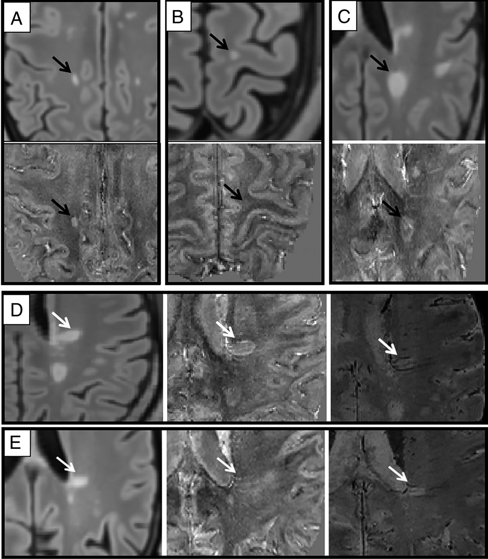
  • FIG 2.
    • Download figure
    • Open in new tab
    • Download powerpoint
    FIG 2.

    Examples of different MS lesion phenotypes. A, B, and C, MS lesions (black arrow) on FLAIR (upper image) and on QSM (lower image) classified, respectively, in HYPER, ISO-HYPO, and PRLs groups. D and E, MS lesions (white arrows) on FLAIR (left), QSM (center), and SMWI (right), which were relocated, respectively, from PRLs to HYPER and from HYPER to ISO-HYPO groups; particularly in D, the hyperintense rim on QSM was determined to be composed of 2 veins (visible as hypointense signal in SMWI). E, The hyperintensity on QSM located at the center of the lesion was a central vein, as shown on SMWI.
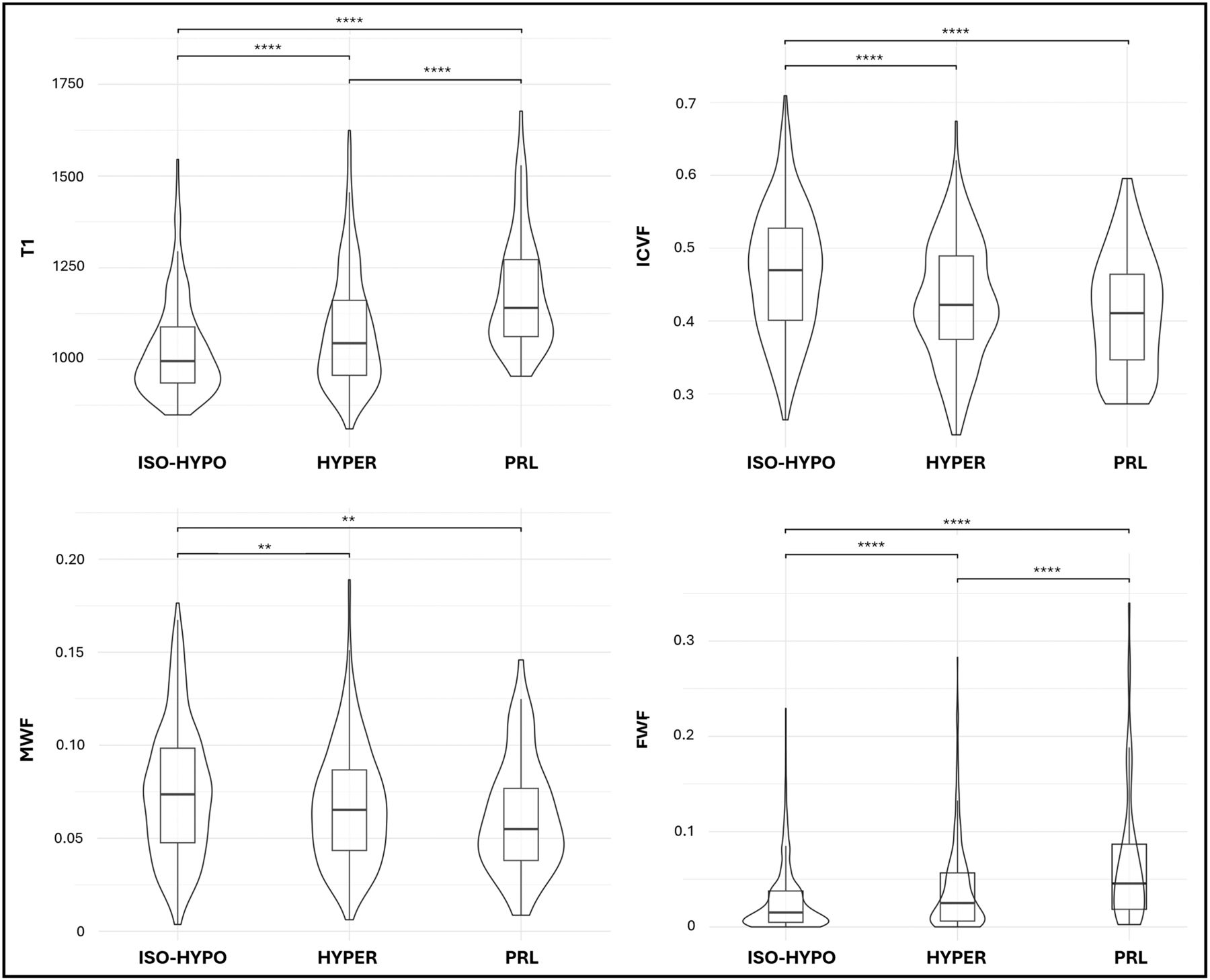
  • FIG 3.
    • Download figure
    • Open in new tab
    • Download powerpoint
    FIG 3.

    Quantitative MR imaging metrics of myelin and axonal loss in different MS lesion phenotypes. Comparisons were performed using ANOVA with Bonferroni post hoc comparisons, controlling for age, sex, lesion volume, and subject identification as confounding factors. T1-relaxation times are expressed in milliseconds. **P ≤ .05; ****P < .001.

Tables

  • Figures
    • View popup
    Table 1:

    Demographic and clinical features

    Patients with MS (n = 53)Healthy Controls (n = 24)P Value
    Female (No.) (%)34 (64.2%)16 (66.7%).830
    Mean age (yr)43.64 (SD, 12.58)38.12 (SD, 13.2).642
    RRMS (No.) (%)40 (75.5%)NANA
    Mean disease duration (yr)9.91 (SD, 10.56)NANA
    EDSS (median) (range)2.0 (0–7.0)NANA
    ARMSS (mean) (SD)3.99 (SD, 2.32)NANA
    Treatment
     No therapy (No.) (%)9 (17%)NANA
     Low-efficacy DMT (No.) (%)10 (19%)NANA
     High-efficacy DMT (No.) (%)34 (64%)NANA
    • Note:—RRMS indicates relapsing-remitting MS; NA, not applicable; DMT, disease-modifying therapy.

    • View popup
    Table 2:

    Comparison of lesion volume and quantitative MR imaging values of the 3 lesion phenotypes

    ISO-HYPO (Mean) (SD)HYPER (Mean) (SD)PRL (Mean) (SD)ISO-HYPO vs HYPER (P Value)ISO-HYPO vs PRL (P Value)HYPER vs PRL (P Value)
    Lesion volume (μL)311.44 (1069.32)820.93 (1733.99)955.35 (891.77)<.001a<.001a.455
    ICVF0.47 (0.09)0.42 (0.08)0.41 (0.08)<.001a<.001a.188
    ODI0.23 (0.07)0.21 (0.06)0.22 (0.05).060.506.703
    FWF0.03 (0.03)0.04 (0.05)0.07 (0.07).006a<.001a<.001a
    MWF0.075 (0.035)0.068 (0.031)0.062 (0.030).016a.022a.336
    T1-relaxometry (ms)1027.05 (131.35)1077.18 (157.63)1181.43 (157.01).001a<.001a<.001a
    QSM values (ppb)–9.10 (23.7)9.06 (25.1)7.75 (23.5)<.001a<.001a.567
    Distance from ventricular CSF (mm)14.6 (9.8)12.5 (9.7)10.5 (7.4).059.018a.177
    • Note:—ppb indicates parts per billion.

    • ↵a Statistically significant.

    • View popup
    Table 3:

    Pearson correlation among age, EDSS, and ARMSS with the volume of the different MS lesion phenotypes

    ISO-HYPOHYPERPRLHYPER + PRL
    rP ValuerP ValuerP ValuerP Value
    Age0.045.763−0.060.689−0.015.920−0.082.583
    EDSS−0.180.2320.282.057−0.160.2890.228.127
    ARMSS−0.268.0790.382.011a−0.167.2770.342.023a
    • ↵aStatistically significant.

PreviousNext
Back to top
Advertisement
Print
Download PDF
Email Article

Thank you for your interest in spreading the word on American Journal of Neuroradiology.

NOTE: We only request your email address so that the person you are recommending the page to knows that you wanted them to see it, and that it is not junk mail. We do not capture any email address.

Enter multiple addresses on separate lines or separate them with commas.
Multiparametric Characterization and Spatial Distribution of Different MS Lesion Phenotypes
(Your Name) has sent you a message from American Journal of Neuroradiology
(Your Name) thought you would like to see the American Journal of Neuroradiology web site.
CAPTCHA
This question is for testing whether or not you are a human visitor and to prevent automated spam submissions.
Cite this article
Francesco Tazza, Giacomo Boffa, Simona Schiavi, Caterina Lapucci, Gian Franco Piredda, Emilio Cipriano, Domenico Zacà, Luca Roccatagliata, Tom Hilbert, Tobias Kober, Matilde Inglese, Mauro Costagli
Multiparametric Characterization and Spatial Distribution of Different MS Lesion Phenotypes
American Journal of Neuroradiology May 2024, DOI: 10.3174/ajnr.A8271

Citation Manager Formats

  • BibTeX
  • Bookends
  • EasyBib
  • EndNote (tagged)
  • EndNote 8 (xml)
  • Medlars
  • Mendeley
  • Papers
  • RefWorks Tagged
  • Ref Manager
  • RIS
  • Zotero
0 Responses
Respond to this article
Share
Bookmark this article
Multiparametric Characterization and Spatial Distribution of Different MS Lesion Phenotypes
Francesco Tazza, Giacomo Boffa, Simona Schiavi, Caterina Lapucci, Gian Franco Piredda, Emilio Cipriano, Domenico Zacà, Luca Roccatagliata, Tom Hilbert, Tobias Kober, Matilde Inglese, Mauro Costagli
American Journal of Neuroradiology May 2024, DOI: 10.3174/ajnr.A8271
del.icio.us logo Twitter logo Facebook logo Mendeley logo
  • Tweet Widget
  • Facebook Like
  • Google Plus One
Purchase

Jump to section

  • Article
    • Abstract
    • ABBREVIATIONS:
    • MATERIALS AND METHODS
    • RESULTS
    • DISCUSSION
    • CONCLUSIONS
    • Footnotes
    • References
  • Figures & Data
  • Info & Metrics
  • Responses
  • References
  • PDF

Related Articles

  • No related articles found.
  • PubMed
  • Google Scholar

Cited By...

  • No citing articles found.
  • Crossref
  • Google Scholar

This article has not yet been cited by articles in journals that are participating in Crossref Cited-by Linking.

More in this TOC Section

  • Automated Lesion Segmentation Software in MS
  • DL Image Reconstruction in T2-Weighted TSE at 7T
  • 7T MRI vasculitis imaging
Show more Ultra-High-Field MRI/Imaging of Epilepsy/Demyelinating Diseases/Inflammation/Infection

Similar Articles

Advertisement

Indexed Content

  • Current Issue
  • Accepted Manuscripts
  • Article Preview
  • Past Issues
  • Editorials
  • Editor's Choice
  • Fellows' Journal Club
  • Letters to the Editor
  • Video Articles

Cases

  • Case Collection
  • Archive - Case of the Week
  • Archive - Case of the Month
  • Archive - Classic Case

More from AJNR

  • Trainee Corner
  • Imaging Protocols
  • MRI Safety Corner
  • Book Reviews

Multimedia

  • AJNR Podcasts
  • AJNR Scantastics

Resources

  • Turnaround Time
  • Submit a Manuscript
  • Submit a Video Article
  • Submit an eLetter to the Editor/Response
  • Manuscript Submission Guidelines
  • Statistical Tips
  • Fast Publishing of Accepted Manuscripts
  • Graphical Abstract Preparation
  • Imaging Protocol Submission
  • Evidence-Based Medicine Level Guide
  • Publishing Checklists
  • Author Policies
  • Become a Reviewer/Academy of Reviewers
  • News and Updates

About Us

  • About AJNR
  • Editorial Board
  • Editorial Board Alumni
  • Alerts
  • Permissions
  • Not an AJNR Subscriber? Join Now
  • Advertise with Us
  • Librarian Resources
  • Feedback
  • Terms and Conditions
  • AJNR Editorial Board Alumni

American Society of Neuroradiology

  • Not an ASNR Member? Join Now

© 2025 by the American Society of Neuroradiology All rights, including for text and data mining, AI training, and similar technologies, are reserved.
Print ISSN: 0195-6108 Online ISSN: 1936-959X

Powered by HighWire