
Article Figures & Data
Figures
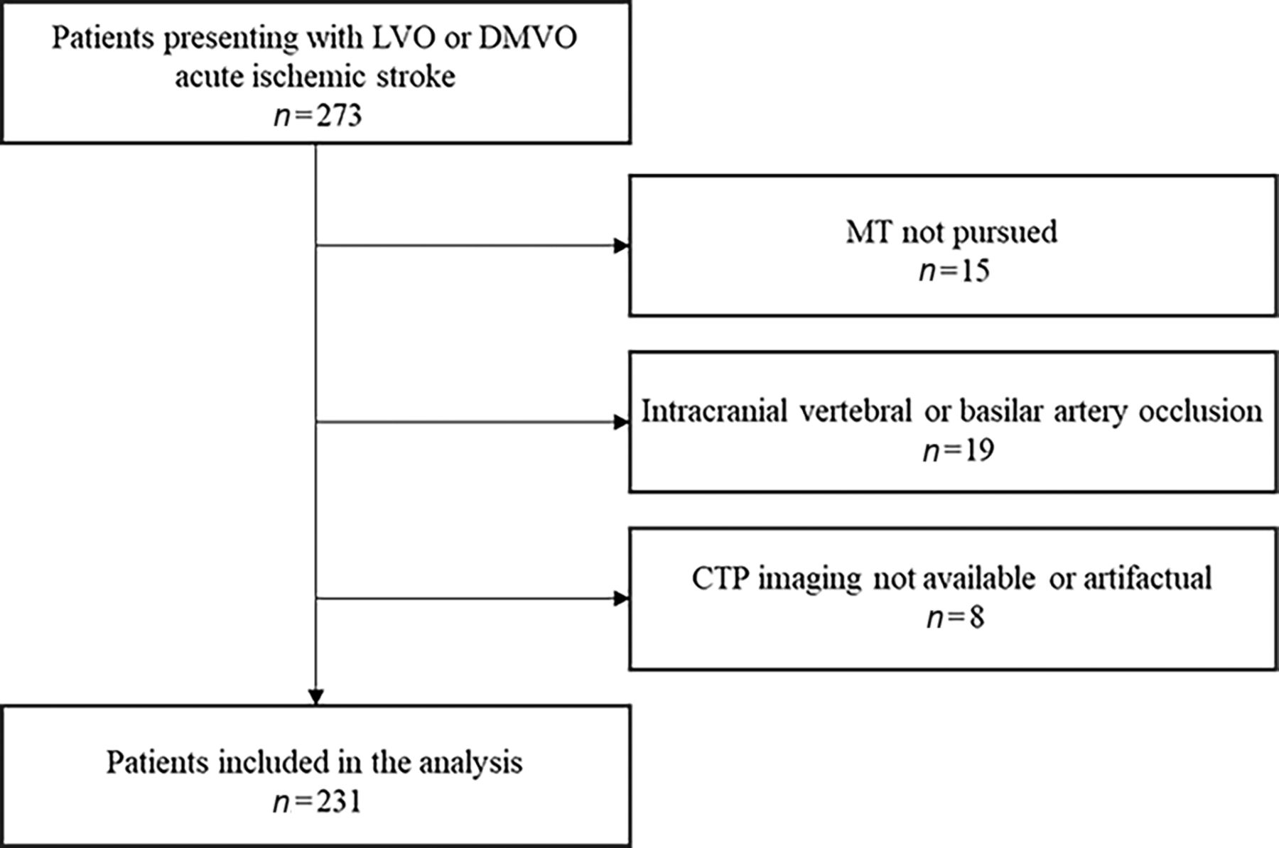
- FIG 1.
Flow chart of study design and patient selection.
- FIG 2.
Representative imaging demonstrating HIR ≥ 0.54 (A) versus HIR < 0.54 (B) CTP profiles. A, Tmax maps illustrating HIR calculated at 0.7 by RAPID software in a 51-year-old patient who presented with an initial NIHSS score of 9, ASPECTS of 10, and right MCA M1 occlusion and underwent MT with TICI 3 reperfusion. The 24-hour post-MT NIHSS score worsened to 17, indicating the occurrence of END. B, Tmax maps illustrating HIR calculated at 0.1 by RAPID software in a 54-year-old patient who presented with an initial NIHSS score of 10, ASPECTS of 9, and right MCA M1 occlusion and underwent MT with TICI 2b reperfusion. The 24-hour post-MT NIHSS score improved to 4.


Tables
- Table 1:
Patient characteristics of the entire cohort as stratified according to HIR statusa
All Patients HIR < 0.54 HIR ≥ 0.54 (n = 231) (n = 183) (n = 48) P Value Characteristics Age (yr) 70 (58–78) 70 (59–79) 67 (56–75) .323 Sex, female 119 (51.5) 96 (52.5) 23 (47.9) .628 LKW to presentation 0–6 hr 141 (61) 111 (60.7) 30 (62.6) .869 Initial NIHSS score 11 (5–18) 9 (4–16) 17 (11–21.5) <.001 NIHSS score at 24 hr post-MT 8 (2–14) 6 (2–13) 13 (7.5–20) <.001 Presence of END 26 (11.3) 20 (10.9) 6 (12.5) .798 Use of rtPA 76 (32.9) 62 (33.9) 14 (29.2) .607 ASPECTS 9 (8–10) 9 (8–10) 8 (7–9) .009 Occlusion location <.001 LVO 125 (54.1) 88 (48.1) 37 (77.1) DMVO 106 (45.9) 95 (51.9) 11 (22.9) LA grade 3–4 50 (21.6) 42 (23) 8 (16.7) .433 Poor collateral grade (0–1) 86 (37.2) 52 (28.4) 34 (70.8) <.001 Core volume (rCBF <30%, mL) 8 (0–23.5) 5 (0–13) 53 (22–76) <.001 rCBF <38% (mL) 17 (6–40) 12 (5–24) 76 (44–106.5) <.001 Tmax > 6 sec (mL) 87 (50–137.5) 71 (45.5–113) 147.5 (97.5–209.5) <.001 Tmax > 10 sec (mL) 25 (9.5–53) 18 (6–35.5) 91.5 (62.5–131.5) <.001 Mismatch volume (mL) 64 (40–112.5) 60 (39.5–102) 89.5 (46–122.5) .048 Pre-MT SBP (mm Hg) 151.5 (137–170) 151 (135–170.5) 152 (141–163) .802 TICI score 1.000 TICI 0–2a 24 (10.4) 19 (10.4) 5 (10.4) TICI 2b–3 207 (89.6) 164 (89.6) 43 (89.6) No. of passes 1 (1–2) 1 (1–2) 2 (1–3.5) .030 Stroke etiology .153 LAA 28 (12.1) 20 (10.9) 8 (16.7) Cardioembolic 96 (41.6) 74 (40.4) 22 (45.8) Cryptogenic 103 (44.6) 87 (47.5) 16 (33.3) Other determined 4 (1.7) 2 (1.1) 2 (4.2) Note:—LAA indicates large artery atherosclerosis.
↵a Data are No. (%) or median (25th–75th quartile).
- Table 2:
Multivariable linear regression models for factors associated with NIHSS score at 24 hours post-MT in the entire cohort, patients with LVOs, and DMVOsa
B 95% CI for B β P Value All patients Initial NIHSS score 0.418 0.315–0.522 0.427 <.001 LA (grade 3–4) 2.685 0.716–4.654 0.138 .008 HIR ≥ 0.54 3.237 1.156–5.319 0.163 .002 Use of rtPA −2.214 (−3.924)–(−0.505) −0.129 .011 Pre-MT SBP 0.029 0.0001–0.058 0.101 .047 No. of passes 1.325 0.731–1.919 0.242 <.001 TICI score 2b–3 −2.803 −5.715–0.109 −0.103 .059 Stroke etiology (cryptogenic) 1.586 −0.019–3.1901 0.098 .053 LVOs Initial NIHSS score 0.373 0.226–0.520 0.366 <.001 HIR ≥ 0.54 3.968 1.194–6.742 0.210 .005 Use of rtPA −3.802 (−6.428)–(−1.176) −0.206 .005 Pre-MT SBP 0.045 0.003–0.087 0.154 .035 No. of passes 1.304 0.558–2.050 0.250 .001 Stroke etiology (cryptogenic) 2.430 0.022–4.837 0.140 .048 DMVOs LA (grade 3–4) 3.280 0.912–5.648 0.202 .007 No. of passes 1.088 0.181–1.995 0.181 .019 TICI score 2b–3 −5.978 (−9.64)–(−2.316) −0.245 .002 Stroke etiology (LAA) 3.708 0.184–7.232 0.144 .039 Note:—LAA indicates large artery atherosclerosis.
↵a Models are adjusted for age, sex, initial NIHSS score, ASPECTS, CTA collateral score, rCBF <38%, HIR, LA, rtPA use, pre-MT SBP, time from LKW to presentation, TICI score, number of passes during MT, and stroke etiology. We used P < .1 as a criterion for backward steps. All patients: Adjusted R2 = 0.452, F = 24.180, P <.001. LVOs group: Adjusted R2 = 0.414, F = 15.262, P <.001. DMVOs group: Adjusted R2 = 0.516, F = 22.994, P <.001.
All Patients END Absent END Present (n = 231) (n = 205) (n = 26) P Value Characteristics Age (yr) 70 (58–78) 69 (57–78) 73.5 (65–80) .095 Sex, female 119 (51.5) 104 (50.7) 15 (57.7) .538 LKW to presentation 0–6 hr 141 (61) 128 (62.4) 13 (50) .286 Initial NIHSS score 11 (5–18) 12 (6–19) 6 (3–12) .005 NIHSS score at 24 hr post-MT 8 (2–14) 6 (2–13) 18 (12–24) <.001 Use of rtPA 76 (32.9) 72 (35.1) 4 (15.4) .048 ASPECTS 9 (8–10) 9 (8–10) 9 (8–10) .663 Occlusion location .835 LVO 125 (54.1) 110 (53.7) 15 (57.7) DMVO 106 (45.9) 95 (46.3) 11 (42.3) LA grade 3–4 50 (21.6) 43 (21) 7 (26.9) .613 CTA collateral score 0–1 86 (37.2) 172 (35.1) 14 (53.8) .084 rCBF <38% (mL) 17 (6–40) 17 (6–45) 19 (6.5–32) .836 Core volume (rCBF <30% mL) 8 (0–23.5) 8 (0–26) 6.5 (0–16) .451 Tmax > 6 sec (mL) 87 (50–137.5) 89 (52–139) 66.5 (43–116) .278 Tmax > 10 sec (mL) 25 (9–53) 28 (11–57) 18.5 (8–27) .141 Mismatch volume (mL) 64 (40–112.5) 66 (40–113) 60.5 (35–103) .479 HIR ≥ 0.54 48 (20.8) 42 (20.5) 6 (23.1) .798 Pre-MT SBP (mm Hg) 151.5 (137–170) 151 (135–167) 167.5 (146–189) .010 TICI score <.001 TICI 0–2a 24 (10.4) 14 (6.8) 10 (38.5) TICI 2b–3 207 (89.6) 191 (93.2) 16 (61.5) No. of passes 1 (1–2) 1 (1–2) 2 (2–4) <.001 Stroke etiology .379 LAA 28 (12.1) 23 (11.2) 5 (19.2) Cardioembolic 96 (41.6) 88 (42.9) 8 (30.8) Cryptogenic 103 (44.6) 90 (43.9) 13 (50) Other determined 4 (1.7) 4 (2) 0 (0) Note:—LAA indicates large artery atherosclerosis.
↵a Data are No. (%) or median (25th–75th quartile).
- Table 4:
Multivariable logistic regression models for factors associated with END in the entire cohort, patients with LVOs and DMVOsa
Adjusted OR 95% CI P Value All patients Age, per year 1.043 1.001–1.088 .044 Initial NIHSS score 0.842 0.771–0.919 <.001 ASPECTS 1.087 0.922–1.281 .320 HIR ≥ 0.54 3.100 0.838–11.598 .093 CTA collateral grade 0–1 3.013 1.058–8.582 .039 Pre-MT SBP (per mm Hg) 1.022 1.004–1.041 .018 TICI score 2b–3 0.096 0.028–0.328 <.001 LVOs Initial NIHSS score 0.836 0.751–0.932 .001 Pre-MT SBP (per mm Hg) 1.028 1.005–1.051 .016 Use of rtPA 5.195 0.606–44.535 .133 HIR ≥ 0.54 5.263 1.170–23.674 .030 DMVOs TICI 2b–3 0.020 0.003–0.124 <.001 Stroke etiology (LAA) 11.901 1.684–84.124 .013 Note:—LAA indicates large artery atherosclerosis.
↵a Models are adjusted for age, sex, initial NIHSS score, ASPECTS, collateral status, rCBF <38%, HIR, LA, rtPA use, pre-MT SBP, time from LKW to presentation, TICI score, number of passes during MT and stroke etiology. We used P < .1 as a criterion for backward steps. All patients: Hosmer–Lemeshow goodness of fit χ2 = 15.563, P = .049; LVOs group: Hosmer–Lemeshow goodness of fit χ2 = 6.235, P = .621; DMVOs group: Hosmer–Lemeshow goodness of fit χ2 = 0.002, P = .962.


