Advertisement

Main menu

  • Home
  • Content
    • Current Issue
    • Accepted Manuscripts
    • Article Preview
    • Past Issue Archive
    • Video Articles
    • AJNR Case Collection
    • Case of the Week Archive
    • Case of the Month Archive
    • Classic Case Archive
  • Special Collections
    • AJNR Awards
    • Low-Field MRI
    • Alzheimer Disease
    • ASNR Foundation Special Collection
    • Photon-Counting CT
    • View All
  • Multimedia
    • AJNR Podcasts
    • AJNR SCANtastic
    • Trainee Corner
    • MRI Safety Corner
    • Imaging Protocols
  • For Authors
    • Submit a Manuscript
    • Submit a Video Article
    • Submit an eLetter to the Editor/Response
    • Manuscript Submission Guidelines
    • Statistical Tips
    • Fast Publishing of Accepted Manuscripts
    • Graphical Abstract Preparation
    • Imaging Protocol Submission
    • Author Policies
  • About Us
    • About AJNR
    • Editorial Board
    • Editorial Board Alumni
  • More
    • Become a Reviewer/Academy of Reviewers
    • Subscribers
    • Permissions
    • Alerts
    • Feedback
    • Advertisers
    • ASNR Home

User menu

  • Alerts
  • Log in

Search

  • Advanced search
American Journal of Neuroradiology

American Journal of Neuroradiology

ASHNR American Society of Functional Neuroradiology ASHNR American Society of Pediatric Neuroradiology ASSR
  • Alerts
  • Log in

Advanced Search

  • Home
  • Content
    • Current Issue
    • Accepted Manuscripts
    • Article Preview
    • Past Issue Archive
    • Video Articles
    • AJNR Case Collection
    • Case of the Week Archive
    • Case of the Month Archive
    • Classic Case Archive
  • Special Collections
    • AJNR Awards
    • Low-Field MRI
    • Alzheimer Disease
    • ASNR Foundation Special Collection
    • Photon-Counting CT
    • View All
  • Multimedia
    • AJNR Podcasts
    • AJNR SCANtastic
    • Trainee Corner
    • MRI Safety Corner
    • Imaging Protocols
  • For Authors
    • Submit a Manuscript
    • Submit a Video Article
    • Submit an eLetter to the Editor/Response
    • Manuscript Submission Guidelines
    • Statistical Tips
    • Fast Publishing of Accepted Manuscripts
    • Graphical Abstract Preparation
    • Imaging Protocol Submission
    • Author Policies
  • About Us
    • About AJNR
    • Editorial Board
    • Editorial Board Alumni
  • More
    • Become a Reviewer/Academy of Reviewers
    • Subscribers
    • Permissions
    • Alerts
    • Feedback
    • Advertisers
    • ASNR Home
  • Follow AJNR on Twitter
  • Visit AJNR on Facebook
  • Follow AJNR on Instagram
  • Join AJNR on LinkedIn
  • RSS Feeds

AJNR Awards, New Junior Editors, and more. Read the latest AJNR updates

Research ArticleBRAIN TUMOR IMAGING

Differentiating Low-Grade from High-Grade Intracranial Ependymomas: Comparison of Dynamic Contrast-Enhanced MRI and Diffusion-Weighted Imaging

Julio Arevalo-Perez, Elena Yllera-Contreras, Kyung K. Peck, Vaios Hatzoglou, Onur Yildirim, Marc K. Rosenblum and Andrei I. Holodny
American Journal of Neuroradiology May 2024, DOI: https://doi.org/10.3174/ajnr.A8226
Julio Arevalo-Perez
aFrom the Department of Radiology (J.A.-P., E.Y.-C., V.H., O.Y., A.I.H.), Memorial Sloan Kettering Cancer Center, New York, New York
  • Find this author on Google Scholar
  • Find this author on PubMed
  • Search for this author on this site
  • ORCID record for Julio Arevalo-Perez
Elena Yllera-Contreras
aFrom the Department of Radiology (J.A.-P., E.Y.-C., V.H., O.Y., A.I.H.), Memorial Sloan Kettering Cancer Center, New York, New York
  • Find this author on Google Scholar
  • Find this author on PubMed
  • Search for this author on this site
  • ORCID record for Elena Yllera-Contreras
Kyung K. Peck
bDepartment of Medical Physics (K.K.P.), Memorial Sloan Kettering Cancer Center, New York, New York
  • Find this author on Google Scholar
  • Find this author on PubMed
  • Search for this author on this site
  • ORCID record for Kyung K. Peck
Vaios Hatzoglou
aFrom the Department of Radiology (J.A.-P., E.Y.-C., V.H., O.Y., A.I.H.), Memorial Sloan Kettering Cancer Center, New York, New York
  • Find this author on Google Scholar
  • Find this author on PubMed
  • Search for this author on this site
Onur Yildirim
aFrom the Department of Radiology (J.A.-P., E.Y.-C., V.H., O.Y., A.I.H.), Memorial Sloan Kettering Cancer Center, New York, New York
  • Find this author on Google Scholar
  • Find this author on PubMed
  • Search for this author on this site
  • ORCID record for Onur Yildirim
Marc K. Rosenblum
cDepartment of Pathology (M.K.R.), Memorial Sloan Kettering Cancer Center, New York, New York
  • Find this author on Google Scholar
  • Find this author on PubMed
  • Search for this author on this site
Andrei I. Holodny
aFrom the Department of Radiology (J.A.-P., E.Y.-C., V.H., O.Y., A.I.H.), Memorial Sloan Kettering Cancer Center, New York, New York
  • Find this author on Google Scholar
  • Find this author on PubMed
  • Search for this author on this site
  • ORCID record for Andrei I. Holodny
  • Article
  • Figures & Data
  • Info & Metrics
  • Responses
  • References
  • PDF
Loading

Article Figures & Data

Figures

  • Tables
  • FIG 1.
    • Download figure
    • Open in new tab
    • Download powerpoint
    FIG 1.

    Histopathologic features of low-grade (A) and high-grade (B) ependymomas. A, Modestly cellular low-grade ependymoma, with broad perivascular pseudorosettes, devoid of mitotic activity. B, Densely cellular high-grade ependymoma, with a narrower perivascular pseudorosette, microvascular proliferation, and mitotic activity.
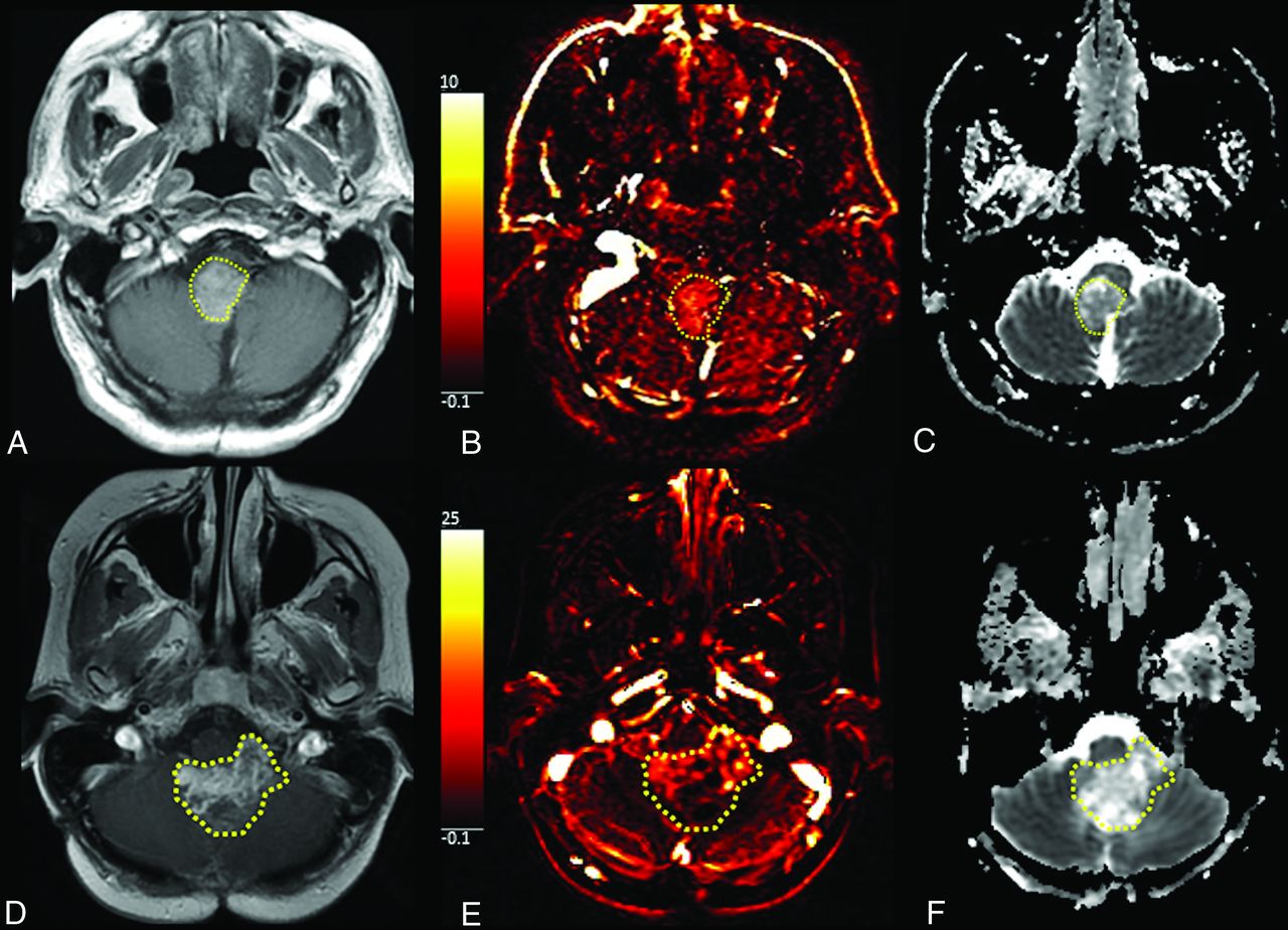
  • FIG 2.
    • Download figure
    • Open in new tab
    • Download powerpoint
    FIG 2.

    Examples of low-grade ependymomas (A, B, and C). A, T1-weighted postcontrast axial image demonstrates an enhancing mass in the inferior fourth ventricle and along the lower pons and dorsal medulla. B, Vp perfusion map shows mild elevation of Vp, 5.43. C, ADC map shows mildly elevated ADC values (579 mm2/s). Examples of high-grade ependymomas (D, E, and F). D, T1-weighted postcontrast axial image shows a heterogeneously enhancing partially cystic mass centered within the fourth ventricle with mass effect. E, Vp map shows areas of cystic changes and necrosis but also foci of elevated Vp (19.39), suggesting high vascularity. F, ADC map shows overall increased values with scattered areas of low ADC (514 mm2/s). The highlighted areas show the enhancing tumor in the inferior fourth ventricle; low-grade ependymoma, images A, B and C and high-grade ependymoma, images D, E and F.
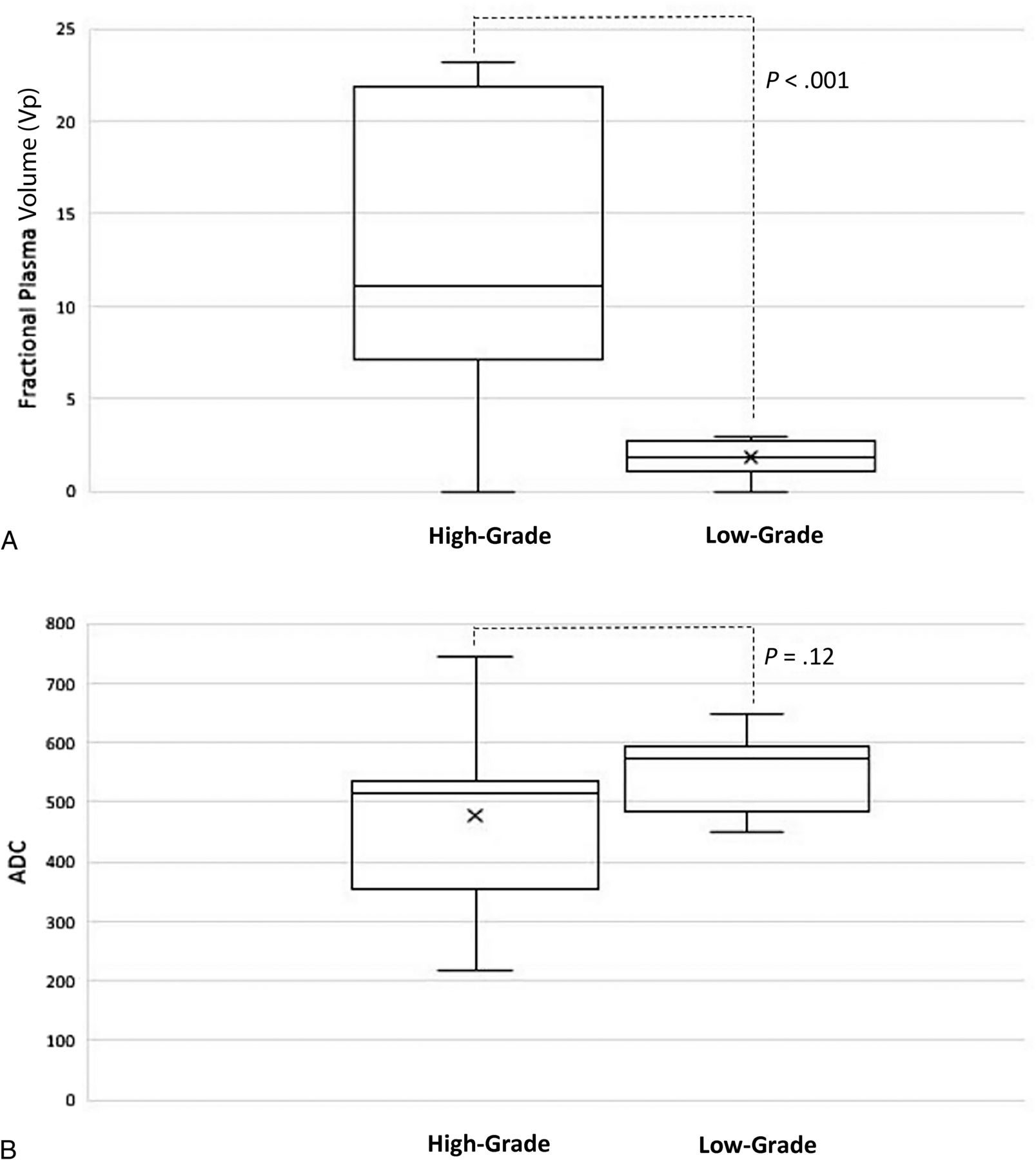
  • FIG 3.
    • Download figure
    • Open in new tab
    • Download powerpoint
    FIG 3.

    Boxplot of normalized Vp and ADC between low-grade and high-grade ependymomas.

  • FIG 4.
    • Download figure
    • Open in new tab
    • Download powerpoint
    FIG 4.

    ROC curve of Vp (blue) and ADC (green) values to differentiate low- and high-grade of ependymoma (reference line: red).

Tables

  • Figures
  • Demographic data, location of tumor, field strength of scanner, and normalized VPmax and ADCmin values

    HistopathologyAgeLocationSexField StrengthrVPmaxrADCmin
    Low-grade ependymoma21Posterior FossaM3T2.570.8
    Low-grade ependymoma72SupratentorialM3T0.951.02
    Low-grade ependymoma51Posterior FossaF3T1.090.91
    Low-grade ependymoma70Posterior FossaM3T1.80.9
    Low-grade ependymoma20Posterior FossaM3T1.530.73
    Low-grade ependymoma57Posterior FossaM3T2.750.85
    Low-grade ependymoma36Posterior FossaM3T1.410.89
    Low-grade ependymoma58Posterior FossaM1.5T2.980.93
    Low-grade ependymoma9SupratentorialF3T2.120.92
    Low-grade ependymoma13Posterior FossaM3T2.930.85
    Anaplastic ependymoma16Posterior FossaM1.5T16.290.73
    Anaplastic ependymoma14Posterior FossaM3T21.560.79
    Anaplastic ependymoma59SupratentorialM1.5T5.430.29
    Anaplastic ependymoma29SupratentorialF3T23.190.53
    Anaplastic ependymoma32SupratentorialF3T11.110.9
    Anaplastic ependymoma58SupratentorialM1.5T7.071.15
    Anaplastic ependymoma6Posterior FossaF3T21.90.99
    Anaplastic ependymoma42SupratentorialF1.5T22.50.83
    Anaplastic ependymoma10Posterior FossaF3T10.040.51
    Anaplastic ependymoma31Posterior FossaF3T10.40.83
    • Note:—rVPmax indicates relative maximum plama volume, rADCmin, relative ADCmin.

PreviousNext
Back to top
Advertisement
Print
Download PDF
Email Article

Thank you for your interest in spreading the word on American Journal of Neuroradiology.

NOTE: We only request your email address so that the person you are recommending the page to knows that you wanted them to see it, and that it is not junk mail. We do not capture any email address.

Enter multiple addresses on separate lines or separate them with commas.
Differentiating Low-Grade from High-Grade Intracranial Ependymomas: Comparison of Dynamic Contrast-Enhanced MRI and Diffusion-Weighted Imaging
(Your Name) has sent you a message from American Journal of Neuroradiology
(Your Name) thought you would like to see the American Journal of Neuroradiology web site.
CAPTCHA
This question is for testing whether or not you are a human visitor and to prevent automated spam submissions.
Cite this article
Julio Arevalo-Perez, Elena Yllera-Contreras, Kyung K. Peck, Vaios Hatzoglou, Onur Yildirim, Marc K. Rosenblum, Andrei I. Holodny
Differentiating Low-Grade from High-Grade Intracranial Ependymomas: Comparison of Dynamic Contrast-Enhanced MRI and Diffusion-Weighted Imaging
American Journal of Neuroradiology May 2024, DOI: 10.3174/ajnr.A8226

Citation Manager Formats

  • BibTeX
  • Bookends
  • EasyBib
  • EndNote (tagged)
  • EndNote 8 (xml)
  • Medlars
  • Mendeley
  • Papers
  • RefWorks Tagged
  • Ref Manager
  • RIS
  • Zotero
0 Responses
Respond to this article
Share
Bookmark this article
Differentiating Low-Grade from High-Grade Intracranial Ependymomas: Comparison of Dynamic Contrast-Enhanced MRI and Diffusion-Weighted Imaging
Julio Arevalo-Perez, Elena Yllera-Contreras, Kyung K. Peck, Vaios Hatzoglou, Onur Yildirim, Marc K. Rosenblum, Andrei I. Holodny
American Journal of Neuroradiology May 2024, DOI: 10.3174/ajnr.A8226
del.icio.us logo Twitter logo Facebook logo Mendeley logo
  • Tweet Widget
  • Facebook Like
  • Google Plus One
Purchase

Jump to section

  • Article
    • Abstract
    • ABBREVIATIONS:
    • MATERIALS AND METHODS
    • RESULTS
    • DISCUSSION
    • CONCLUSIONS
    • Footnotes
    • References
  • Figures & Data
  • Info & Metrics
  • Responses
  • References
  • PDF

Related Articles

  • No related articles found.
  • PubMed
  • Google Scholar

Cited By...

  • No citing articles found.
  • Crossref
  • Google Scholar

This article has not yet been cited by articles in journals that are participating in Crossref Cited-by Linking.

More in this TOC Section

  • Brain Tumor Subtyping Image-Based search via MRI
  • Neuroimaging of Erdheim-Chester Disease
  • CE MRI for Brain Metastasis Detection
Show more Brain Tumor Imaging

Similar Articles

Advertisement

Indexed Content

  • Current Issue
  • Accepted Manuscripts
  • Article Preview
  • Past Issues
  • Editorials
  • Editor's Choice
  • Fellows' Journal Club
  • Letters to the Editor
  • Video Articles

Cases

  • Case Collection
  • Archive - Case of the Week
  • Archive - Case of the Month
  • Archive - Classic Case

More from AJNR

  • Trainee Corner
  • Imaging Protocols
  • MRI Safety Corner
  • Book Reviews

Multimedia

  • AJNR Podcasts
  • AJNR Scantastics

Resources

  • Turnaround Time
  • Submit a Manuscript
  • Submit a Video Article
  • Submit an eLetter to the Editor/Response
  • Manuscript Submission Guidelines
  • Statistical Tips
  • Fast Publishing of Accepted Manuscripts
  • Graphical Abstract Preparation
  • Imaging Protocol Submission
  • Evidence-Based Medicine Level Guide
  • Publishing Checklists
  • Author Policies
  • Become a Reviewer/Academy of Reviewers
  • News and Updates

About Us

  • About AJNR
  • Editorial Board
  • Editorial Board Alumni
  • Alerts
  • Permissions
  • Not an AJNR Subscriber? Join Now
  • Advertise with Us
  • Librarian Resources
  • Feedback
  • Terms and Conditions
  • AJNR Editorial Board Alumni

American Society of Neuroradiology

  • Not an ASNR Member? Join Now

© 2025 by the American Society of Neuroradiology All rights, including for text and data mining, AI training, and similar technologies, are reserved.
Print ISSN: 0195-6108 Online ISSN: 1936-959X

Powered by HighWire