
Article Figures & Data
Figures
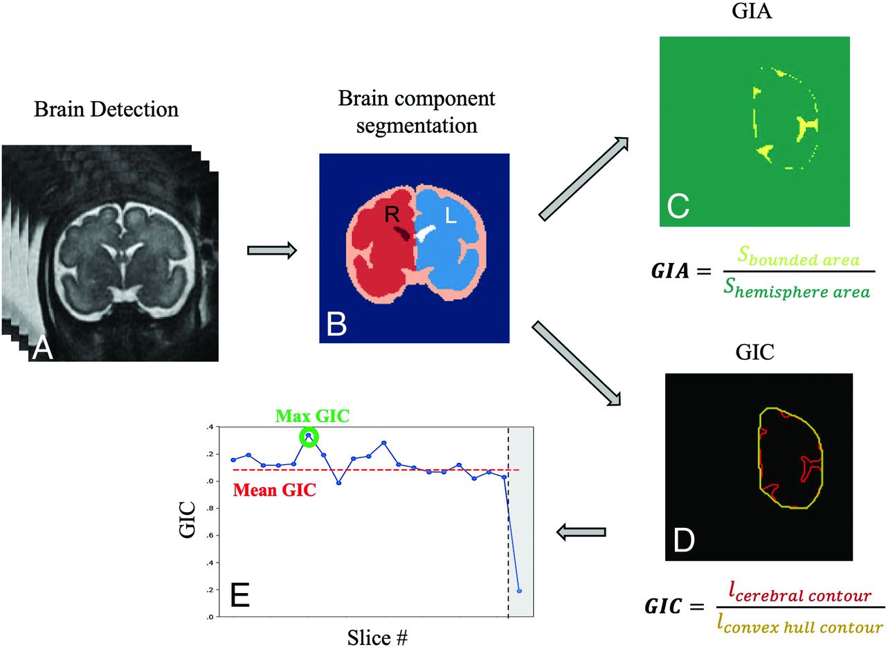
- FIG 1.
Image-analysis pipeline: A, Brain detection. B, Brain component segmentation. C, GIA calculation based on the bounded area (yellow) between the cerebral cortex and its convex hull and the hemisphere area (green, surrounded by the yellow area). D, GIC calculation based on contour extraction of the cerebral hemisphere (red) and its convex hull (yellow). E, Example of GIC values in all slices of a single fetus. Outliers are marked in gray and were not included in the mean GIC.
- FIG 2.
T2-weighted MR imaging of coronal views for control fetuses demonstrating gyrification development of the right hemisphere (red, hemisphere contour) at the Sylvian fissure level with gestation.
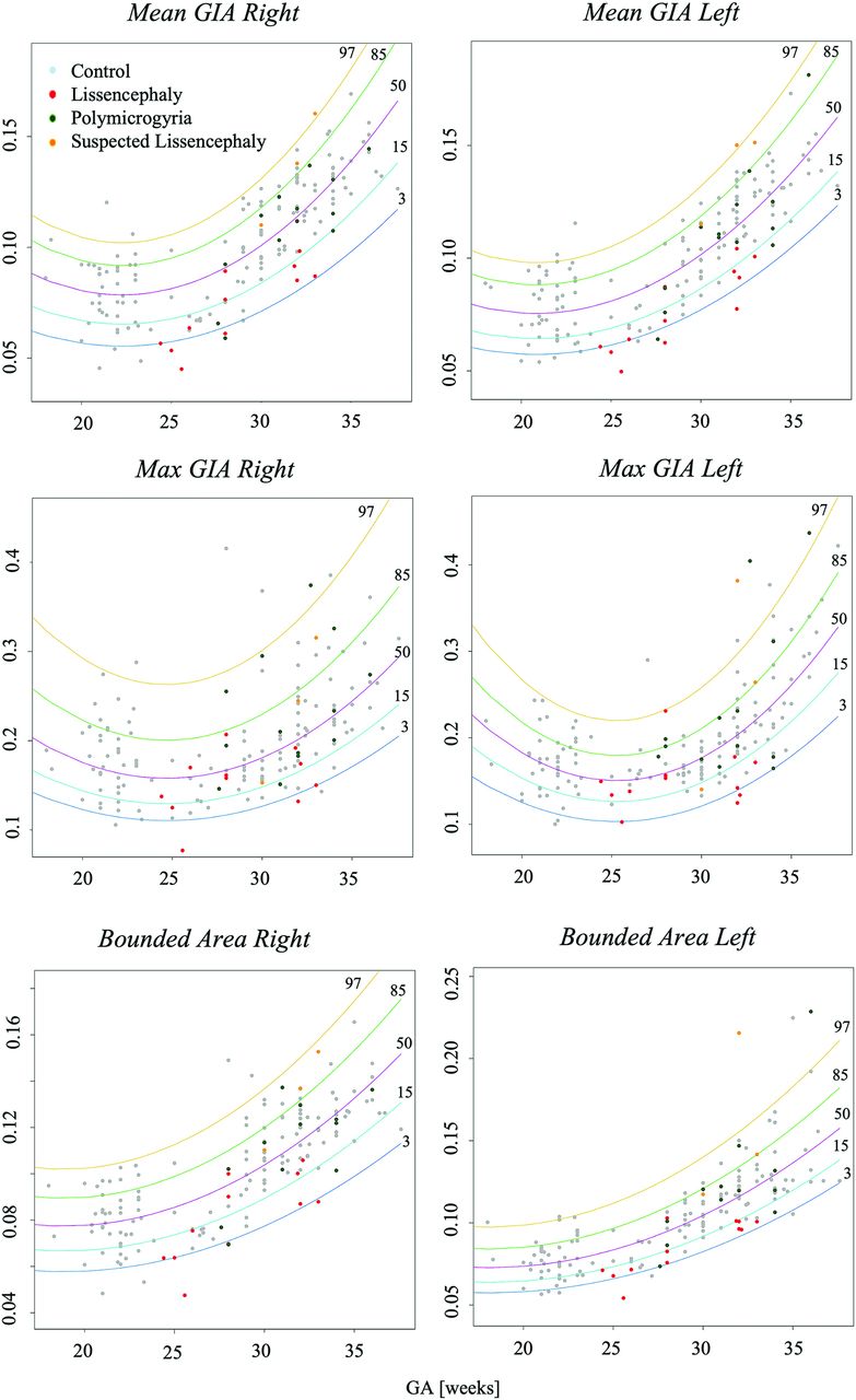
- FIG 3.
Development curves with GA of area-based gyrification parameters. Curve percentile lines 3, 15, 50, 85, and 97 are presented as blue, light blue, purple, green, and yellow, respectively. Note control fetuses (gray), PMG (green), LIS (red), and suspected LIS (orange).
- FIG 4.
Development curves with GA of contour-based gyrification parameters. Curve percentile lines 3, 15, 50, 85, and 97 are presented as blue, light blue, purple, green, and yellow, respectively. Note control fetuses (gray), PMG (green), LIS (red), and suspected LIS (orange).
- FIG 5.
Symmetry indices for GI parameters with GA. All are close to zero, with homogeneous dispersion, indicating no brain asymmetry.
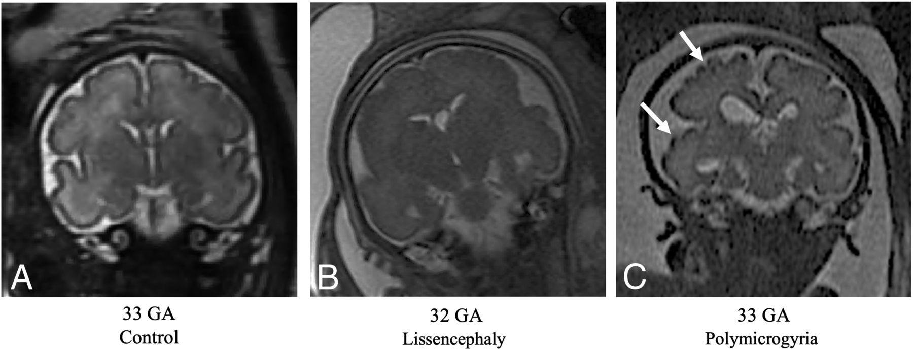
- FIG 6.
T2-weighted MR imaging of coronal views for controls and fetuses with LIS and PMG of equivalent ages. A, Control fetus, 33 weeks’ GA. B, Fetus diagnosed with LIS at 32 weeks’ GA, characterized by undeveloped gyrification patterns. C, Fetus diagnosed with PMG at 33 weeks’ GA, with abnormal excessive gyri (white arrows).


Tables
Vendor/System (Magnetic Field) Sequence No. TE (Milliseconds [SD]) TR (Milliseconds [SD]) Spacing (mm [SD]) GE Healthcare Discovery MR450 (1.5T) FRFSE 40 121.7 [SD, 2.3] 9946.5 [SD, 1984.5] 3.8 [SD, 0.8] Signa (1.5T) FIESTA 10 1.7 [SD, 0.09] 3.9 [SD, 0.2] 4.6 [SD, 0.9] SS-FSE 3 111.7 [SD, 0.6] 2517.4 [SD, 835.9] 3.9 [SD, 0.65 Siemens Magnetom Aera (1.5T) HASTE 4 94 [SD, 0] 1200 [SD, 0] 3.05 [SD, 0.7] Magnetom Prisma (3T) HASTE 2 96 [SD, 16.9] 2000 [SD, 0] 3.3 [SD, 1.8] TRUFI 4 2.5 [SD, 0.01] 4.9 [SD, 0.02] 3.15 [SD, 0.3] Skyra (3T) HASTE 31 92.4 [SD, 21.7] 1839.1 [SD, 325.1] 3.5 [SD, 0.6] TRUFI 7 2.5 [SD, 0.04] 4.9 [SD, 0.08] 3.9 [SD, 0.7] Magnetom Vida (3T) TRUFI 1 2.5 [SD, 0] 4.9 [SD, 0] 3 [SD, 0] Note:—SS-FSE indicates single-shot, fast spin-echo; TRUFI, true fast imaging with steady-state free precession; FRFSE, fast recovery fast spin echo; HASTE, Half-Fourier acquisition single-shot turbo spin-echo.
- Table 2:
Comparisons between LIS or PMG with control fetuses—the adjusted P values and CIs
Parameter LIS PMG P Value (CI Lower, CI Upper) P Value (CI Lower, CI Upper) Mean GIC right .002a (0.001–0.007) <.001a (0.002–0.008) Mean GIC left .002a (0.001–0.007) <.001a (0.002–0.008) Max GIC right <.001a (0.004–0.01) <.001a (0.003–0.008) Max GIC left <.001a (0.004–0.009) .001a (0.002–0.007) Bounded volume right .01 a (0.0003–0.001) .994 (–0.0004–0.0004) Bounded volume left <.001a (0.0003–0.009) .366 (–0.0006–0.0002) Mean GIA right <.001a (0.0006–0.001) .46 (–0.0002–0.0006) Mean GIA left <.001a (0.0006–0.001) .46 (–0.0003–0.0005) Max GIA right .02a (0.0002–0.002) .88 (–0.001–0.001) Max GIA left <.001a (0.0008–0.003) .994 (–0.001–0.001) a P value < .05.


Jump to section
Related Articles
Cited By...
- No citing articles found.