
Article Figures & Data
Figures
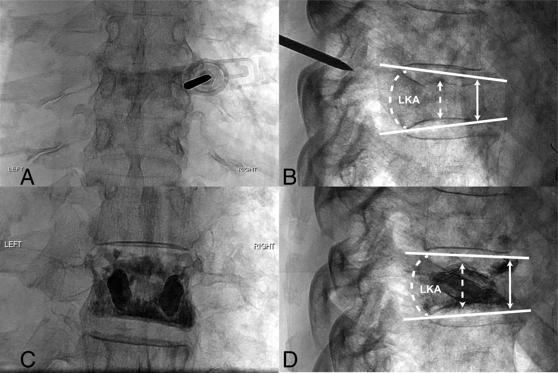
- FIG 1.
Sample radiographic measurements. A 72-year-old man with a history of osteoporosis presented with severe midthoracic back pain. Anterior-posterior (A) and lateral (B) projections of fluoroscopic images of the thoracic spine with the needle at the level of T8 demonstrate a preprocedural T8 vertebral compression fracture, as shown by an increased LKA formed by solid white lines along the superior and inferior endplates and decreased midvertebral (dashed arrow) and anterior-vertebral (solid arrow) body heights. Following vertebral augmentation with SJ, anterior-posterior (C) and lateral (D) projections show postprocedural changes with improvement in LKA and VH at T8.
- FIG 2.
Pain score comparison. A, Distribution of patient-reported pain scores on a scale between 0 and 10 at rest for 3 procedure types pre- (red) and postprocedure (blue). B, The same pain score distribution reported at worst. Shown are median and interquartile ranges.
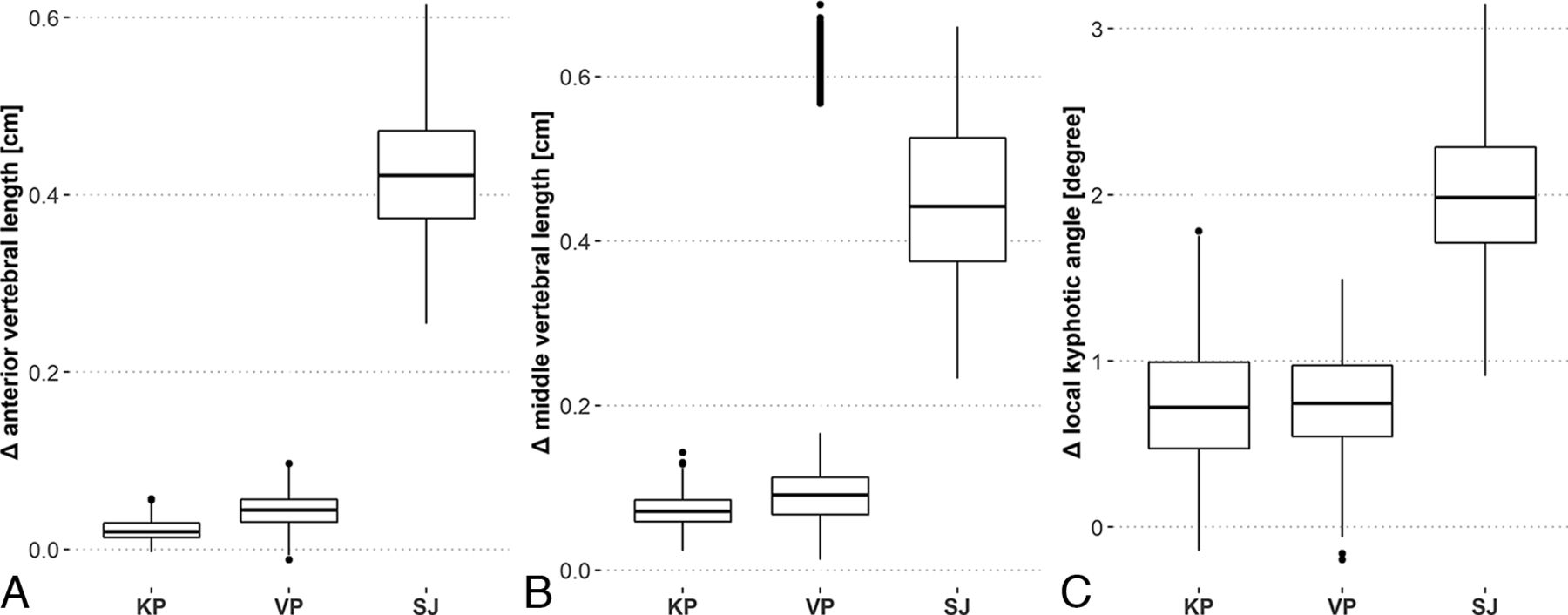
- FIG 3.
Vertebral augmentation radiologic changes. A, Distribution of changes in radiographic vertebral augmentation measurements for anterior vertebral length for the 3 procedures B, The same distribution of changes for middle vertebral length. C, The same distribution of changes for local kyphotic angle. Shown are median and interquartile ranges.


Tables
Kyphoplasty Vertebroplasty SJ No. of patients 67 74 61 No. of total procedures 79 84 67 Age (mean) (yr) 64.2 (SD, 12.3) 63.5 (SD, 12.8) 68.3 (SD, 10.6) No. of female (%) 37 (55.2) 38 (51.4) 35 (57.3) No. of African American (%) 12 (17.9) 15 (20.3) 14 (22.9) No. of European (%) 46 (68.6) 49 (66.2) 37 (60.7) No. of other race (%) 9 (13.4) 10 (13.5) 10 (16.4) BMI (mean) 28.2 (SD, 7.3) 26.8 (SD, 5.8) 28.7 (SD, 7.1) No. of pathologic fracturesa 46 59 19 No. of structural fracturesa 19 15 42 No. of treatment sessions 1 Procedure 58 64 57 2 Procedures 7 10 2 3 Procedures 1 0 2 4 Procedures 1 0 0 Median follow-up period (days) 39.5 70 94 ↵a Not all patients had recorded fracture type.
KP VP SJ P Valuea Anterior vertebral length (mm) Pre (mean) 18.4 20.6 13.0 <.001 Post (mean) 18.6 21.0 17.3 Δ 95% CI (0.03–0.4) (0.09–0.79) (3.1–5.6) % Increase (mean) 1.1 2.2 32.4 % CI (0.14–2.59) (0.4–3.9) (23.5–43.6) Middle vertebral length (mm) Pre (mean) 15.9 17.9 12.7 <.001 Post (mean) 16.6 19.5 17.2 Δ 95% CI (0.4–1.1) (0.4–6.5) (3.0–6.0) % Increase (mean) 4.5 5.0 35.6 % CI (2.3–7.1) (2.1–36.8) (24.0–46.4) LKA Pre (mean) 7.6° 6.8° 8.9° <.001 Post (mean) 6.9° 6.1° 6.9° Δ Angle Δ 95% CI (0.09°–1.38°) (0.15°–1.33°) (1.24°–2.80°) ↵a The P value calculated with Kruskal-Wallis χ2 tests across procedures.


Jump to section
Related Articles
Cited By...
- No citing articles found.