
Article Figures & Data
Figures
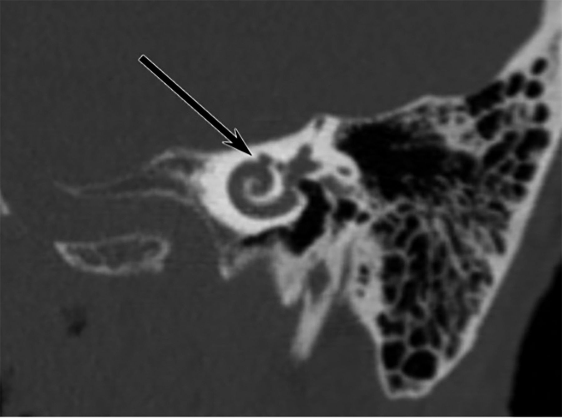
- FIG 1.
Cochlear-facial dehiscence. Oblique reformatted CT scan along the basal turn of the cochlea (modified Stenvers reformat) shows a dehiscence (arrow) between the middle turn of the cochlea and the labyrinthine segment of the facial nerve canal.
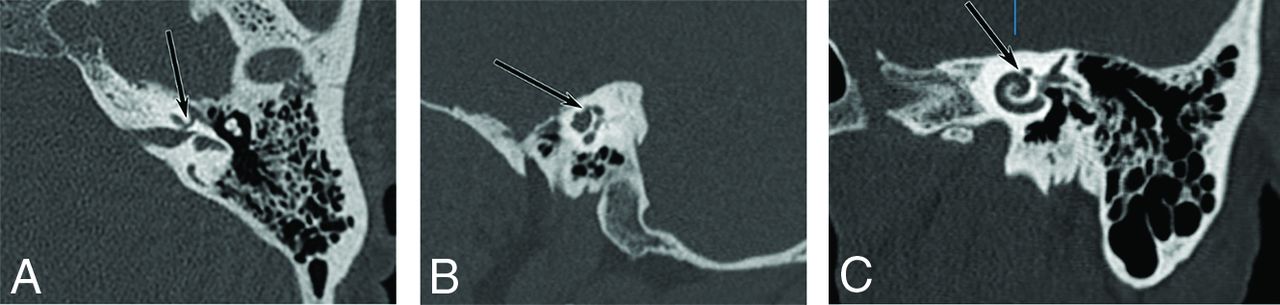
- FIG 2.
The importance of multiplanar oblique reformats for the diagnosis of cochlear-facial dehiscence. Axial CT image (A) shows an apparent dehiscence (arrow) between the middle turn of the cochlea and the labyrinthine segment of the facial nerve. Sagittal reformatted image (B) appears to confirm the dehiscence (arrow). However, multiple oblique reformats along the basal turn of the cochlea (C) reveal that the bone between the cochlea and the facial canal (arrow) is thin but intact.
- FIG 3.
JVD. Axial CT image shows dehiscence between a diverticulum of the jugular bulb (arrow) and the vestibular aqueduct (arrowhead).
- FIG 4.
Cochlear-carotid plate. Sagittal reformatted CT image shows thin-but-intact bone (arrow) between the petrous segment of the ICA and the basal turn of the cochlea. None of the 1204 temporal bones in this study demonstrated a true dehiscence at this location.


Tables
- Table 1:
Asymptomatic patient characteristics and indications for temporal bone CT imaging
Asymptomatic Patients Median age (range) (yr) 47 (15–93) No. Male (%) 325 (65.0%) Imaging indication Trauma 368 (73.6%) Mass 45 (9.0%) Infection 37 (7.4%) Pain 20 (4.0%) CSF leak/cephalocele 15 (3.0%) Cranial nerve palsy 5 (1.0%) Otosclerosis 4 (0.8%) Surgical planning 2 (0.4%) Other 4 (0.8%) - Table 2:
Symptomatic patient characteristics and indications for temporal bone CT imaginga
Symptomatic Patients Median age (range) (yr) 50 (14–87) No. of males (%) 43 (42.2%) Imaging indication Dizziness 52 (51.0%) Hearing loss 16 (15.7%) Suspected SSCD 14 (13.7%) Tinnitus 7 (6.9%) Vertigo 6 (5.9%) Otosclerosis 3 (2.9%) Multiple auditory/vestibular symptoms 3 (2.9%) Mass involving otic capsule 1 (1.0%) ↵a Symptomatic patients with multiple symptoms were classified on the basis of their primary symptom, so that there is no overlap between imaging indications.


Jump to section
Related Articles
Cited By...
- No citing articles found.