Advertisement

Main menu

  • Home
  • Content
    • Current Issue
    • Accepted Manuscripts
    • Article Preview
    • Past Issue Archive
    • Video Articles
    • AJNR Case Collection
    • Case of the Week Archive
    • Case of the Month Archive
    • Classic Case Archive
  • Special Collections
    • AJNR Awards
    • Low-Field MRI
    • Alzheimer Disease
    • ASNR Foundation Special Collection
    • Photon-Counting CT
    • View All
  • Multimedia
    • AJNR Podcasts
    • AJNR SCANtastic
    • Trainee Corner
    • MRI Safety Corner
    • Imaging Protocols
  • For Authors
    • Submit a Manuscript
    • Submit a Video Article
    • Submit an eLetter to the Editor/Response
    • Manuscript Submission Guidelines
    • Statistical Tips
    • Fast Publishing of Accepted Manuscripts
    • Graphical Abstract Preparation
    • Imaging Protocol Submission
    • Author Policies
  • About Us
    • About AJNR
    • Editorial Board
    • Editorial Board Alumni
  • More
    • Become a Reviewer/Academy of Reviewers
    • Subscribers
    • Permissions
    • Alerts
    • Feedback
    • Advertisers
    • ASNR Home

User menu

  • Alerts
  • Log in

Search

  • Advanced search
American Journal of Neuroradiology

American Journal of Neuroradiology

ASHNR American Society of Functional Neuroradiology ASHNR American Society of Pediatric Neuroradiology ASSR
  • Alerts
  • Log in

Advanced Search

  • Home
  • Content
    • Current Issue
    • Accepted Manuscripts
    • Article Preview
    • Past Issue Archive
    • Video Articles
    • AJNR Case Collection
    • Case of the Week Archive
    • Case of the Month Archive
    • Classic Case Archive
  • Special Collections
    • AJNR Awards
    • Low-Field MRI
    • Alzheimer Disease
    • ASNR Foundation Special Collection
    • Photon-Counting CT
    • View All
  • Multimedia
    • AJNR Podcasts
    • AJNR SCANtastic
    • Trainee Corner
    • MRI Safety Corner
    • Imaging Protocols
  • For Authors
    • Submit a Manuscript
    • Submit a Video Article
    • Submit an eLetter to the Editor/Response
    • Manuscript Submission Guidelines
    • Statistical Tips
    • Fast Publishing of Accepted Manuscripts
    • Graphical Abstract Preparation
    • Imaging Protocol Submission
    • Author Policies
  • About Us
    • About AJNR
    • Editorial Board
    • Editorial Board Alumni
  • More
    • Become a Reviewer/Academy of Reviewers
    • Subscribers
    • Permissions
    • Alerts
    • Feedback
    • Advertisers
    • ASNR Home
  • Follow AJNR on Twitter
  • Visit AJNR on Facebook
  • Follow AJNR on Instagram
  • Join AJNR on LinkedIn
  • RSS Feeds

AJNR Awards, New Junior Editors, and more. Read the latest AJNR updates

Review ArticleReview Articles
Open Access

Update from the 5th Edition of the WHO Classification of Nasal, Paranasal, and Skull Base Tumors: Imaging Overview with Histopathologic and Genetic Correlation

A. Agarwal, A.A. Bhatt, G. Bathla, S. Kanekar, N. Soni, J. Murray, K. Vijay, P. Vibhute and P.H. Rhyner
American Journal of Neuroradiology August 2023, DOI: https://doi.org/10.3174/ajnr.A7960
A. Agarwal
aFrom the Departments of Radiology (A.A., J.M., P.V., P.H.R.)
  • Find this author on Google Scholar
  • Find this author on PubMed
  • Search for this author on this site
  • ORCID record for A. Agarwal
A.A. Bhatt
bRadiology & Otolaryngology (A.A.B.), Mayo Clinic, Jacksonville, Florida
  • Find this author on Google Scholar
  • Find this author on PubMed
  • Search for this author on this site
  • ORCID record for A.A. Bhatt
G. Bathla
cDepartment of Radiology (G.B.), Mayo Clinic, Rochester, Minnesota
  • Find this author on Google Scholar
  • Find this author on PubMed
  • Search for this author on this site
  • ORCID record for G. Bathla
S. Kanekar
dPenn State University Health System (S.K.), Hershey, Pennsylvania
  • Find this author on Google Scholar
  • Find this author on PubMed
  • Search for this author on this site
  • ORCID record for S. Kanekar
N. Soni
eDepartment of Radiology (N.S.), University of Rochester Medical Center, Rochester, New York
  • Find this author on Google Scholar
  • Find this author on PubMed
  • Search for this author on this site
  • ORCID record for N. Soni
J. Murray
aFrom the Departments of Radiology (A.A., J.M., P.V., P.H.R.)
  • Find this author on Google Scholar
  • Find this author on PubMed
  • Search for this author on this site
  • ORCID record for J. Murray
K. Vijay
fDepartment of Radiology (K.V.), University of Texas Southwestern Medical Center, Dallas, Texas
  • Find this author on Google Scholar
  • Find this author on PubMed
  • Search for this author on this site
P. Vibhute
aFrom the Departments of Radiology (A.A., J.M., P.V., P.H.R.)
  • Find this author on Google Scholar
  • Find this author on PubMed
  • Search for this author on this site
  • ORCID record for P. Vibhute
P.H. Rhyner
aFrom the Departments of Radiology (A.A., J.M., P.V., P.H.R.)
  • Find this author on Google Scholar
  • Find this author on PubMed
  • Search for this author on this site
  • ORCID record for P.H. Rhyner
  • Article
  • Figures & Data
  • Info & Metrics
  • Responses
  • References
  • PDF
Loading

Article Figures & Data

Figures

  • Tables
  • FIG 1.
    • Download figure
    • Open in new tab
    • Download powerpoint
    FIG 1.

    IDH2-mutated SNUC. A large, infiltrative sinonasal mass with homogeneous, mild T2 hyperintensity (A), avid contrast enhancement, and erosion of the skull base with intracranial extension (B). A large intratumoral cyst (B, arrow) with enhancement of the cyst walls. Radiographic differentials included SNUC and olfactory neuroblastoma. Histopathology shows sheet-like and nested growth of cells with hyperchromatic nuclei, with diffuse positivity on cytokeratin (Oscar) stain (C) distinguishing carcinomas (like SNUC) from nonepithelial malignancies. Immunohistochemistry for IDH1/2 (D) shows strong and diffuse granular cytoplasmic staining, confirming an IDH-mutation.

  • FIG 2.
    • Download figure
    • Open in new tab
    • Download powerpoint
    FIG 2.

    DICER1-mutant tumors. Nasal chondromesenchymal hamartomas with pathogenic germline variation in DICER1 presenting with extensive soft-tissue “masses” in the frontal and ethmoid sinuses involving the anterior skull base with calcific/ossific bodies within the matrix (A and B, arrows). Embryonal bladder rhabdomyosarcoma in the same patient, which is also one of the hallmark tumors associated with a DICER mutation. Bladder rhabdomyosarcoma is seen as a heterogeneous, solid, avidly-enhancing mass within the bladder lumen (C, arrow). Multiple thin-walled pulmonary cysts (D, arrow) with tiny septal nodules are indeterminate but may represent type Ir (regressed) pleuropulmonary blastomas in the setting of the DICER1 mutation. The histopathology of a bladder lesion reveals embryonal rhabdomyosarcoma with diffuse anaplasia (E) and extensive cartilaginous differentiation and diffusely positive staining for desmin (muscle marker) (F).
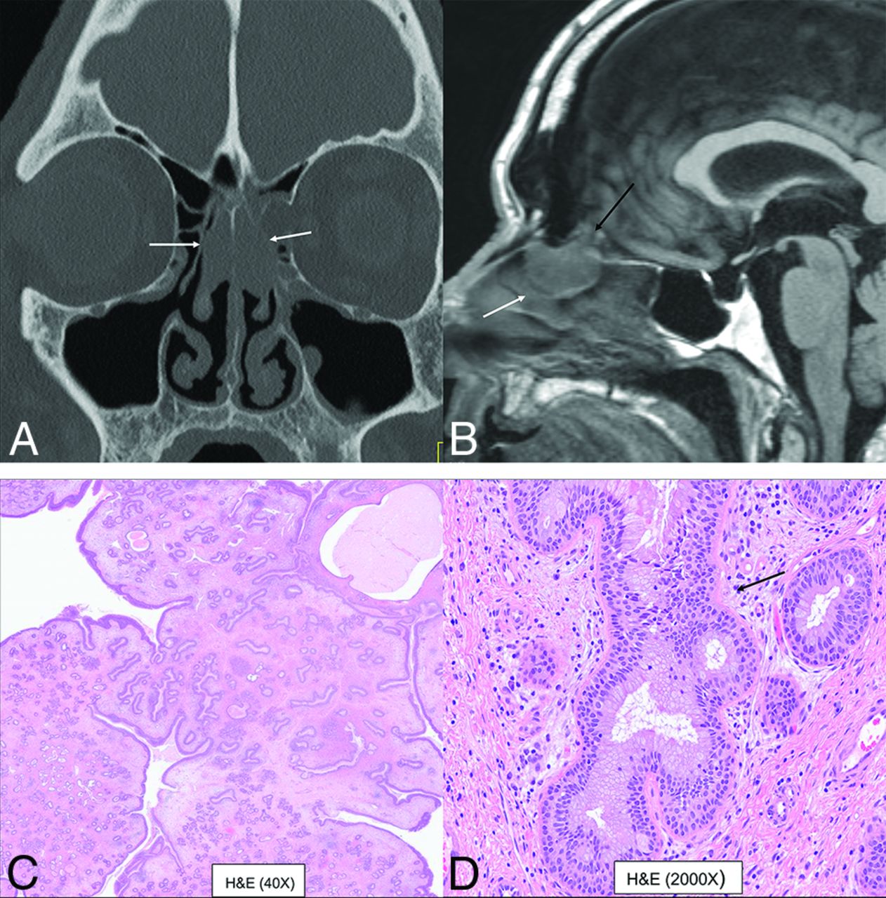
  • FIG 3.
    • Download figure
    • Open in new tab
    • Download powerpoint
    FIG 3.

    Bilateral REAH. Smooth polypoid lesions noted within the nasal cavity bilaterally (A and B, white arrows). The lesion expands the olfactory cleft (B, black arrow) with smooth remodeling, without erosive changes. This cannot be differentiated from seromucinous hamartoma with both exhibiting a crescentic (half-moon) shape on sagittal images (B). Histopathology revealed prominent glandular structures of varying sizes (C). Glands are lined with ciliated respiratory epithelium with bland nuclei and mucinous gland metaplasia (D, black arrow).

  • FIG 4.
    • Download figure
    • Open in new tab
    • Download powerpoint
    FIG 4.

    Sinonasal papilloma, oncocytic type, in the left frontal sinus in a 78-year-old man. Smooth polypoid lesions are noted within the nasal cavity with intrinsic T1 hyperintensity (A, arrow). The lesions obstruct the frontoethmoidal recess with trapped proteinaceous content in the frontal sinus. Lesions show a “cerebriform” pattern on the T2-weighted image (B, arrow) with avid enhancement (C, arrow). Histopathology reveals characteristic oncocytic cuboidal-to-columnar cells and intraepithelial microcysts with mucin and/or neutrophilic microabscesses (D, black arrows).

  • FIG 5.
    • Download figure
    • Open in new tab
    • Download powerpoint
    FIG 5.

    NUT sinonasal carcinoma in a 22-year-old man. Left nasal cavity mass (A and B, white arrows) with hyperostosis of the septum (black arrows). Metastatic disease with lesions in the lymph nodes, liver, and lung (C, white arrows). H&E stain (D) shows sheets and nests of high-grade tumor cells with zones of tumor necrosis. The tumor exhibits positive staining on NUT immunohistochemistry (E) with preserved INI1 staining (F) (ruling out SMARCB1 deficiency). Genetic analysis revealed NUT-BRD4 fusion, the defining feature of NUT carcinomas.
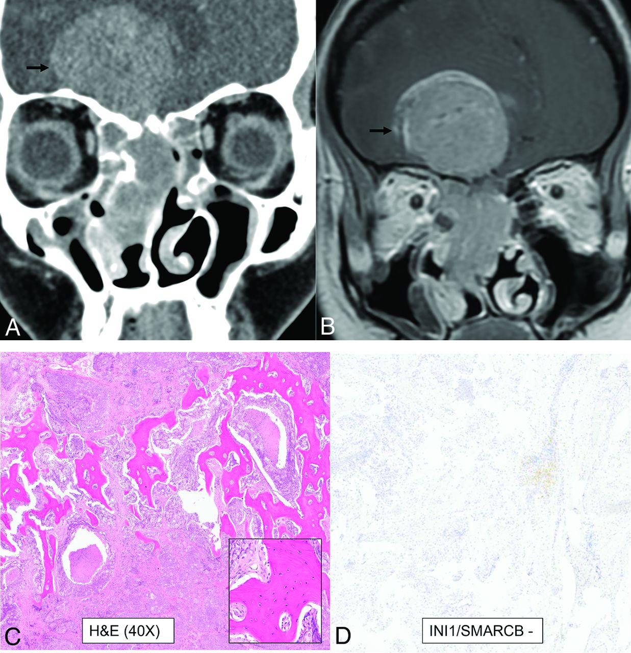
  • FIG 6.
    • Download figure
    • Open in new tab
    • Download powerpoint
    FIG 6.

    SWI/SNF complex–deficient sinonasal carcinoma in a 30-year-old woman. A large infiltrative sinonasal mass with intracranial extension (A and B, black arrows) and marked peritumoral edema in the frontal lobes. Peritumoral cysts were noted in the intracranial component. Radiologic differentials included olfactory neuroblastoma, SNUC, SWI/SNF complex–deficient (SMARCB-deficient) tumor, NUT carcinoma, and high-grade neuroendocrine tumor. H&E stain shows a basaloid low-power appearance (C) with hyaline-appearing cytoplasm showing a “pink” cell appearance, in which the tumor cells are somewhat plasmacytoid (C, inset). Complete loss of INI1/SMARCB1 (D) expression by immunohistochemistry defines this tumor, as seen here. Immunostaining for NUT and synaptophysin (a neuroendocrine marker) were negative.
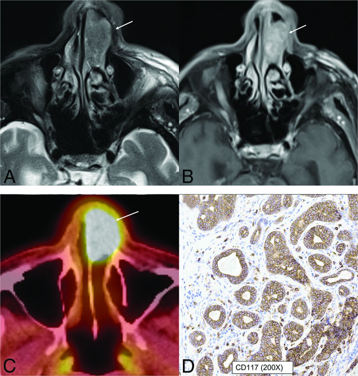
  • FIG 7.
    • Download figure
    • Open in new tab
    • Download powerpoint
    FIG 7.

    HPV-associated multiphenotypic sinonasal carcinoma in an 85-year-old man. A well-circumscribed, low T2-signal mass (A, arrow) with avid contrast enhancement (B, arrow) seen within the left nasal cavity with smooth expansion and high FDG uptake on PET/CT (C, arrow). CD117 (c-kit) (D) highlights the tumor ducts in a staining pattern that is the inverse of the myoepithelial cell markers. Findings of in situ hybridization for HPV (E6/E7) RNA was positive.

  • FIG 8.
    • Download figure
    • Open in new tab
    • Download powerpoint
    FIG 8.

    Biphenotypic sinonasal sarcoma (low-grade sinonasal sarcoma with neural and myogenic features) in a 68-year-old woman. Marked intralesional hyperostotic changes are noted within the nasal component of the tumor (A and B, white arrows). The tumor is well-marginated but locally aggressive with intracranial extension (B, black arrows). H&E stain (C) shows the tumor growing as fascicles of uniform spindle cells with minimal mitotic activity or atypia and overt rhabdomyoblastic differentiation in the form of strap cells (C, arrow). Diffusely positive staining of S-100 (nerve sheath tumor marker) seen as diffuse brown staining (D). Fluorescence in site hybridization testing was positive for PAX3 gene rearrangement, supporting the above diagnosis.

  • FIG 9.
    • Download figure
    • Open in new tab
    • Download powerpoint
    FIG 9.

    Radiographic differentials of an invasive sinonasal mass with intracranial extension are broad with significant overlap in imaging features, including previously described characteristic findings like intratumoral cyst in olfactory neuroblastoma. The final diagnosis is now based primarily on immunohistochemistry and genetics rather than histology. The prognosis of these tumors is also widely variable depending on the underlying molecular and genetic expression. Ca indicates carcinoma.

  • FIG 10.
    • Download figure
    • Open in new tab
    • Download powerpoint
    FIG 10.

    DEK-AFF2 carcinoma. A smooth homogeneous density mass (A, arrow) noted within the posterior ethmoid and nasal cavity with heterogeneous enhancement on MRI (B, arrow). The mass blocks the sphenoethmoidal recess with trapped secretions in the sphenoid sinus. The carcinoma demonstrates classic features of nonkeratinizing SCCa, with blunted papillae and invasion into the stroma as interconnecting ribbons on the H&E stains (C) with infiltrate of inflammatory cells, including lymphocytes and neutrophils. The tumor is diffusely positive for the squamous immunohistochemical marker p40 (D). To evaluate fusion-driven tumor, we performed targeted RNA sequencing, and DEK-AFF2 fusion was found, with breakpoints of DEK (exon 7) and AFF2 (exon 6).

Tables

  • Figures
  • Summary of changes including deletion, addition, and reclassification of SNT tumors in the 5th edition of the WHO Classification 2022

    WHO Classification 4th Edition 2017 (Chapter 1)WHO Classification 5th Edition 2022 (Chapter 2)
    Respiratory epithelial lesionsHamartomas (new category)
    REAHREAH
    Seromucinous hamartoma (new entity in 4th edition)Seromucinous hamartoma
    Nasal chondromesenchymal hamartoma (reclassification from “other” category)
    Sinonasal papillomasSubcategory under respiratory epithelial lesions in 5th edition (2022)Respiratory epithelial lesions (includes papillomas, carcinomas, adenocarcinomas with removal of hamartomatous lesions)
    Sinonasal papillomas
    CarcinomasSubcategory under respiratory epithelial lesions in 5th edition(2022)Carcinomas
    Keratinizing SCCaKeratinizing SCCa
    Nonkeratinizing SCCaNonkeratinizing SCCa (includes emerging entity DEK-AFF2 carcinoma)
    Spindle cell (sarcomatoid) SCCaNUT carcinoma
    Lymphoepithelial carcinomaSWI/SNF complex-deficient sinonasal carcinoma (new entity)
    Sinonasal undifferentiated carcinomaSinonasal lymphoepithelial carcinoma
    NUT carcinoma (new entity in 4th edition)Sinonasal undifferentiated carcinoma (includes emerging entity IDH-mutated carcinoma)
    Neuroendocrine carcinomaAdenocarcinomas separated as a new subcategoryTeratocarcinosarcoma
    AdenocarcinomaHPV-associated multiphenotypic sinonasal carcinoma (new entity)
     Intestinal-type adenocarcinoma
     Non–intestinal-type adenocarcinoma
    Adenocarcinomas (new subcategory, was previously under carcinomas)
     Intestinal-type sinonasal adenocarcinoma
     Non-intestinal-type sinonasal adenocarcinoma
    Malignant soft-tissue tumorsCategories removed with dedicated chapters on soft-tissue tumors in the new editionMesenchymal tumors of the SNT (new category with few retained entities from soft-tissue tumors, occurring exclusively or primarily in the SNT)
    Borderline/low-grade malignant soft-tissue tumorsSNT angiofibroma
    Benign soft-tissue tumorsSinonasal glomangiopericytoma
    Neuroectodermal/melanocytic tumorsBiphenotypic sinonasal sarcoma
    Hematolymphoid tumorsChordoma
    Other tumorsSinonasal ameloblastoma
    MeningiomaAdamantinomatous craniopharyngioma (reclassified from chapter on “nasopharynx”)
    Sinonasal ameloblastomaMeningioma of the SNT, ear
    Chondromesenchymal hamartoma (moved to hamartomas)Olfactory neuroblastoma
PreviousNext
Back to top
Advertisement
Print
Download PDF
Email Article

Thank you for your interest in spreading the word on American Journal of Neuroradiology.

NOTE: We only request your email address so that the person you are recommending the page to knows that you wanted them to see it, and that it is not junk mail. We do not capture any email address.

Enter multiple addresses on separate lines or separate them with commas.
Update from the 5th Edition of the WHO Classification of Nasal, Paranasal, and Skull Base Tumors: Imaging Overview with Histopathologic and Genetic Correlation
(Your Name) has sent you a message from American Journal of Neuroradiology
(Your Name) thought you would like to see the American Journal of Neuroradiology web site.
CAPTCHA
This question is for testing whether or not you are a human visitor and to prevent automated spam submissions.
Cite this article
A. Agarwal, A.A. Bhatt, G. Bathla, S. Kanekar, N. Soni, J. Murray, K. Vijay, P. Vibhute, P.H. Rhyner
Update from the 5th Edition of the WHO Classification of Nasal, Paranasal, and Skull Base Tumors: Imaging Overview with Histopathologic and Genetic Correlation
American Journal of Neuroradiology Aug 2023, DOI: 10.3174/ajnr.A7960

Citation Manager Formats

  • BibTeX
  • Bookends
  • EasyBib
  • EndNote (tagged)
  • EndNote 8 (xml)
  • Medlars
  • Mendeley
  • Papers
  • RefWorks Tagged
  • Ref Manager
  • RIS
  • Zotero
0 Responses
Respond to this article
Share
Bookmark this article
Update from the 5th Edition of the WHO Classification of Nasal, Paranasal, and Skull Base Tumors: Imaging Overview with Histopathologic and Genetic Correlation
A. Agarwal, A.A. Bhatt, G. Bathla, S. Kanekar, N. Soni, J. Murray, K. Vijay, P. Vibhute, P.H. Rhyner
American Journal of Neuroradiology Aug 2023, DOI: 10.3174/ajnr.A7960
del.icio.us logo Twitter logo Facebook logo Mendeley logo
  • Tweet Widget
  • Facebook Like
  • Google Plus One
Purchase

Jump to section

  • Article
    • Abstract
    • ABBREVIATIONS:
    • CONCLUSIONS
    • Footnotes
    • References
  • Figures & Data
  • Info & Metrics
  • Responses
  • References
  • PDF

Related Articles

  • No related articles found.
  • PubMed
  • Google Scholar

Cited By...

  • No citing articles found.
  • Crossref
  • Google Scholar

This article has not yet been cited by articles in journals that are participating in Crossref Cited-by Linking.

More in this TOC Section

  • An Atlas of Neonatal Neurovascular Imaging Anatomy as Depicted with Microvascular Imaging: The Intracranial Arteries
  • An Atlas of Neonatal Neurovascular Imaging Anatomy as Depicted with Microvascular Imaging: The Intracranial Veins
  • Clinical Translation of Hyperpolarized 13C Metabolic Probes for Glioma Imaging
Show more Review Articles

Similar Articles

Advertisement

Indexed Content

  • Current Issue
  • Accepted Manuscripts
  • Article Preview
  • Past Issues
  • Editorials
  • Editor's Choice
  • Fellows' Journal Club
  • Letters to the Editor
  • Video Articles

Cases

  • Case Collection
  • Archive - Case of the Week
  • Archive - Case of the Month
  • Archive - Classic Case

More from AJNR

  • Trainee Corner
  • Imaging Protocols
  • MRI Safety Corner
  • Book Reviews

Multimedia

  • AJNR Podcasts
  • AJNR Scantastics

Resources

  • Turnaround Time
  • Submit a Manuscript
  • Submit a Video Article
  • Submit an eLetter to the Editor/Response
  • Manuscript Submission Guidelines
  • Statistical Tips
  • Fast Publishing of Accepted Manuscripts
  • Graphical Abstract Preparation
  • Imaging Protocol Submission
  • Evidence-Based Medicine Level Guide
  • Publishing Checklists
  • Author Policies
  • Become a Reviewer/Academy of Reviewers
  • News and Updates

About Us

  • About AJNR
  • Editorial Board
  • Editorial Board Alumni
  • Alerts
  • Permissions
  • Not an AJNR Subscriber? Join Now
  • Advertise with Us
  • Librarian Resources
  • Feedback
  • Terms and Conditions
  • AJNR Editorial Board Alumni

American Society of Neuroradiology

  • Not an ASNR Member? Join Now

© 2025 by the American Society of Neuroradiology All rights, including for text and data mining, AI training, and similar technologies, are reserved.
Print ISSN: 0195-6108 Online ISSN: 1936-959X

Powered by HighWire