
Article Figures & Data
Figures
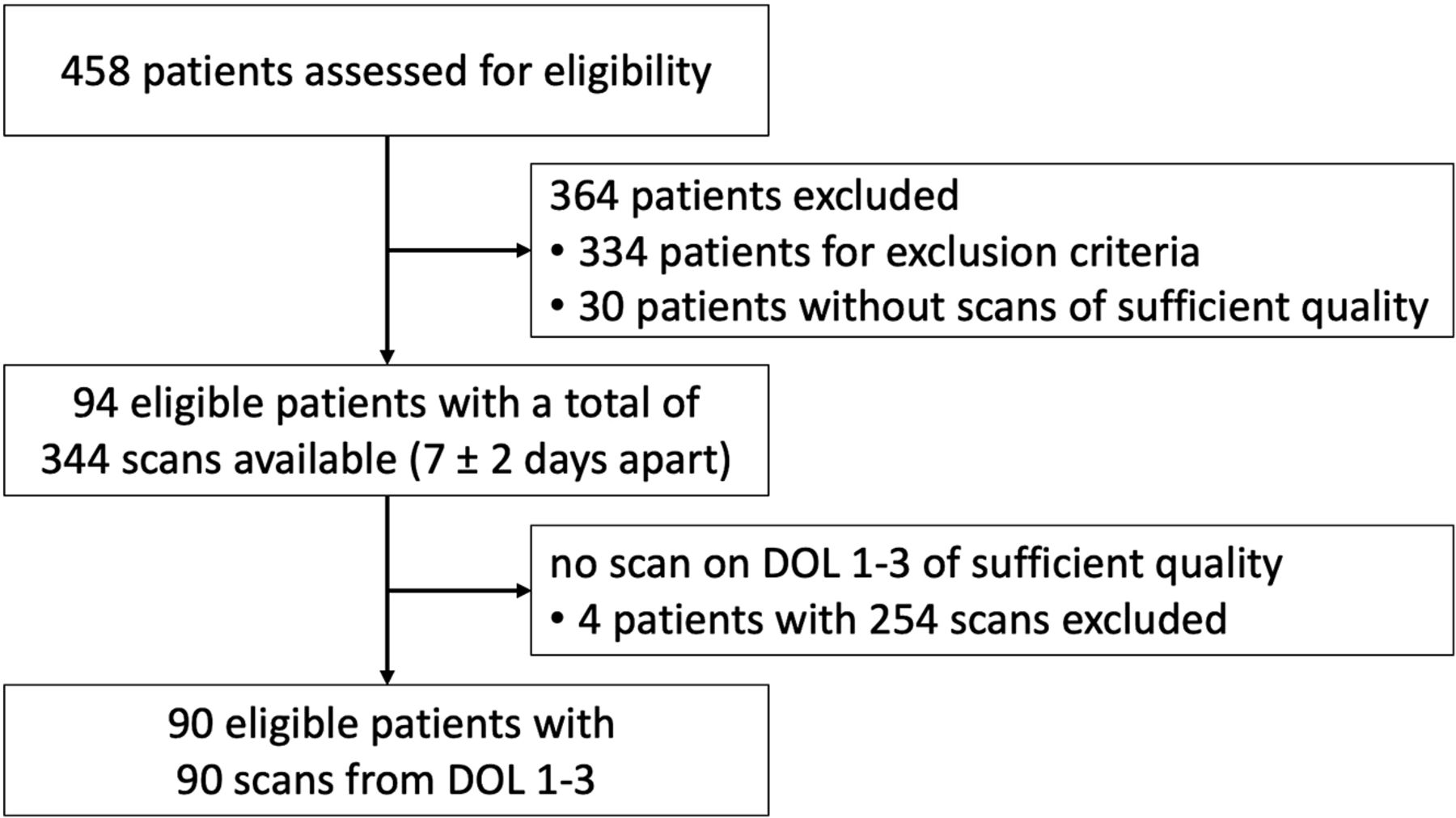
- FIG 1.
Flowchart of patient recruitment.
- FIG 2.
Ultrasound identification of the structures evaluated for the USBD in an infant with a PMA of 26 weeks. A, Midcoronal view at the level of the foramina of Monro: interopercular opening* (1), height of the insular cortex—curved measurement (2) and straight measurement* (3), depth of the Sylvian fissure (4), thickness of the corpus callosum (5), height of the cingulate gyrus (6), depth of cingulate sulcus* (7). B, Midsagittal view: circumferent length of corpus callosum (8), thickness of corpus callosum (midsagittal) at the genu (9a) and at the body (9b), and height of cingulate gyrus (10). Asterisks indicate structures selected for the USBD (bold lines, A).
- FIG 3.
Progression of opercularization with lengthening of insular cortex and closure of interopercular opening in the midcoronal view at different PMAs. A, PMA 22 + 6/7 weeks. B, PMA 30 + 0/7 weeks. C, PMA 34 + 2/7 weeks.
- FIG 4.
Scatterplot of measurements of selected structures for the USBD and trajectories with 25th and 75th percentiles, regression curves, and selected cutoff values for USBD marked as lines. A, Interopercular opening. B, Height of insular cortex. C, Depth of cingulate sulcus in the midcoronal plane.
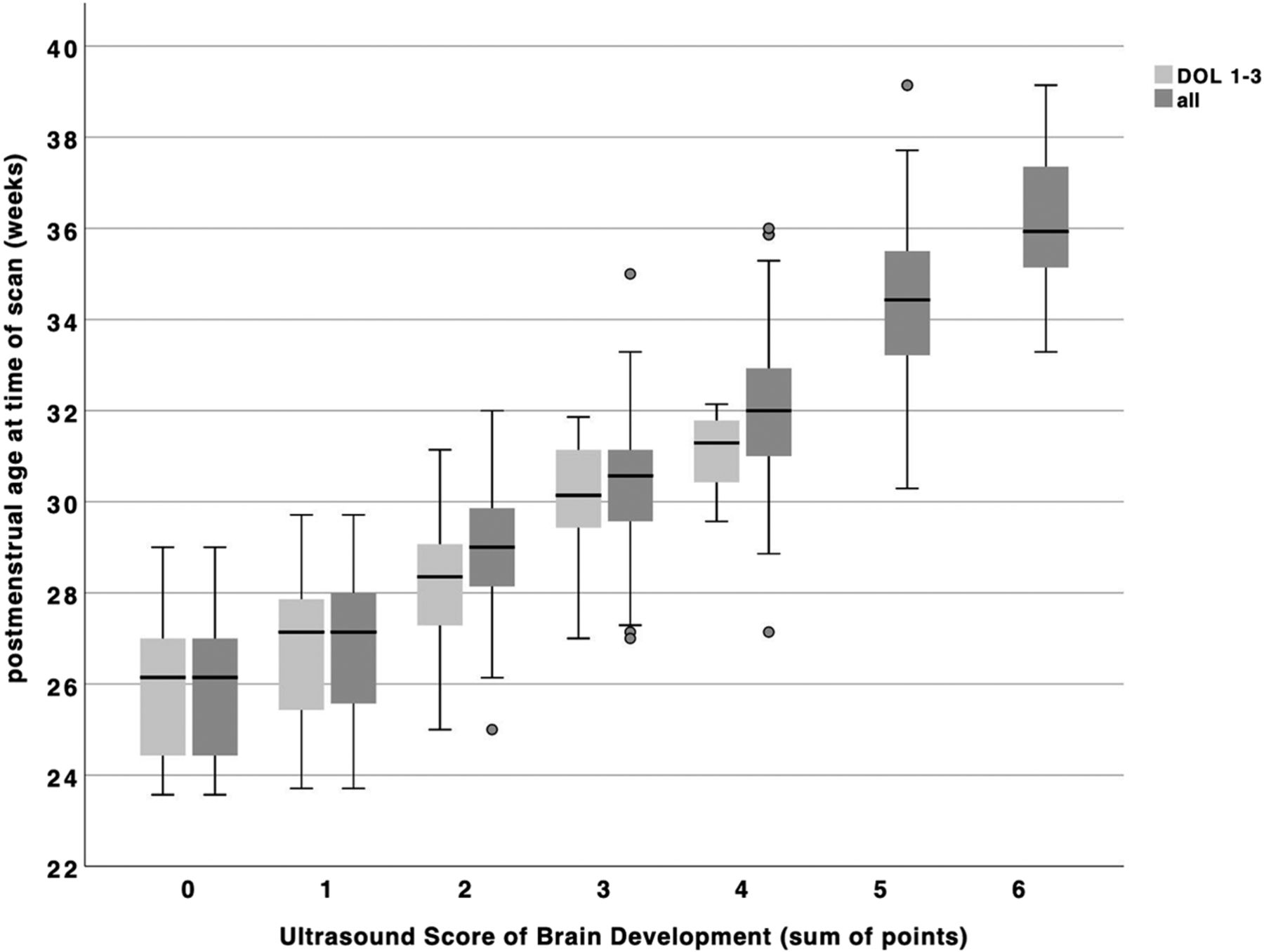
- FIG 5.
Boxplot of USBD score points by postmenstrual age grouped by day of scan: light gray including only scorable scans from DOL 1–3 (n = 88); dark gray including all scorable scans (n = 312).


Tables
Variable Patients (n = 94) Sex (No.) Male 48 Female 46 PMA at birth (weeks)a Mean 27.8 (SD, 2.2) Range (min–max) 23 + 3/7 to 32 + 0/7 PMA at time of scan (weeks) Mean 30.9 (SD, 3.4) Range (min–max) 23 + 4/7 to 39 + 1/7 Birth weight (g) Mean 1034 (SD, 349) Range (min–max) 340–1810 Birth weight percentile < 3rd (No.) (%) 3 (3.2%) 3rd to 10th (No.) (%) 16 (17.0%) Head circumference at birth (cm) Mean 25.3 (SD, 2.7) Range (min–max) 18.4–31.0 Head circumference at birth percentile < 3rd (No.) (%) 2 (2.1%) 3rd to 10th (No.) (%) 21 (22.3%) Note:—Min indicates minimum; max, maximum.
↵a PMA at birth was determined by ultrasound measurement of fetal crown rump length in the first trimester.
No. R β P Value Interopercular openinga 343 –0.74 –0.06 <.001 Height of insular cortex, curved measurement 343 0.84 0.09 <.001 Height of insular cortex, straight measurementa 343 0.85 0.09 <.001 Depth of Sylvian fissure 341 0.19 0.06 <.001 Thickness of CC (midcoronal) 342 0.26 0.003 <.001 Height of cingulate gyrus (midcoronal) 313 0.72 0.06 <.001 Depth of cingulate sulcusa 315 0.83 0.05 <.001 Circumferent length of CC 311 0.68 0.17 <.001 Thickness of CC (midsagittal) Genu 335 0.32 0.01 <.001 Body 339 0.18 0.002 <.001 Height of cingulate gyrus (midsagittal) 260 0.44 0.02 <.001 Total USBD (all scans) 312 0.88 0.43 <.001 Total USBD (DOL 1–3) 87 0.76 0.41 <.001 Note:—No. indicates the number of scans in which the structure could be measured; CC, corpus callosum.
↵a Structures selected for the USBD.
Score Points 0 1 2 Interopercular opening (cm) ≥ 0.55 0.15–0.54 < 0.15 Height of insular cortex (cm) ≤ 1.25 1.26–1.74 ≥ 1.75 Depth of cingulate sulcus (cm) < 0.10 0.11–0.44 ≥ 0.45 ↵a Score points from each structure add to a total score, which can range from a minimum of 0 points to a maximum of 6 points (a higher total score reflects a higher PMA).
Score No. of Scans with Score Assigned PMA (Weeks)(mean) PMA (Weeks)(range) 0 20 25.8 (SD, 1.4) 23 + 4/7 to 29 + 0/7 1 25 26.9 (SD, 1.6) 23 + 5/7 to 29 + 5/7 2 53 28.9 (SD, 1.3) 25 + 0/7 to 32 + 0/7 3 56 30.4 (SD, 1.5) 27 + 0/7 to 35 + 0/7 4 67 32.1 (SD, 1.6) 27 + 1/7 to 36 + 0/7 5 67 34.4 (SD, 1.9) 30 + 2/7 to 39 + 1/7 6 24 36.1 (SD, 1.6) 33 + 2/7 to 39 + 1/7


Jump to section
Related Articles
Cited By...
- No citing articles found.