
Article Figures & Data
Figures
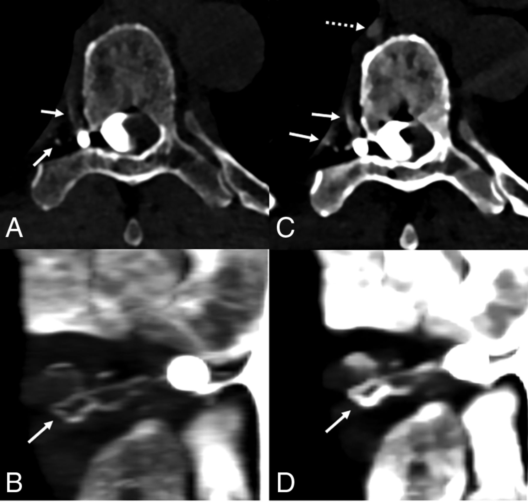
- Fig 1.
A 70-year-old man with 18 months of orthostatic headaches and brain MR imaging demonstrating diffuse pachymeningeal enhancement (not shown). Axial and coronal T3D reconstructions from a right lateral decubitus PCD CTM (A and B) at a section thickness of 0.2 mm demonstrate exquisite delineation of tiny veins draining a right T6 CVF (A and B, arrows). Axial and coronal 40-keV reconstructions at the identical time point and window/level settings, both at a section thickness of 0.4 mm (minimum allowable), demonstrate improved conspicuity of the iodine signal within the same veins (C and D, solid arrows) and within the more distal azygous vein (C, dashed arrow), though with slightly diminished anatomic characterization. PCD CT confers excellent spatial resolution and spectral information, which have a complementary role and can both be useful in imaging CVFs. The patient underwent transvenous Onyx embolization of the CVF.
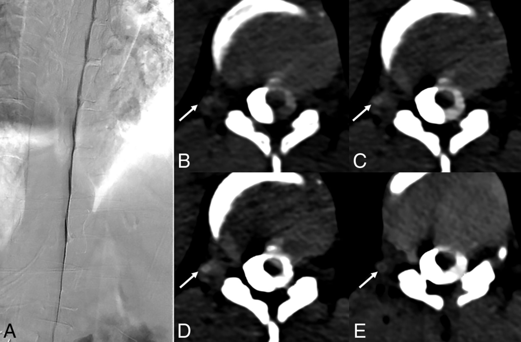
- Fig 2.
A 65-year-old woman with several years of orthostatic headaches and brain MR imaging demonstrating diffuse pachymeningeal enhancement and brain sag (not shown). Representative image from a right lateral decubitus DSM (A) shows multiple meningeal diverticula that remained stable during dynamic imaging (A, dashed arrows), with no evidence of a CVF. Axial images from a right lateral decubitus PCD CTM at the same time point (B–D) reconstructed at a T3D (B), 55 keV (C), and 40 keV (D) demonstrate a right T3 CVF (B–D, arrows), which is most apparent at 40 keV. The CVF is not apparent during imaging 30 seconds later, even at 40 keV (E, arrow). Additional images at adjacent slices, both from the earlier time point at 40 keV (F and G), show that this venous enhancement, though discontinuous, does, in fact, arise from a prominent meningeal diverticulum (F and G, arrows). In this case, the high degree of temporal resolution and spectral imaging conferred by PCD CT were necessary to confidently make the diagnosis. The patient underwent successful transvenous Onyx embolization of the right T3 CVF.
- Fig 3.
A 36-year-old woman with several years of orthostatic headaches, tinnitus, and MR imaging demonstrating brain sag without pachymeningeal enhancement (not shown). A representative image from a right lateral decubitus DSM (A) shows no evidence of a CVF. Axial images from a right lateral decubitus PCD CTM at the same time point (B–D) reconstructed at a T3D (B), 55 keV (C), and 40 keV (D) demonstrate a subtle right T11 CVF (B–D, arrows), most apparent at 40-keV. An axial 40-keV image obtained 20 seconds later (E) no longer shows this subtle venous opacification (E, arrow). The patient underwent Onyx embolization of this right T11 fistula.
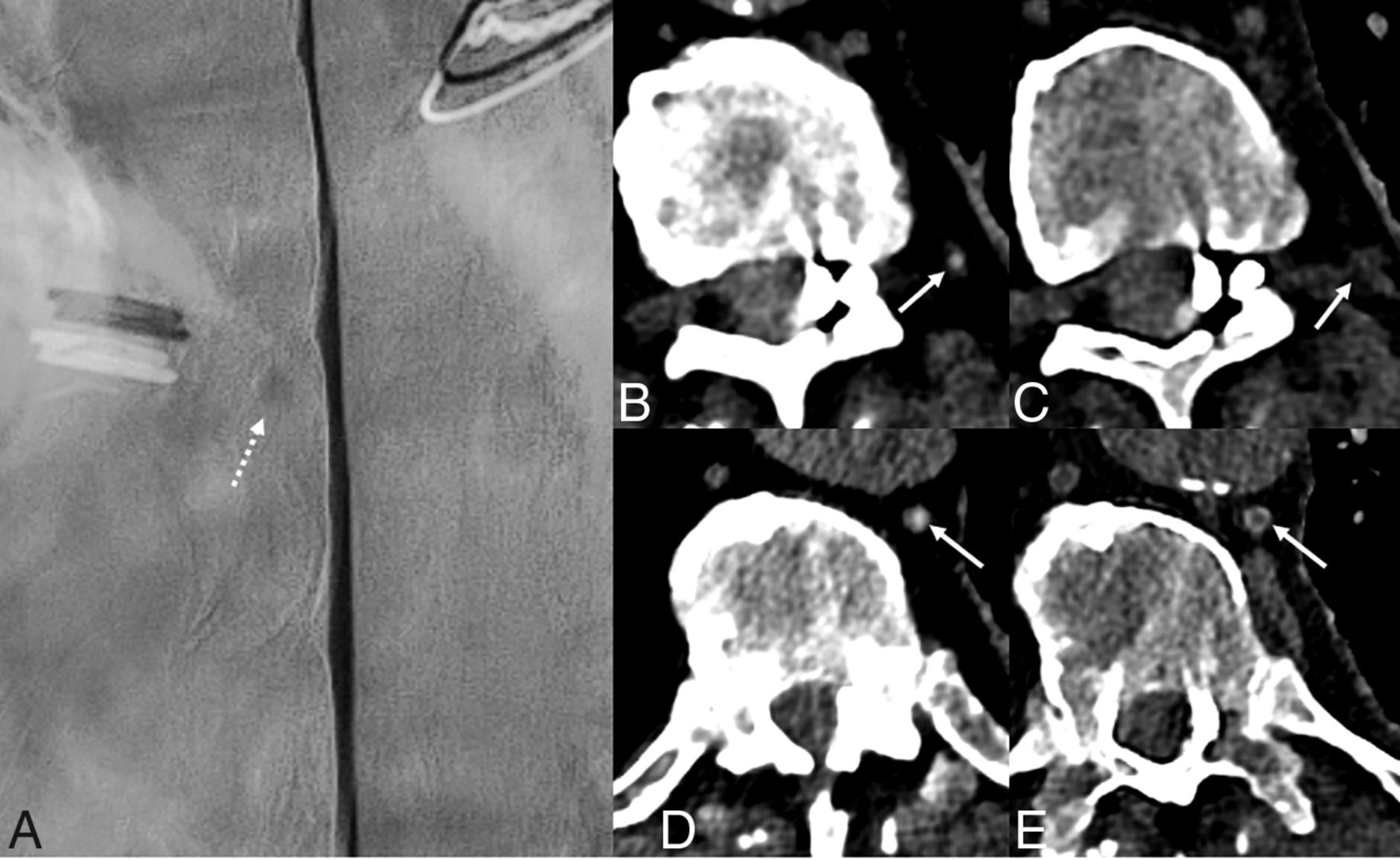
- Fig 4.
An 80-year-old man with several years of orthostatic headaches but normal brain MR imaging findings (not shown). Representative image from a left lateral decubitus DSM centered at T10 (A) shows a small meningeal diverticulum (A, dashed arrow) but no evidence of a CVF. Axial 40-keV images at 2 adjacent slices from a left lateral decubitus PCD CTM (B and D) demonstrate opacification of a paraspinal vein (B, arrow) and the azygous and hemiazygous vein (D, arrow), consistent with a CVF. Axial 40-keV images at the same slices obtained 30 seconds later (C and E) no longer show contrast within these veins (C and E, arrows). The patient underwent transvenous Onyx embolization of the left T10 CVF.
- Fig 5.
A 55-year-old woman with SIH according to the ICHD-3 criteria, including dural enhancement and venous distention on brain MR imaging (not shown). Coronal and axial images from a right lateral decubitus dynamic CTM (A and B) on an EID scanner reconstructed at 0.6 mm show no clear CVF. Subsequent PCD CTM with the patient in the right lateral decubitus position, including 0.2-mm coronal and axial reconstructions (C and D), shows a clear right T11 CVF (C and D, arrows).
- Fig 6.
A 65-year-old woman with SIH confirmed by the ICHD-3 criteria, as well as brain MR imaging showing dural enhancement and venous distention (not shown). Sagittal and axial images during decubitus dynamic EID CTM (A and B, 0.625-mm section thickness) show no evidence of a CVF. Subsequent right lateral decubitus PCD CTM (C and D, 0.2-mm section thickness) demonstrates a clear right T6 CVF (C and D, arrows).


Jump to section
Related Articles
Cited By...
- Optimization of Photon-Counting CT Myelography for the Detection of CSF-Venous Fistulas Using Convolutional Neural Network Denoising: A Comparative Analysis of Reconstruction Techniques
- Evaluation of Spontaneous Intracranial Hypotension Probabilistic Brain MRI Scoring Systems in Normal Patients
- Additional Diagnostic Value of Conebeam CT Myelography Performed after Digital Subtraction Myelography for Detecting CSF-Venous Fistulas
- Photon-Counting CT Myelography for the Detection of Spinal CSF Leaks
- CSF-Venous Fistulas Arising Intraosseously within Bone Remodeled by Meningeal Diverticula
- Spontaneous Intracranial Hypotension in Children: A Multi-Institutional Review of Spinal CSF Leaks Localized on Advanced Myelography
- Spinal CSF Leaks: The Neuroradiologist Transforming Care
- Myelographic Techniques for the Localization of CSF-Venous Fistulas: Updates in 2024
- Direct comparison of digital subtraction myelography versus CT myelography in lateral decubitus position: evaluation of diagnostic yield for cerebrospinal fluid-venous fistulas
- Diagnostic Yield of Decubitus CT Myelography for Detection of CSF-Venous Fistulas
- Photon-Counting CT in the Head and Neck: Current Applications and Future Prospects
- Back to the Future: Dynamic Contrast-Enhanced Photon-Counting Detector CT for the Detection of Pituitary Adenoma in Cushing Disease
- Benefits of Photon-Counting CT Myelography for Localization of Dural Tears in Spontaneous Intracranial Hypotension
- Application of a Denoising High-Resolution Deep Convolutional Neural Network to Improve Conspicuity of CSF-Venous Fistulas on Photon-Counting CT Myelography
- Diagnostic Performance of Decubitus Photon-Counting Detector CT Myelography for the Detection of CSF-Venous Fistulas