Advertisement

Main menu

  • Home
  • Content
    • Current Issue
    • Accepted Manuscripts
    • Article Preview
    • Past Issue Archive
    • Video Articles
    • AJNR Case Collection
    • Case of the Week Archive
    • Case of the Month Archive
    • Classic Case Archive
  • Special Collections
    • AJNR Awards
    • Low-Field MRI
    • Alzheimer Disease
    • ASNR Foundation Special Collection
    • Photon-Counting CT
    • View All
  • Multimedia
    • AJNR Podcasts
    • AJNR SCANtastic
    • Trainee Corner
    • MRI Safety Corner
    • Imaging Protocols
  • For Authors
    • Submit a Manuscript
    • Submit a Video Article
    • Submit an eLetter to the Editor/Response
    • Manuscript Submission Guidelines
    • Statistical Tips
    • Fast Publishing of Accepted Manuscripts
    • Graphical Abstract Preparation
    • Imaging Protocol Submission
    • Author Policies
  • About Us
    • About AJNR
    • Editorial Board
    • Editorial Board Alumni
  • More
    • Become a Reviewer/Academy of Reviewers
    • Subscribers
    • Permissions
    • Alerts
    • Feedback
    • Advertisers
    • ASNR Home

User menu

  • Alerts
  • Log in

Search

  • Advanced search
American Journal of Neuroradiology

American Journal of Neuroradiology

ASHNR American Society of Functional Neuroradiology ASHNR American Society of Pediatric Neuroradiology ASSR
  • Alerts
  • Log in

Advanced Search

  • Home
  • Content
    • Current Issue
    • Accepted Manuscripts
    • Article Preview
    • Past Issue Archive
    • Video Articles
    • AJNR Case Collection
    • Case of the Week Archive
    • Case of the Month Archive
    • Classic Case Archive
  • Special Collections
    • AJNR Awards
    • Low-Field MRI
    • Alzheimer Disease
    • ASNR Foundation Special Collection
    • Photon-Counting CT
    • View All
  • Multimedia
    • AJNR Podcasts
    • AJNR SCANtastic
    • Trainee Corner
    • MRI Safety Corner
    • Imaging Protocols
  • For Authors
    • Submit a Manuscript
    • Submit a Video Article
    • Submit an eLetter to the Editor/Response
    • Manuscript Submission Guidelines
    • Statistical Tips
    • Fast Publishing of Accepted Manuscripts
    • Graphical Abstract Preparation
    • Imaging Protocol Submission
    • Author Policies
  • About Us
    • About AJNR
    • Editorial Board
    • Editorial Board Alumni
  • More
    • Become a Reviewer/Academy of Reviewers
    • Subscribers
    • Permissions
    • Alerts
    • Feedback
    • Advertisers
    • ASNR Home
  • Follow AJNR on Twitter
  • Visit AJNR on Facebook
  • Follow AJNR on Instagram
  • Join AJNR on LinkedIn
  • RSS Feeds

AJNR Awards, New Junior Editors, and more. Read the latest AJNR updates

Research ArticlePediatric Neuroimaging

Diagnostic Utility of 3D Gradient-Echo MR Imaging Sequences through the Filum Compared with Spin-Echo T1 in Children with Concern for Tethered Cord

F. Rafiee, W.A. Mehan, S. Rincon, S. Rohatgi, O. Rapalino and K. Buch
American Journal of Neuroradiology February 2023, DOI: https://doi.org/10.3174/ajnr.A7791
F. Rafiee
aFrom the Department of Radiology, Massachusetts General Hospital, Harvard Medical School, Boston, Massachusetts
  • Find this author on Google Scholar
  • Find this author on PubMed
  • Search for this author on this site
  • ORCID record for F. Rafiee
W.A. Mehan
aFrom the Department of Radiology, Massachusetts General Hospital, Harvard Medical School, Boston, Massachusetts
  • Find this author on Google Scholar
  • Find this author on PubMed
  • Search for this author on this site
  • ORCID record for W.A. Mehan
S. Rincon
aFrom the Department of Radiology, Massachusetts General Hospital, Harvard Medical School, Boston, Massachusetts
  • Find this author on Google Scholar
  • Find this author on PubMed
  • Search for this author on this site
  • ORCID record for S. Rincon
S. Rohatgi
aFrom the Department of Radiology, Massachusetts General Hospital, Harvard Medical School, Boston, Massachusetts
  • Find this author on Google Scholar
  • Find this author on PubMed
  • Search for this author on this site
  • ORCID record for S. Rohatgi
O. Rapalino
aFrom the Department of Radiology, Massachusetts General Hospital, Harvard Medical School, Boston, Massachusetts
  • Find this author on Google Scholar
  • Find this author on PubMed
  • Search for this author on this site
  • ORCID record for O. Rapalino
K. Buch
aFrom the Department of Radiology, Massachusetts General Hospital, Harvard Medical School, Boston, Massachusetts
  • Find this author on Google Scholar
  • Find this author on PubMed
  • Search for this author on this site
  • ORCID record for K. Buch
  • Article
  • Figures & Data
  • Info & Metrics
  • Responses
  • References
  • PDF
Loading

Article Figures & Data

Figures

  • Tables
  • FIG 1.
    • Download figure
    • Open in new tab
    • Download powerpoint
    FIG 1.

    Different acquisitions of T1-weighted MR imaging on the axial planes of the lumbar region at the level of the L4–L5 intervertebral disc in an 18-year-old patient. A, T1 FSE sequence shows the dark-signal thecal sac containing the barely visible hypo- to iso-signal intensity cauda equina nerve roots and a round midline intradural lesion (black arrow) with bright signal intensity located posteriorly in the spinal canal at the expected anatomic site of the filum terminale, consistent with a FIL. Phase wrap-around artifacts are noted on both sides of the image. B, A radiofrequency spoiled 3D gradient-echo sequence known as VIBE of the same patient study at the same level shows a similar bright-signal intradural structure (black arrow), the FIL. Note the smaller AP and RL dimensions of the FIL compared with the T1 FSE sequence (A). The cauda equina nerve roots are well-demarcated as opposed to the T1 FSE sequence (A). VIBE/LAVA can underrate the AP and RL diameters of the FILs.

  • FIG 2.
    • Download figure
    • Open in new tab
    • Download powerpoint
    FIG 2.

    Different acquisitions of T1-weighted MR imaging on the axial plane through the lumbar region at the L4–L5 intervertebral disc in a 10-year-old boy. A, A T1 FSE sequence shows a tiny, round, midline intradural lesion (black arrow) with bright signal intensity located posteriorly in the spinal canal at the expected anatomic site of the filum terminale, consistent with a FIL. B, VIBE of the same patient at the same level shows iso-signal intensity nerve roots of the cauda equina traversing the spinal canal. Although the window level and width are adjusted for better detection of bright lesions, the VIBE sequence failed to reveal the FIL. Note the smaller AP and RL dimensions of the FIL compared with the case in Fig 3. The VIBE sequence is less sensitive in the detection of small FILs and can miss such lesions.
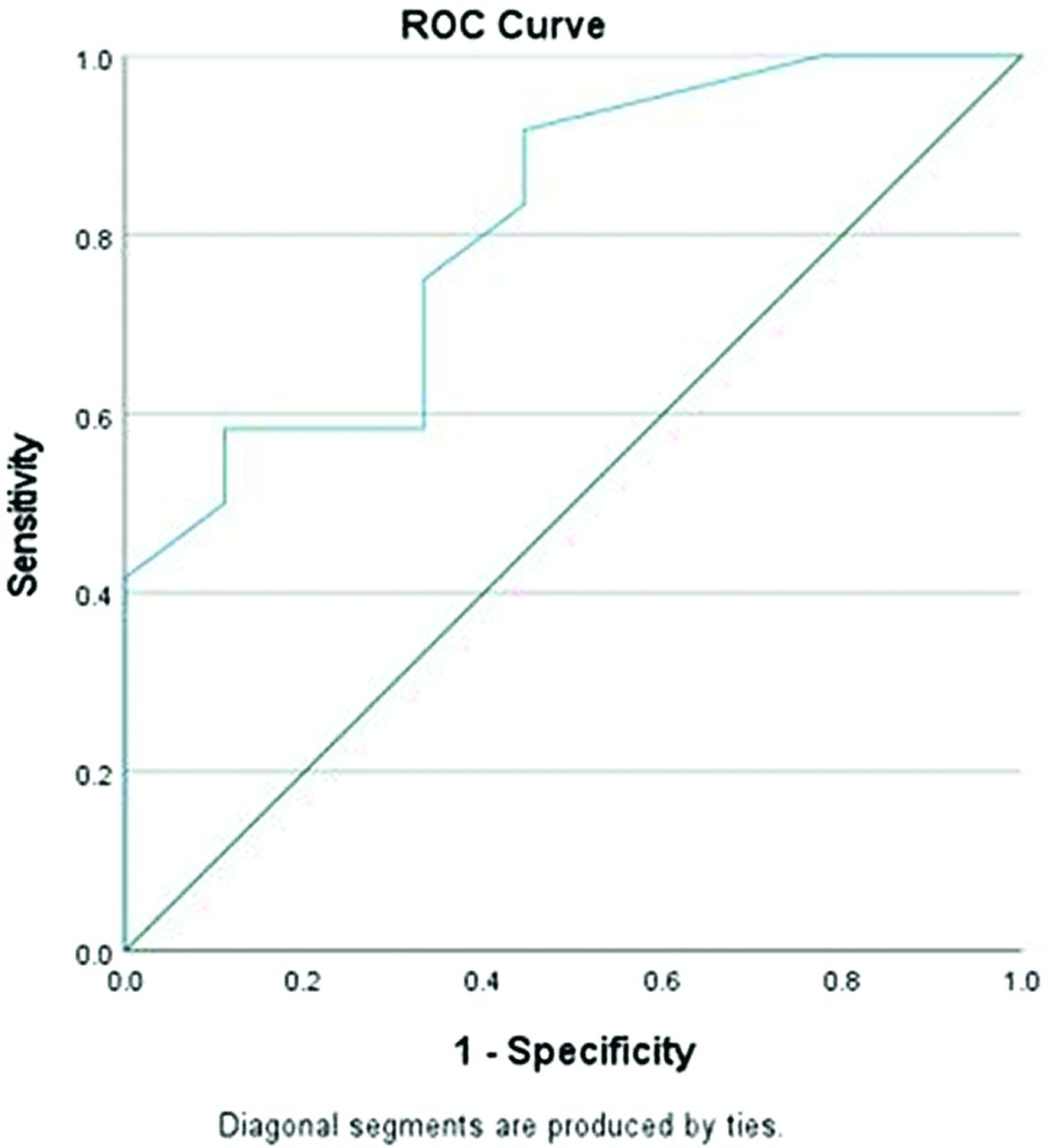
  • FIG 3.
    • Download figure
    • Open in new tab
    • Download powerpoint
    FIG 3.

    ROC curve for FIL AP diameter (area under the curve = 0.810, P value = .017).

  • FIG 4.
    • Download figure
    • Open in new tab
    • Download powerpoint
    FIG 4.

    ROC curve for FIL RL diameter (area under the curve = 0.815, P value = .016).

Tables

  • Figures
    • View popup
    Table 1:

    Presenting sign and/or symptoms

    Indication for MR ImagingTotalFIL +veFIL –ve
    Sacral abnormality16 (24.2%)9 (40.9%)7 (16%)
    Urinary dysfunction10 (15.1%)5 (22.7%)5 (11.3%)
    Bowel dysfunction8 (12.1%)2 (9%)6 (13.6)
    Scoliosis7 (10.6%)2 (9%)5 (11.3%)
    Lower-extremity weakness6 (9%)1 (4.5%)5 (11.3%)
    Back pain6 (9%)1 (4.5%)5 (11.3%)
    Othera13 (19.5%)2 (9%)11 (25%)
    Total662244
    • Note:—FIL +ve indicates positive detection of a FIL; FIL –ve, negative detection of a FIL.

    • ↵a Other indications for MR imaging include abnormal gait, Chiari I malformation, abnormal findings on spine sonography, and vertebral defects, anal atresia, cardiac defects, tracheo-esophageal fistula, renal anomalies, and limb abnormalities (VACTERL).

    • View popup
    Table 2:

    Number, size, and extent of FIL each sequence detected

    SequenceFIL +veFIL +ve Mean APFIL +ve Mean RLFIL +ve Mean CC
    +T1 FSE and +VIBE/LAVA, measured in T1 FSE21/22 (95.4)5.453.8
    +T1 FSE and +VIBE/LAVA, measured in VIBE/LAVA12/22 (54.5)4.94.62.2
    +T1 FSE and −VIBE/LAVA, measured in T1 FSE9/21 (42.9)1.51.62.5
    • Note:—RL indicates right-left dimension; CC, craniocaudal dimension; FIL +ve, positive detection of a FIL; FIL –ve, negative detection of a FIL.

PreviousNext
Back to top
Advertisement
Print
Download PDF
Email Article

Thank you for your interest in spreading the word on American Journal of Neuroradiology.

NOTE: We only request your email address so that the person you are recommending the page to knows that you wanted them to see it, and that it is not junk mail. We do not capture any email address.

Enter multiple addresses on separate lines or separate them with commas.
Diagnostic Utility of 3D Gradient-Echo MR Imaging Sequences through the Filum Compared with Spin-Echo T1 in Children with Concern for Tethered Cord
(Your Name) has sent you a message from American Journal of Neuroradiology
(Your Name) thought you would like to see the American Journal of Neuroradiology web site.
CAPTCHA
This question is for testing whether or not you are a human visitor and to prevent automated spam submissions.
Cite this article
F. Rafiee, W.A. Mehan, S. Rincon, S. Rohatgi, O. Rapalino, K. Buch
Diagnostic Utility of 3D Gradient-Echo MR Imaging Sequences through the Filum Compared with Spin-Echo T1 in Children with Concern for Tethered Cord
American Journal of Neuroradiology Feb 2023, DOI: 10.3174/ajnr.A7791

Citation Manager Formats

  • BibTeX
  • Bookends
  • EasyBib
  • EndNote (tagged)
  • EndNote 8 (xml)
  • Medlars
  • Mendeley
  • Papers
  • RefWorks Tagged
  • Ref Manager
  • RIS
  • Zotero
0 Responses
Respond to this article
Share
Bookmark this article
Diagnostic Utility of 3D Gradient-Echo MR Imaging Sequences through the Filum Compared with Spin-Echo T1 in Children with Concern for Tethered Cord
F. Rafiee, W.A. Mehan, S. Rincon, S. Rohatgi, O. Rapalino, K. Buch
American Journal of Neuroradiology Feb 2023, DOI: 10.3174/ajnr.A7791
del.icio.us logo Twitter logo Facebook logo Mendeley logo
  • Tweet Widget
  • Facebook Like
  • Google Plus One
Purchase

Jump to section

  • Article
    • Abstract
    • ABBREVIATIONS:
    • MATERIALS AND METHODS
    • RESULTS
    • DISCUSSION
    • CONCLUSIONS
    • Footnotes
    • References
  • Figures & Data
  • Info & Metrics
  • Responses
  • References
  • PDF

Related Articles

  • No related articles found.
  • PubMed
  • Google Scholar

Cited By...

  • No citing articles found.
  • Crossref
  • Google Scholar

This article has not yet been cited by articles in journals that are participating in Crossref Cited-by Linking.

More in this TOC Section

  • Frontal Paraventricular Cysts
  • Sodium MRI in Pediatric Brain Tumors
  • FRACTURE MR in Congenital Vertebral Anomalies
Show more Pediatric Neuroimaging

Similar Articles

Advertisement

Indexed Content

  • Current Issue
  • Accepted Manuscripts
  • Article Preview
  • Past Issues
  • Editorials
  • Editor's Choice
  • Fellows' Journal Club
  • Letters to the Editor
  • Video Articles

Cases

  • Case Collection
  • Archive - Case of the Week
  • Archive - Case of the Month
  • Archive - Classic Case

More from AJNR

  • Trainee Corner
  • Imaging Protocols
  • MRI Safety Corner
  • Book Reviews

Multimedia

  • AJNR Podcasts
  • AJNR Scantastics

Resources

  • Turnaround Time
  • Submit a Manuscript
  • Submit a Video Article
  • Submit an eLetter to the Editor/Response
  • Manuscript Submission Guidelines
  • Statistical Tips
  • Fast Publishing of Accepted Manuscripts
  • Graphical Abstract Preparation
  • Imaging Protocol Submission
  • Evidence-Based Medicine Level Guide
  • Publishing Checklists
  • Author Policies
  • Become a Reviewer/Academy of Reviewers
  • News and Updates

About Us

  • About AJNR
  • Editorial Board
  • Editorial Board Alumni
  • Alerts
  • Permissions
  • Not an AJNR Subscriber? Join Now
  • Advertise with Us
  • Librarian Resources
  • Feedback
  • Terms and Conditions
  • AJNR Editorial Board Alumni

American Society of Neuroradiology

  • Not an ASNR Member? Join Now

© 2025 by the American Society of Neuroradiology All rights, including for text and data mining, AI training, and similar technologies, are reserved.
Print ISSN: 0195-6108 Online ISSN: 1936-959X

Powered by HighWire