
Article Figures & Data
Figures
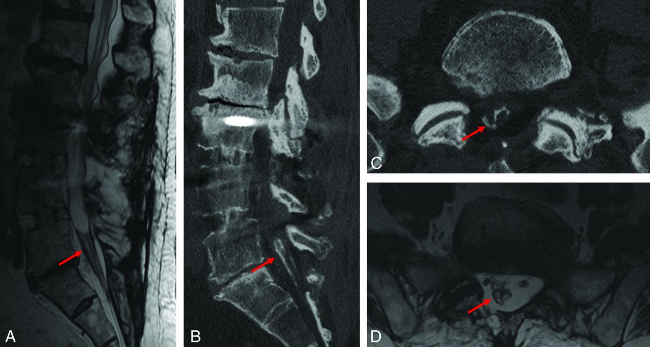
- FIG 1.
A 38-year-old male patient presenting 7 years following gunshot injury to the lumbar spine. Imaging from the time of the injury was unavailable for review. Sagittal T2-weighted MR imaging (A) demonstrates low signal thickening of the thecal sac as well as contour deformity of the thecal sac and its contents. Intermediate-signal tissue is present at the conus medullaris, relating to a posttraumatic scar. CT image (B) demonstrates peripheral and weblike ossification within the thecal sac (white arrows), which is not definitively identified on MR imaging (white arrows). The weblike ossification is better demonstrated on axial CT images (C and D), which also show multiple foci of metallic debris related to the prior gunshot injury (red arrows).
- FIG 2.
CT images from a 66-year-old female patient with back pain and stiffness. Sagittal (A and B) and axial (C) CT demonstrate features of spinal ankylosis relating to inflammatory spondyloathropathy (blue arrows). AO is evident by thick peripheral ossification of the meninges most prominent at the tip of the thecal sac; however, it can also be seen in the thoracic spine (red arrows).
- FIG 3.
A 78-year-old man status post multiple lumbar surgeries. T2-weighted MR imaging of the lumbar spine including sagittal (A) and axial (D) reformats demonstrates low signal thickening of the thecal sac and surrounding the nerve roots (red arrows), which manifests as weblike ossification on sagittal (B) and axial (C) CT images (red arrows). MR images (A and D) depict the associated thecal sac distortion.
- FIG 4.
Comparison of CT scans of the lumbar spine of the same patient performed in 2008 with follow-up in 2018 demonstrates progression of ossification (red arrows), which potentially contributes to progression of pain and neurologic dysfunction.
- FIG 5.
A 16-year-old female patient with a history of remote tethered cord release and posterolateral fusion. MR imaging T2-weighted images in the sagittal (A) and axial (B) planes demonstrate low signal thickening of the thecal sac with peripheralization and clumping of the nerve roots. C, H&E stains of the resection specimen from the thecal sac at the lumbar spine demonstrate meningothelial cells within the resected leptomeninges (black arrow), with ossification along the margin of the specimen (white arrowhead) as well as scattered foci of calcification (black arrowheads), consistent with AO.
- FIG 6.
In this series of 41 patients, 4 patterns of ossification that can be seen in AO were identified. Ossification can occur as a combination of patterns, which was the most common in this series. CT images demonstrate the 4 patterns. A, The central pattern demonstrates central ossification within the thecal sac and can have the appearance of a dagger on the sagittal plane. B, Nerve root encasement appears as circumferential ossification surrounding single or multiple roots of the cauda equina. C, The peripheral pattern involves the walls of the thecal sac and can be circumferential or discontinuous. D, The weblike pattern appears as ossification filling the thecal sac and insinuating between the nerve roots. In all panels the red arrows point out the areas of ossification.
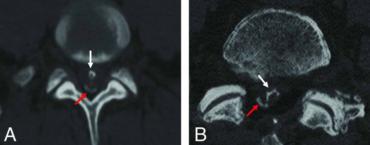
- FIG 7.
In some patients, a combined pattern of ossification was observed. Axial CT images in 2 unique patients demonstrate 2 patterns coexisting. A, The red arrow demonstrates the peripheral pattern of ossification coupled with a central pattern as shown by the white arrow. B. In this patient, the red arrow also indicates a region of peripheral ossification, coupled with weblike ossification as indicated by the white arrow.


Tables
CT Protocol kV(peak) 120 mAs AutomA and SmartmAs with maximum dose 400 mA Section thickness 0.625-mm section thickness, Bone Plus Algorithm Reformations Multiplanar reformations, 2-mm reformats in sagittal and coronal planes and to disc levels and in soft-tissue windows - Table 2:
MR imaging protocol used in spine imaging of the 41 patients included in the case series
2D Sagittal T2 2D Coronal T2 2D Axial T2 2D Sagittal T1 2D Sagittal T2 FLEX TR (ms) 3500 4000 3500 620 5500 TE (ms) 110 104 110 10 110 Flip angle 180° 90° 180° 90° 142° FOV (mm2) 260 280 280 280 260 Matrix 512 × 256 512 × 256 416 × 224 512 × 256 416 × 224 NEX 1.5 1 1.5 1 1 Receiver bandwidth (MHz) ±83.33 ±195.31 ±83.33 ±195.31 ±244.14 Section thickness (mm) 3.5 3.5 3.5 3.5 3.5 Echo-train length 14 12 12 3 16 Acquisition time (min) 3–4 3–4 4–5 3–4 3–4 Note:– FLEX indicates 2-point Dixon fat-suppression.
Demographics and Characteristics Total patients 41 Age (yr) 63 (SD, 18.8); range, 16–92 Etiology Postsurgical 32 Idiopathic 7 Inflammatory 1 Posttraumatic 1 Sex Male 18 Female 23 Location Lumbar spine 36 Thoracic spine 5 Follow-up duration 3.9 (SD 3.7) years; range, 8 months to 14 years No follow-up 18 patients Patients with MR imaging correlation 25


Jump to section
Related Articles
Cited By...
- No citing articles found.