Advertisement

Main menu

  • Home
  • Content
    • Current Issue
    • Accepted Manuscripts
    • Article Preview
    • Past Issue Archive
    • Video Articles
    • AJNR Case Collection
    • Case of the Week Archive
    • Case of the Month Archive
    • Classic Case Archive
  • Special Collections
    • AJNR Awards
    • Low-Field MRI
    • Alzheimer Disease
    • ASNR Foundation Special Collection
    • Photon-Counting CT
    • View All
  • Multimedia
    • AJNR Podcasts
    • AJNR SCANtastic
    • Trainee Corner
    • MRI Safety Corner
    • Imaging Protocols
  • For Authors
    • Submit a Manuscript
    • Submit a Video Article
    • Submit an eLetter to the Editor/Response
    • Manuscript Submission Guidelines
    • Statistical Tips
    • Fast Publishing of Accepted Manuscripts
    • Graphical Abstract Preparation
    • Imaging Protocol Submission
    • Author Policies
  • About Us
    • About AJNR
    • Editorial Board
    • Editorial Board Alumni
  • More
    • Become a Reviewer/Academy of Reviewers
    • Subscribers
    • Permissions
    • Alerts
    • Feedback
    • Advertisers
    • ASNR Home

User menu

  • Alerts
  • Log in

Search

  • Advanced search
American Journal of Neuroradiology

American Journal of Neuroradiology

ASHNR American Society of Functional Neuroradiology ASHNR American Society of Pediatric Neuroradiology ASSR
  • Alerts
  • Log in

Advanced Search

  • Home
  • Content
    • Current Issue
    • Accepted Manuscripts
    • Article Preview
    • Past Issue Archive
    • Video Articles
    • AJNR Case Collection
    • Case of the Week Archive
    • Case of the Month Archive
    • Classic Case Archive
  • Special Collections
    • AJNR Awards
    • Low-Field MRI
    • Alzheimer Disease
    • ASNR Foundation Special Collection
    • Photon-Counting CT
    • View All
  • Multimedia
    • AJNR Podcasts
    • AJNR SCANtastic
    • Trainee Corner
    • MRI Safety Corner
    • Imaging Protocols
  • For Authors
    • Submit a Manuscript
    • Submit a Video Article
    • Submit an eLetter to the Editor/Response
    • Manuscript Submission Guidelines
    • Statistical Tips
    • Fast Publishing of Accepted Manuscripts
    • Graphical Abstract Preparation
    • Imaging Protocol Submission
    • Author Policies
  • About Us
    • About AJNR
    • Editorial Board
    • Editorial Board Alumni
  • More
    • Become a Reviewer/Academy of Reviewers
    • Subscribers
    • Permissions
    • Alerts
    • Feedback
    • Advertisers
    • ASNR Home
  • Follow AJNR on Twitter
  • Visit AJNR on Facebook
  • Follow AJNR on Instagram
  • Join AJNR on LinkedIn
  • RSS Feeds

AJNR Awards, New Junior Editors, and more. Read the latest AJNR updates

Research ArticlePediatric Neuroimaging

Can MRI Differentiate between Infectious and Immune-Related Acute Cerebellitis? A Retrospective Imaging Study

G. Orman, S.F. Kralik, N.K. Desai, A. Meoded, H. Sangi-Haghpeykar, G. Jallo, E. Boltshauser and T.A.G.M. Huisman
American Journal of Neuroradiology September 2021, DOI: https://doi.org/10.3174/ajnr.A7301
G. Orman
aFrom the Edward B. Singleton Department of Radiology (G.O., S.F.K., N.K.D., A.M., H.S.-H., T.A.G.M.H.), Texas Children's Hospital, Houston, Texas
  • Find this author on Google Scholar
  • Find this author on PubMed
  • Search for this author on this site
  • ORCID record for G. Orman
S.F. Kralik
aFrom the Edward B. Singleton Department of Radiology (G.O., S.F.K., N.K.D., A.M., H.S.-H., T.A.G.M.H.), Texas Children's Hospital, Houston, Texas
  • Find this author on Google Scholar
  • Find this author on PubMed
  • Search for this author on this site
  • ORCID record for S.F. Kralik
N.K. Desai
aFrom the Edward B. Singleton Department of Radiology (G.O., S.F.K., N.K.D., A.M., H.S.-H., T.A.G.M.H.), Texas Children's Hospital, Houston, Texas
  • Find this author on Google Scholar
  • Find this author on PubMed
  • Search for this author on this site
  • ORCID record for N.K. Desai
A. Meoded
aFrom the Edward B. Singleton Department of Radiology (G.O., S.F.K., N.K.D., A.M., H.S.-H., T.A.G.M.H.), Texas Children's Hospital, Houston, Texas
  • Find this author on Google Scholar
  • Find this author on PubMed
  • Search for this author on this site
  • ORCID record for A. Meoded
H. Sangi-Haghpeykar
aFrom the Edward B. Singleton Department of Radiology (G.O., S.F.K., N.K.D., A.M., H.S.-H., T.A.G.M.H.), Texas Children's Hospital, Houston, Texas
bDepartment of Obstetrics and Gynecology (H.S.-H.), Baylor College of Medicine, Houston, Texas
  • Find this author on Google Scholar
  • Find this author on PubMed
  • Search for this author on this site
  • ORCID record for H. Sangi-Haghpeykar
G. Jallo
cInstitute for Brain Protection Sciences and Department of Neurosurgery (G.J.), Johns Hopkins All Children’s, St. Petersburg, Florida
  • Find this author on Google Scholar
  • Find this author on PubMed
  • Search for this author on this site
  • ORCID record for G. Jallo
E. Boltshauser
dDepartment of Pediatric Neurology (E.B.), University Children’s Hospital Zürich, Zürich, Switzerland
  • Find this author on Google Scholar
  • Find this author on PubMed
  • Search for this author on this site
  • ORCID record for E. Boltshauser
T.A.G.M. Huisman
aFrom the Edward B. Singleton Department of Radiology (G.O., S.F.K., N.K.D., A.M., H.S.-H., T.A.G.M.H.), Texas Children's Hospital, Houston, Texas
  • Find this author on Google Scholar
  • Find this author on PubMed
  • Search for this author on this site
  • ORCID record for T.A.G.M. Huisman
  • Article
  • Figures & Data
  • Info & Metrics
  • Responses
  • References
  • PDF
Loading

Article Figures & Data

Figures

  • Tables
  • FIG 1.
    • Download figure
    • Open in new tab
    • Download powerpoint
    FIG 1.

    Unilateral cerebellitis in a 6-year-old boy who presented with ataxia with increased WBC on blood tests (infectious acute cerebellitis). Lumbar puncture was not performed. MR imaging acquired at presentation (upper row, A–F) shows unilateral left cerebellar gray matter and diffuse T2- and FLAIR hyperintensity (A and B, thick arrow), causing a shift of midline structures and displacement of the vermis (A, thin arrows). DWI (C) and an ADC map (D) show vasogenic (C and D, thick arrows) and cytotoxic edema (C and D, thin arrows). Leptomeningeal contrast enhancement is appreciated on postcontrast T1-weighted imaging (E, thick arrows). Sagittal T2-weighted imaging (F) shows the swollen cerebellum with upward (F, thick arrow) and downward (F, dashed arrow) herniation, supratentorial hydrocephalus, and effacement of the fourth ventricle (F, asterisk) and posterior subarachnoid spaces (F, thin arrow). Last follow-up MR imaging, which was acquired after 68 days, shows near-complete resolution of brain MR imaging findings (lower row, G–L) and atrophy. Note that sagittal T1-weighted imaging (L) shows the complete resolution of swollen cerebellum without any upward (L, thick arrow) and downward herniation (L, dashed arrow), no supratentorial hydrocephalus, and no effacement of the fourth ventricle (L, asterisks) and posterior subarachnoid spaces (L, thin arrow).

  • FIG 2.
    • Download figure
    • Open in new tab
    • Download powerpoint
    FIG 2.

    Bilateral cerebellitis in a 7-year-old boy who presented with ataxia and headache. He had a history of appendectomy 2 weeks before presentation (infectious acute cerebellitis). Blood tests at the time of imaging revealed WBC within normal limits. There were low total protein levels in the CSF, which was tapped 1 day before. The CSF culture had negative findings. A head CT (not shown) demonstrated diffuse swelling of the bilateral cerebellar hemispheres with crowding at the level of the foramen magnum with supratentorial hydrocephalus. Brain MR imaging acquired a day later (upper row, A–E) shows diffuse bilateral hyperintense signal on T2 (A, thick arrows) and FLAIR (B, thick arrows) sequences with corresponding leptomeningeal enhancement (thin arrows) on postcontrast axial (C) and coronal (D) T1-weighted imaging. Sagittal T2-weighted imaging (E) shows the swollen cerebellum with upward (E, thick arrow) and downward (E, dashed arrow) herniation, supratentorial hydrocephalus, and effacement of the fourth ventricle (E, asterisk) and posterior subarachnoid spaces (E, thin arrow). Follow-up MR imaging (lower row, F–J) acquired after 8 days shows improvement of hyperintense signal on axial T2 (F, thick arrows) and FLAIR (G, thick arrows) sequences. No contrast enhancement is seen on postcontrast axial (H) and coronal (I) T1-weighted imaging. Sagittal T1-weighted imaging (J) shows complete resolution of cerebellar edema, no upward (J, thick arrow) and downward (J, dashed arrow) herniation, no supratentorial hydrocephalus, and no effacement of the fourth ventricle (J, asterisk) and posterior subarachnoid spaces (J, thin arrow).

  • FIG 3.
    • Download figure
    • Open in new tab
    • Download powerpoint
    FIG 3.

    Acute disseminated encephalomyelitis in a 5-year-old girl who presented with ataxia and absent lower extremity reflexes (immune-mediated acute cerebellitis). Blood tests and CSF revealed an increased white blood cell count. CSF culture had negative findings. Brain MR imaging at presentation shows bilateral, diffuse, patchy hyperintense signal on axial T2 (A, arrows) and FLAIR (B, arrows) sequences. Corresponding vasogenic edema is seen on axial DWI (C, arrows) and ADC (D, arrows). Axial postcontrast T1-weighted imaging demonstrates bilateral, subtle leptomeningeal contrast enhancement (E, arrows). Consecutive supratentorial slices reveal subtle T2-hyperintense signal in the white matter of the adjacent trigone of the left lateral ventricle (F and G, arrow) without contrast enhancement or diffusion alteration (not shown). Sagittal T1-weighted imaging (H) shows no upward or downward herniation. Follow-up MR imaging was not available.

  • FIG 4.
    • Download figure
    • Open in new tab
    • Download powerpoint
    FIG 4.

    Bilateral cerebellitis in a 6.5-year-old boy (infectious acute cerebellitis) who had a history of gastroenteritis for 2–3 days. He developed marked and prolonged headache after a few days. Neurologic examination showed no cerebellar dysfunction. CSF examination was not performed. MR imaging shows bilateral, diffuse, T2 hyperintense signal and mild edema (A–C, arrows) in the cerebellar white and gray matter. Postcontrast imaging shows mild leptomeningeal contrast enhancement (D–F, arrows).

  • FIG 5.
    • Download figure
    • Open in new tab
    • Download powerpoint
    FIG 5.

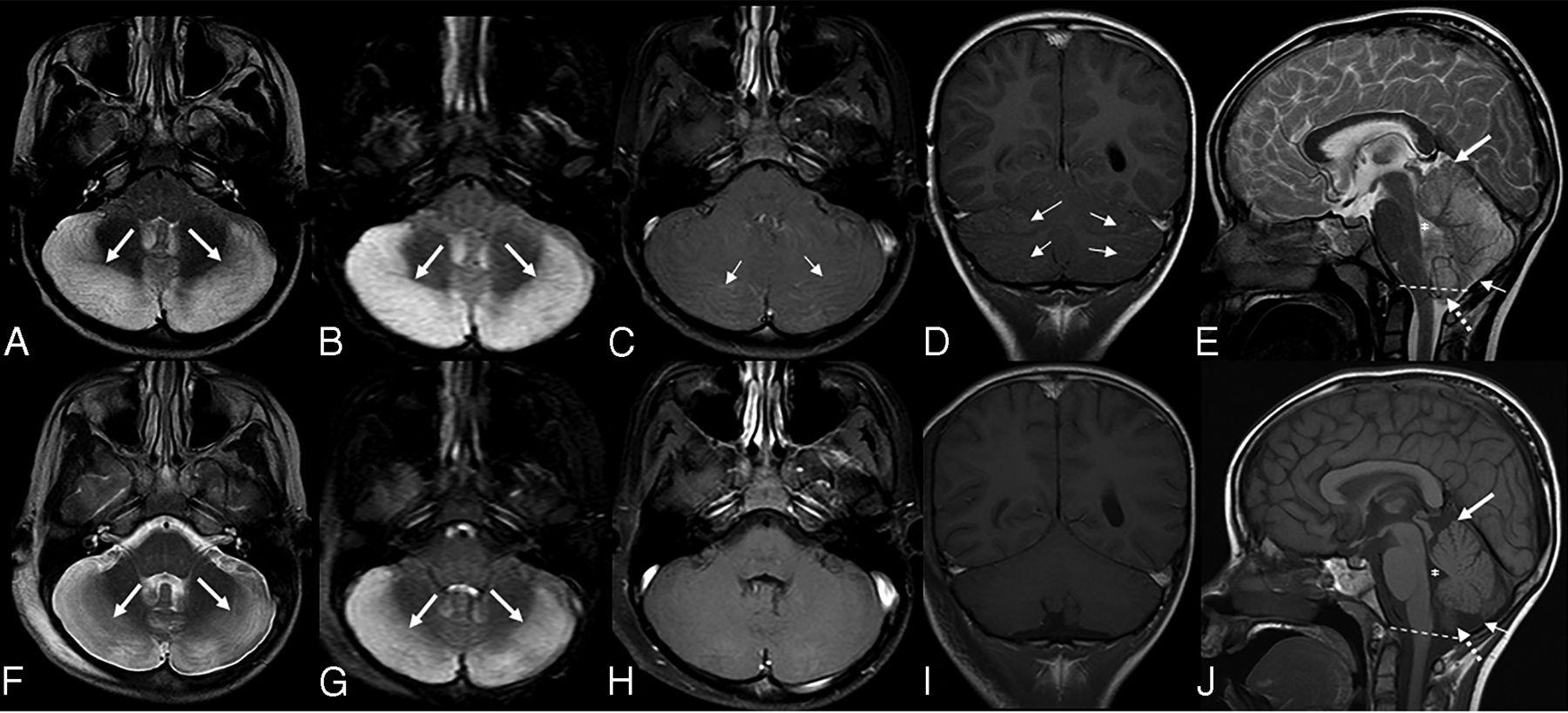
    Bilateral cerebellitis in a 12.5-year-old girl (infectious acute cerebellitis). Initial presentation was nonspecific. The patient was tired with headache for a few days and spontaneously recovered. MR imaging at presentation (upper row) shows bilateral, diffuse, T2-hyperintense signal predominantly on the right side (A–D, arrows) and downward herniation (D, dashed line). A follow-up examination after 3 months shows no abnormal neurologic findings. Follow-up MR imaging after 3 months (lower row) shows complete resolution of previous MR imaging findings (E–H).

  • FIG 6.
    • Download figure
    • Open in new tab
    • Download powerpoint
    FIG 6.

    Upper row A–C, Acute disseminated encephalomyelitis in a 1-year 10-month-old boy (with immune-mediated acute cerebellitis) who presented with acute ataxia and fever with vomiting. A blood test revealed increased white blood cell count, and CSF revealed elevated WBC and normal protein and glucose levels. Blood culture and CSF culture had negative findings. Lower row D–F, Bilateral cerebellitis in a 5-year-old boy (with infectious acute cerebellitis) with a history dominated by intense headache, predominantly at night with some fluctuation. The patient had treatment of symptoms, but there was no clear improvement in headaches. MR imaging was performed about 2 weeks later. He had no cerebellar signs on examination. This figure aims to show the most predominant MR imaging findings for immune-mediated versus infectious acute cerebellitis. Note that there is no downward herniation in immune-mediated acute cerebellitis (A) versus cerebellar tonsil downward herniation in infectious acute cerebellitis (D, arrow). Axial T2 shows subtle hyperintense signal in right middle cerebellar peduncle in immune-mediated acute cerebellitis (B, arrow) versus bilateral diffuse T2-hyperintense signal with diffuse edema in infectious acute cerebellitis (E, arrows). Note that axial FLAIR shows extensive, multifocal, and confluent bilateral hyperintensity involving the cerebral white matter and the gray matter, including the bilateral basal ganglia extensively and bilateral thalami (C, arrows) in immune-mediated acute cerebellitis versus normal supratentorial brain axial T2 signal intensities in infectious acute cerebellitis (F).

Tables

  • Figures
  • Brain MR imaging findings at presentation between infectious (n = 35) and immune-related (n = 8) acute cerebellitis subgroups

    T2/FLAIR Hyperintense SignalInfectious (No.) (%)Immune-Related (No.) (%)P Valuea
    Unilateral cerebellar4 (11.42)2 (25.00).31
    Bilateral cerebellar27 (77.14)4 (50.00).19
    Brainstem1 (2.85)3 (37.50).016
    Supratentorial brain0 (0.00)3 (37.50).004
    Cerebellar white matter10 (28.57)1 (12.50).65
    Cerebellar gray matter25 (71.42)5 (62.50).68
    Associated findings
     Involvement of vermis8 (22.85)1 (12.50)1.00
     Effacement of fourth ventricle12 (34.28)1 (12.50).39
     Effacement of posterior fossa subarachnoid spaces11 (31.42)0 (0.00).09
     Downward herniation15 (42.85)0 (0.00).03
     Upward herniation10 (28.57)0 (0.00).16
     Supratentorial hydrocephalus8 (22.85)0 (0.00).31
    Contrast enhancement
     Leptomeningeal12 (34.28)2 (28.57)1.00
     Cortical7 (20.00)1 (14.28)1.00
    Diffusion-weighted imaging
     Vasogenic edema20 (58.82)4 (50.00).71
     Cytotoxic edema6 (17.64)1 (12.50)1.00
    • ↵a P is based on the Fisher exact test.

PreviousNext
Back to top
Advertisement
Print
Download PDF
Email Article

Thank you for your interest in spreading the word on American Journal of Neuroradiology.

NOTE: We only request your email address so that the person you are recommending the page to knows that you wanted them to see it, and that it is not junk mail. We do not capture any email address.

Enter multiple addresses on separate lines or separate them with commas.
Can MRI Differentiate between Infectious and Immune-Related Acute Cerebellitis? A Retrospective Imaging Study
(Your Name) has sent you a message from American Journal of Neuroradiology
(Your Name) thought you would like to see the American Journal of Neuroradiology web site.
CAPTCHA
This question is for testing whether or not you are a human visitor and to prevent automated spam submissions.
Cite this article
G. Orman, S.F. Kralik, N.K. Desai, A. Meoded, H. Sangi-Haghpeykar, G. Jallo, E. Boltshauser, T.A.G.M. Huisman
Can MRI Differentiate between Infectious and Immune-Related Acute Cerebellitis? A Retrospective Imaging Study
American Journal of Neuroradiology Sep 2021, DOI: 10.3174/ajnr.A7301

Citation Manager Formats

  • BibTeX
  • Bookends
  • EasyBib
  • EndNote (tagged)
  • EndNote 8 (xml)
  • Medlars
  • Mendeley
  • Papers
  • RefWorks Tagged
  • Ref Manager
  • RIS
  • Zotero
0 Responses
Respond to this article
Share
Bookmark this article
Can MRI Differentiate between Infectious and Immune-Related Acute Cerebellitis? A Retrospective Imaging Study
G. Orman, S.F. Kralik, N.K. Desai, A. Meoded, H. Sangi-Haghpeykar, G. Jallo, E. Boltshauser, T.A.G.M. Huisman
American Journal of Neuroradiology Sep 2021, DOI: 10.3174/ajnr.A7301
del.icio.us logo Twitter logo Facebook logo Mendeley logo
  • Tweet Widget
  • Facebook Like
  • Google Plus One
Purchase

Jump to section

  • Article
    • Abstract
    • ABBREVIATIONS:
    • MATERIALS AND METHODS
    • RESULTS
    • DISCUSSION
    • CONCLUSIONS
    • References
  • Figures & Data
  • Info & Metrics
  • Responses
  • References
  • PDF

Related Articles

  • No related articles found.
  • PubMed
  • Google Scholar

Cited By...

  • No citing articles found.
  • Crossref
  • Google Scholar

This article has not yet been cited by articles in journals that are participating in Crossref Cited-by Linking.

More in this TOC Section

  • Frontal Paraventricular Cysts
  • Sodium MRI in Pediatric Brain Tumors
  • FRACTURE MR in Congenital Vertebral Anomalies
Show more Pediatric Neuroimaging

Similar Articles

Advertisement

Indexed Content

  • Current Issue
  • Accepted Manuscripts
  • Article Preview
  • Past Issues
  • Editorials
  • Editor's Choice
  • Fellows' Journal Club
  • Letters to the Editor
  • Video Articles

Cases

  • Case Collection
  • Archive - Case of the Week
  • Archive - Case of the Month
  • Archive - Classic Case

More from AJNR

  • Trainee Corner
  • Imaging Protocols
  • MRI Safety Corner
  • Book Reviews

Multimedia

  • AJNR Podcasts
  • AJNR Scantastics

Resources

  • Turnaround Time
  • Submit a Manuscript
  • Submit a Video Article
  • Submit an eLetter to the Editor/Response
  • Manuscript Submission Guidelines
  • Statistical Tips
  • Fast Publishing of Accepted Manuscripts
  • Graphical Abstract Preparation
  • Imaging Protocol Submission
  • Evidence-Based Medicine Level Guide
  • Publishing Checklists
  • Author Policies
  • Become a Reviewer/Academy of Reviewers
  • News and Updates

About Us

  • About AJNR
  • Editorial Board
  • Editorial Board Alumni
  • Alerts
  • Permissions
  • Not an AJNR Subscriber? Join Now
  • Advertise with Us
  • Librarian Resources
  • Feedback
  • Terms and Conditions
  • AJNR Editorial Board Alumni

American Society of Neuroradiology

  • Not an ASNR Member? Join Now

© 2025 by the American Society of Neuroradiology All rights, including for text and data mining, AI training, and similar technologies, are reserved.
Print ISSN: 0195-6108 Online ISSN: 1936-959X

Powered by HighWire