Advertisement

Main menu

  • Home
  • Content
    • Current Issue
    • Accepted Manuscripts
    • Article Preview
    • Past Issue Archive
    • Video Articles
    • AJNR Case Collection
    • Case of the Week Archive
    • Case of the Month Archive
    • Classic Case Archive
  • Special Collections
    • AJNR Awards
    • Low-Field MRI
    • Alzheimer Disease
    • ASNR Foundation Special Collection
    • Photon-Counting CT
    • View All
  • Multimedia
    • AJNR Podcasts
    • AJNR SCANtastic
    • Trainee Corner
    • MRI Safety Corner
    • Imaging Protocols
  • For Authors
    • Submit a Manuscript
    • Submit a Video Article
    • Submit an eLetter to the Editor/Response
    • Manuscript Submission Guidelines
    • Statistical Tips
    • Fast Publishing of Accepted Manuscripts
    • Graphical Abstract Preparation
    • Imaging Protocol Submission
    • Author Policies
  • About Us
    • About AJNR
    • Editorial Board
    • Editorial Board Alumni
  • More
    • Become a Reviewer/Academy of Reviewers
    • Subscribers
    • Permissions
    • Alerts
    • Feedback
    • Advertisers
    • ASNR Home

User menu

  • Alerts
  • Log in

Search

  • Advanced search
American Journal of Neuroradiology

American Journal of Neuroradiology

ASHNR American Society of Functional Neuroradiology ASHNR American Society of Pediatric Neuroradiology ASSR
  • Alerts
  • Log in

Advanced Search

  • Home
  • Content
    • Current Issue
    • Accepted Manuscripts
    • Article Preview
    • Past Issue Archive
    • Video Articles
    • AJNR Case Collection
    • Case of the Week Archive
    • Case of the Month Archive
    • Classic Case Archive
  • Special Collections
    • AJNR Awards
    • Low-Field MRI
    • Alzheimer Disease
    • ASNR Foundation Special Collection
    • Photon-Counting CT
    • View All
  • Multimedia
    • AJNR Podcasts
    • AJNR SCANtastic
    • Trainee Corner
    • MRI Safety Corner
    • Imaging Protocols
  • For Authors
    • Submit a Manuscript
    • Submit a Video Article
    • Submit an eLetter to the Editor/Response
    • Manuscript Submission Guidelines
    • Statistical Tips
    • Fast Publishing of Accepted Manuscripts
    • Graphical Abstract Preparation
    • Imaging Protocol Submission
    • Author Policies
  • About Us
    • About AJNR
    • Editorial Board
    • Editorial Board Alumni
  • More
    • Become a Reviewer/Academy of Reviewers
    • Subscribers
    • Permissions
    • Alerts
    • Feedback
    • Advertisers
    • ASNR Home
  • Follow AJNR on Twitter
  • Visit AJNR on Facebook
  • Follow AJNR on Instagram
  • Join AJNR on LinkedIn
  • RSS Feeds

AJNR Awards, New Junior Editors, and more. Read the latest AJNR updates

Research ArticlePediatric Neuroimaging
Open Access

MR Imaging Differences in the Circle of Willis between Healthy Children and Adults

K.P. Guilliams, N. Gupta, S. Srinivasan, M.M. Binkley, C. Ying, L. Couture, J. Gross, A. Wallace, R.C. McKinstry, K. Vo, J.-M. Lee, H. An and M.S. Goyal
American Journal of Neuroradiology September 2021, DOI: https://doi.org/10.3174/ajnr.A7290
K.P. Guilliams
aFrom the Department of Neurology (K.P.G., M.M.B., J.-M.L., M.S.G.)
bDepartment of Pediatrics (K.P.G., R.C.M.)
cMallinckrodt Institute of Radiology (K.P.G., N.G., S.S., C.Y., L.C., R.C.M., K.V., J.-M.L., H.A., M.S.G.)
  • Find this author on Google Scholar
  • Find this author on PubMed
  • Search for this author on this site
  • ORCID record for K.P. Guilliams
N. Gupta
cMallinckrodt Institute of Radiology (K.P.G., N.G., S.S., C.Y., L.C., R.C.M., K.V., J.-M.L., H.A., M.S.G.)
  • Find this author on Google Scholar
  • Find this author on PubMed
  • Search for this author on this site
  • ORCID record for N. Gupta
S. Srinivasan
cMallinckrodt Institute of Radiology (K.P.G., N.G., S.S., C.Y., L.C., R.C.M., K.V., J.-M.L., H.A., M.S.G.)
  • Find this author on Google Scholar
  • Find this author on PubMed
  • Search for this author on this site
  • ORCID record for S. Srinivasan
M.M. Binkley
aFrom the Department of Neurology (K.P.G., M.M.B., J.-M.L., M.S.G.)
  • Find this author on Google Scholar
  • Find this author on PubMed
  • Search for this author on this site
  • ORCID record for M.M. Binkley
C. Ying
cMallinckrodt Institute of Radiology (K.P.G., N.G., S.S., C.Y., L.C., R.C.M., K.V., J.-M.L., H.A., M.S.G.)
  • Find this author on Google Scholar
  • Find this author on PubMed
  • Search for this author on this site
  • ORCID record for C. Ying
L. Couture
cMallinckrodt Institute of Radiology (K.P.G., N.G., S.S., C.Y., L.C., R.C.M., K.V., J.-M.L., H.A., M.S.G.)
  • Find this author on Google Scholar
  • Find this author on PubMed
  • Search for this author on this site
J. Gross
fDivision of Neuroradiology (J.G.), Midwest Radiology, St. Paul, Minnesota
  • Find this author on Google Scholar
  • Find this author on PubMed
  • Search for this author on this site
  • ORCID record for J. Gross
A. Wallace
gDepartment of Neurointerventional Surgery (A.W.), Ascension Columbia St. Mary’s Hospital, Milwaukee, Wisconsin
  • Find this author on Google Scholar
  • Find this author on PubMed
  • Search for this author on this site
  • ORCID record for A. Wallace
R.C. McKinstry
bDepartment of Pediatrics (K.P.G., R.C.M.)
cMallinckrodt Institute of Radiology (K.P.G., N.G., S.S., C.Y., L.C., R.C.M., K.V., J.-M.L., H.A., M.S.G.)
  • Find this author on Google Scholar
  • Find this author on PubMed
  • Search for this author on this site
  • ORCID record for R.C. McKinstry
K. Vo
cMallinckrodt Institute of Radiology (K.P.G., N.G., S.S., C.Y., L.C., R.C.M., K.V., J.-M.L., H.A., M.S.G.)
  • Find this author on Google Scholar
  • Find this author on PubMed
  • Search for this author on this site
  • ORCID record for K. Vo
J.-M. Lee
aFrom the Department of Neurology (K.P.G., M.M.B., J.-M.L., M.S.G.)
cMallinckrodt Institute of Radiology (K.P.G., N.G., S.S., C.Y., L.C., R.C.M., K.V., J.-M.L., H.A., M.S.G.)
dDepartment of Biomedical Engineering (J.-M.L.)
  • Find this author on Google Scholar
  • Find this author on PubMed
  • Search for this author on this site
  • ORCID record for J.-M. Lee
H. An
cMallinckrodt Institute of Radiology (K.P.G., N.G., S.S., C.Y., L.C., R.C.M., K.V., J.-M.L., H.A., M.S.G.)
  • Find this author on Google Scholar
  • Find this author on PubMed
  • Search for this author on this site
  • ORCID record for H. An
M.S. Goyal
aFrom the Department of Neurology (K.P.G., M.M.B., J.-M.L., M.S.G.)
cMallinckrodt Institute of Radiology (K.P.G., N.G., S.S., C.Y., L.C., R.C.M., K.V., J.-M.L., H.A., M.S.G.)
eNeuroscience (M.S.G.), Washington University School of Medicine, St. Louis, Missouri
  • Find this author on Google Scholar
  • Find this author on PubMed
  • Search for this author on this site
  • ORCID record for M.S. Goyal
  • Article
  • Figures & Data
  • Supplemental
  • Info & Metrics
  • Responses
  • References
  • PDF
Loading

Article Figures & Data

Figures

  • Tables
  • FIG 1.
    • Download figure
    • Open in new tab
    • Download powerpoint
    FIG 1.

    The circle of Willis. 3D volume-rendering of the CoW from a TOF-MRA in a young child. This shows the key segments comprising the vascular ring, including AcomA (blue), the first segment of the anterior cerebral artery (A1, orange), the PcomA (red), and the first segment of the posterior cerebral artery (P1, green). Other arteries feeding the CoW (the ICA and the basilar artery [BA]) and supplied by the CoW (second segment of the anterior cerebral artery [A2], MCA, and second segment of the posterior cerebral artery [P2]) are also labeled.

  • FIG 2.
    • Download figure
    • Open in new tab
    • Download powerpoint
    FIG 2.

    Double-oblique multiplanar reconstruction for vessel-diameter measurement. Two orthogonal planes of reconstruction were aligned along the long axis of each arterial segment to be measured. These were then used to construct a cross-sectional plane through the midportion of the vessel segment on which 2 perpendicular diameters were measured and averaged. Note that partial volume effects at the margins of the artery could result in a slight degree of measurement bias and error. WL indicates window level; WW, window width.

  • FIG 3.
    • Download figure
    • Open in new tab
    • Download powerpoint
    FIG 3.

    Measurement of the CoW-di. A group average of the ratio of each CoW segment to the mean CoW diameter was calculated. For each individual CoW, similar ratios were calculated for each of the 7 segments. The CoW-di was then calculated as the Euclidean distance between the individual CoW ratios and the group-average ratios, thus representing the degree to which an individual CoW deviated in size from the group average. Examples are shown for two 23 -year-old individuals, one with a very low CoW-di (0.29) and another with a high CoW-di (1.48), largely due to a nonvisualized left P1 segment.
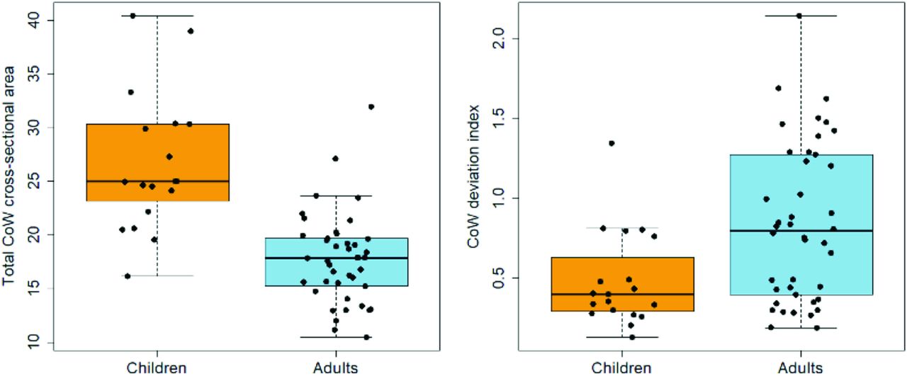
  • FIG 4.
    • Download figure
    • Open in new tab
    • Download powerpoint
    FIG 4.

    Differences in CoW size and symmetry in children versus adults. The total CoW cross-sectional area was measured in healthy children (n = 19) and adults (n = 42) on TOF-MRA images as described in the text. The mean total CoW cross-sectional area was larger in children than in adults (children’s mean CoW-area = 27.7 mm2 versus adults’ mean CoW-area = 17.8 mm2, P < .0001). Note that T2-SPACE measurements replicated these results, though with a smaller effect size as discussed in the text. CoW topology in children also more closely resembled the average topology found in the whole group, despite there being more adults in the whole group (children’s mean CoW-di = 0.48 versus adults’ mean CoW-di = 0.84, P < .001). As described in the text, the difference in CoW topology is, in part, due to a higher prevalence of nonvisualized CoW segments in adults.

  • FIG 5.
    • Download figure
    • Open in new tab
    • Download powerpoint
    FIG 5.

    CoW size tracks with CBF changes during development. Total CoW cross-sectional area based on TOF-MRA and whole-brain CBF was measured in participants ranging from 4 to 74 years of age. There is a gradual decrease in whole-brain CBF from childhood to adulthood that parallels the decrease in total CoW cross-sectional area. Across participants, CoW size also correlated with whole-brain CBF (Pearson r = 0.68, P < 10−7). Smoothing lines are for visualization purposes and are based on LOcally WEighted Scatter-plot Smoother fits with span = 1.

Tables

  • Figures
  • Summary CoW measurements in healthy children versus adults

     Children (0–18 years of age)Adults (19–74 years of age)
    No. of participants2343
    Age (range) (yr)9.5 (4–18)40.7 (19–74)
    Sex (female, male)11, 1221, 22
    Excluded data4 of 231 of 43
    TOF-MRA CoW-area (mean) (mm2)27.7 (SD, 7.9)17.8 (SD, 4.2)
    TOF-MRA CoW-di (mean)0.48 (SD, 0.30)0.84 (SD, 0.49)
    T2-SPACE CoW-area (mean) (mm2)16.6 (SD, 4.1)14.2 (SD, 4.0)
    T2-SPACE CoW-di (mean)0.68 (SD, 0.29)0.93 (SD, 0.49)
    Nonvisualized segments5/133 (3.8%)31/294 (10.5%)
    Whole-brain CBF (mean) (L/min)1.10 (SD, 0.24)0.69 (SD, 0.21)
PreviousNext
Back to top
Advertisement
Print
Download PDF
Email Article

Thank you for your interest in spreading the word on American Journal of Neuroradiology.

NOTE: We only request your email address so that the person you are recommending the page to knows that you wanted them to see it, and that it is not junk mail. We do not capture any email address.

Enter multiple addresses on separate lines or separate them with commas.
MR Imaging Differences in the Circle of Willis between Healthy Children and Adults
(Your Name) has sent you a message from American Journal of Neuroradiology
(Your Name) thought you would like to see the American Journal of Neuroradiology web site.
CAPTCHA
This question is for testing whether or not you are a human visitor and to prevent automated spam submissions.
Cite this article
K.P. Guilliams, N. Gupta, S. Srinivasan, M.M. Binkley, C. Ying, L. Couture, J. Gross, A. Wallace, R.C. McKinstry, K. Vo, J.-M. Lee, H. An, M.S. Goyal
MR Imaging Differences in the Circle of Willis between Healthy Children and Adults
American Journal of Neuroradiology Sep 2021, DOI: 10.3174/ajnr.A7290

Citation Manager Formats

  • BibTeX
  • Bookends
  • EasyBib
  • EndNote (tagged)
  • EndNote 8 (xml)
  • Medlars
  • Mendeley
  • Papers
  • RefWorks Tagged
  • Ref Manager
  • RIS
  • Zotero
0 Responses
Respond to this article
Share
Bookmark this article
MR Imaging Differences in the Circle of Willis between Healthy Children and Adults
K.P. Guilliams, N. Gupta, S. Srinivasan, M.M. Binkley, C. Ying, L. Couture, J. Gross, A. Wallace, R.C. McKinstry, K. Vo, J.-M. Lee, H. An, M.S. Goyal
American Journal of Neuroradiology Sep 2021, DOI: 10.3174/ajnr.A7290
del.icio.us logo Twitter logo Facebook logo Mendeley logo
  • Tweet Widget
  • Facebook Like
  • Google Plus One
Purchase

Jump to section

  • Article
    • Abstract
    • ABBREVIATIONS:
    • MATERIALS AND METHODS
    • RESULTS
    • DISCUSSION
    • CONCLUSIONS
    • Footnotes
    • References
  • Figures & Data
  • Supplemental
  • Info & Metrics
  • Responses
  • References
  • PDF

Related Articles

  • PubMed
  • Google Scholar

Cited By...

  • No citing articles found.
  • Crossref
  • Google Scholar

This article has not yet been cited by articles in journals that are participating in Crossref Cited-by Linking.

More in this TOC Section

  • Frontal Paraventricular Cysts
  • Sodium MRI in Pediatric Brain Tumors
  • FRACTURE MR in Congenital Vertebral Anomalies
Show more Pediatric Neuroimaging

Similar Articles

Advertisement

Indexed Content

  • Current Issue
  • Accepted Manuscripts
  • Article Preview
  • Past Issues
  • Editorials
  • Editor's Choice
  • Fellows' Journal Club
  • Letters to the Editor
  • Video Articles

Cases

  • Case Collection
  • Archive - Case of the Week
  • Archive - Case of the Month
  • Archive - Classic Case

More from AJNR

  • Trainee Corner
  • Imaging Protocols
  • MRI Safety Corner
  • Book Reviews

Multimedia

  • AJNR Podcasts
  • AJNR Scantastics

Resources

  • Turnaround Time
  • Submit a Manuscript
  • Submit a Video Article
  • Submit an eLetter to the Editor/Response
  • Manuscript Submission Guidelines
  • Statistical Tips
  • Fast Publishing of Accepted Manuscripts
  • Graphical Abstract Preparation
  • Imaging Protocol Submission
  • Evidence-Based Medicine Level Guide
  • Publishing Checklists
  • Author Policies
  • Become a Reviewer/Academy of Reviewers
  • News and Updates

About Us

  • About AJNR
  • Editorial Board
  • Editorial Board Alumni
  • Alerts
  • Permissions
  • Not an AJNR Subscriber? Join Now
  • Advertise with Us
  • Librarian Resources
  • Feedback
  • Terms and Conditions
  • AJNR Editorial Board Alumni

American Society of Neuroradiology

  • Not an ASNR Member? Join Now

© 2025 by the American Society of Neuroradiology All rights, including for text and data mining, AI training, and similar technologies, are reserved.
Print ISSN: 0195-6108 Online ISSN: 1936-959X

Powered by HighWire