
Article Figures & Data
Figures
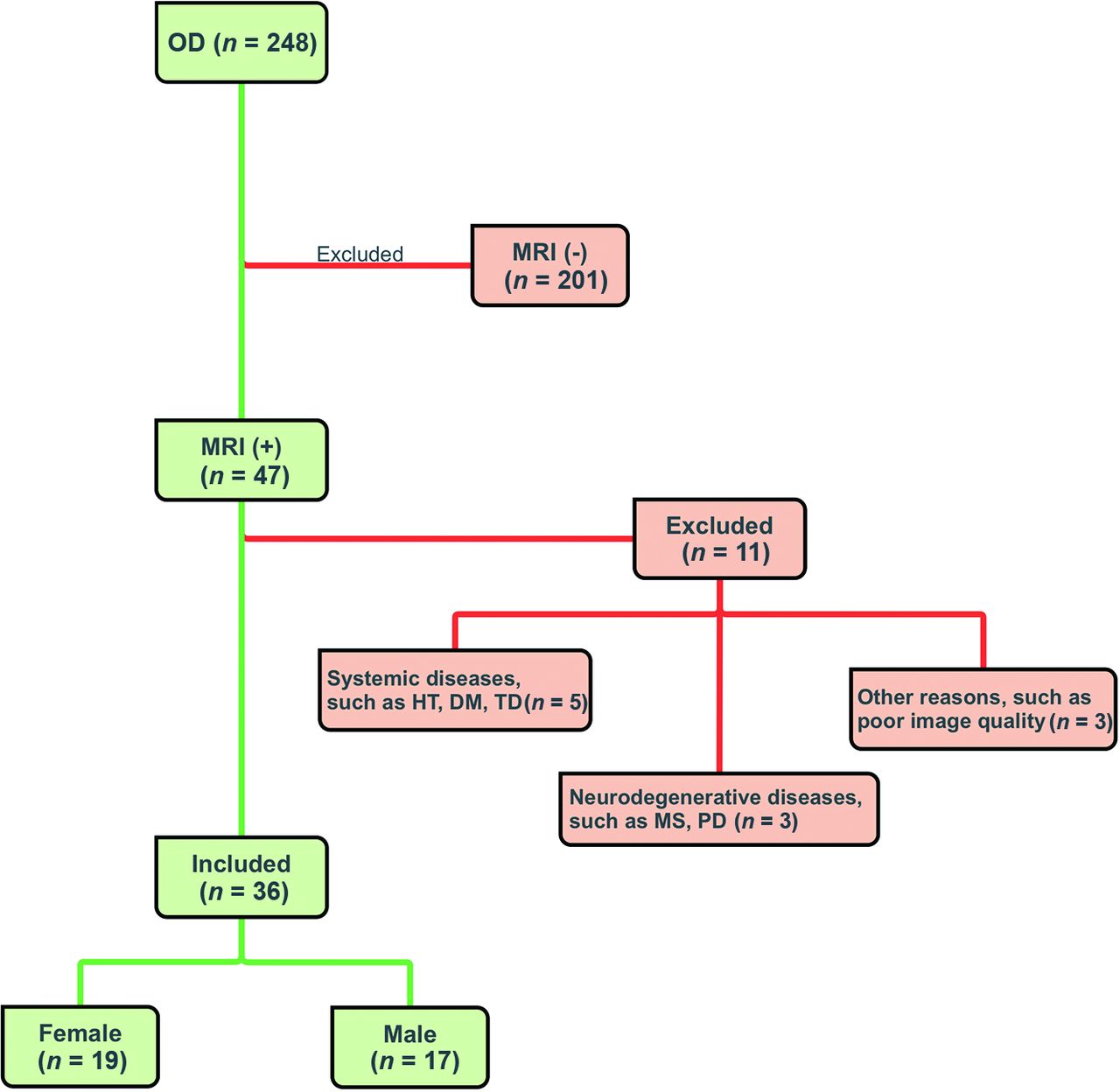
- FIG 1.
CONSORT flow diagram. HT indicates hypertension; DM, diabetes mellitus; TD, thyroid dysfunction; PD, Parkinson disease.
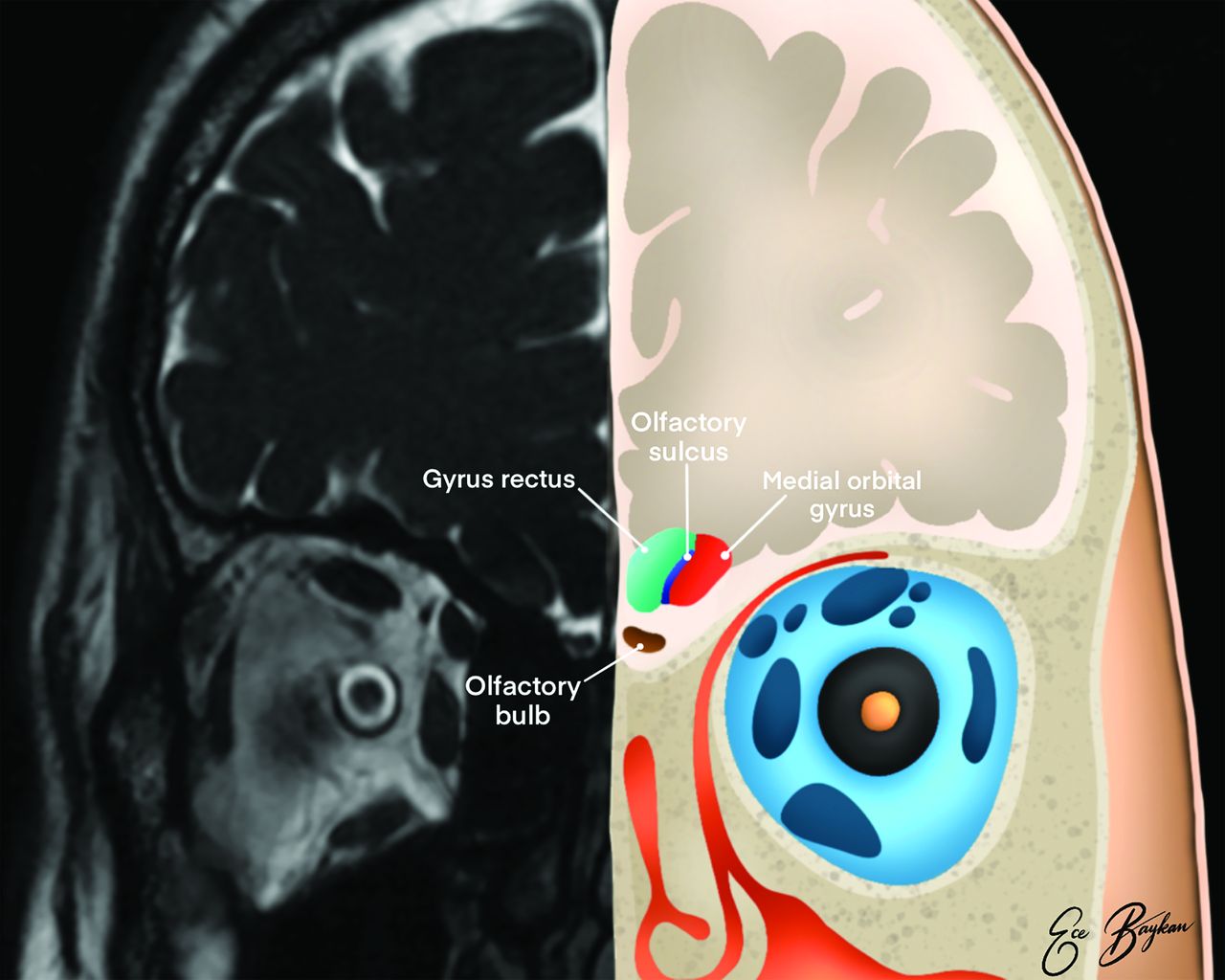
- FIG 2.
Illustration of the olfactory system on coronal plane MR imaging. The medial orbital gyrus (red area), gyrus rectus (green area), olfactory sulcus (blue area), and olfactory bulb (brown area) are shown.
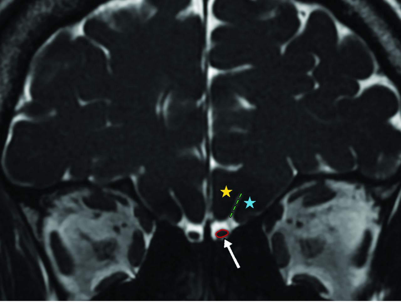
- FIG 3.
Coronal 3D-FIESTA-C MR image of a 41-year-old man showing the right and left olfactory bulbs as hypointense ovoid structures (arrow). The olfactory sulcus (green dashed lines) is seen as a hyperintense line between the medial orbital gyrus (blue star) and gyrus rectus (yellow star). Note the hyperintense CSF surrounding the OBs.
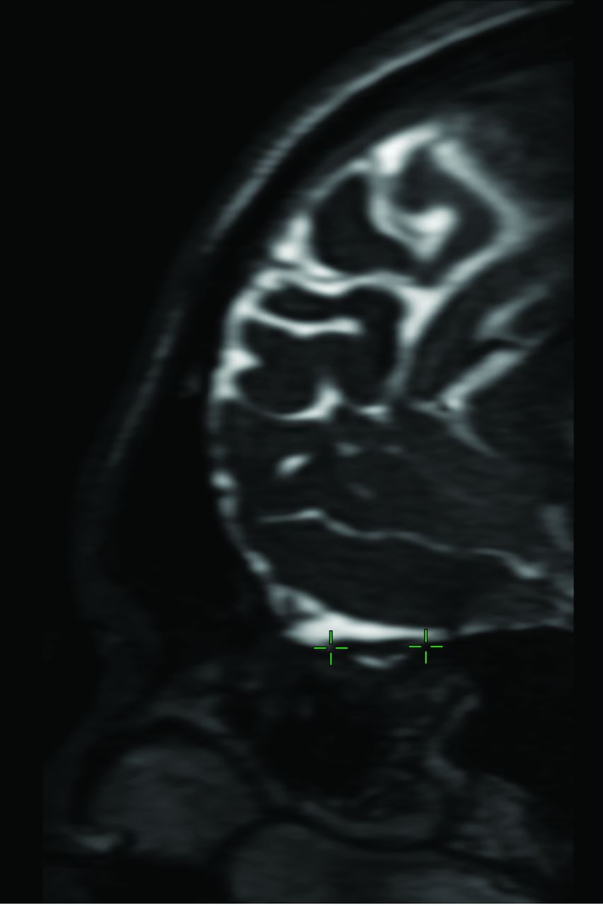
- FIG 4.
Sagittal multiplanar reconstruction of 3D-FIESTA-C MR imaging of a 27-year-old man showing the left olfactory tract (crosshairs).
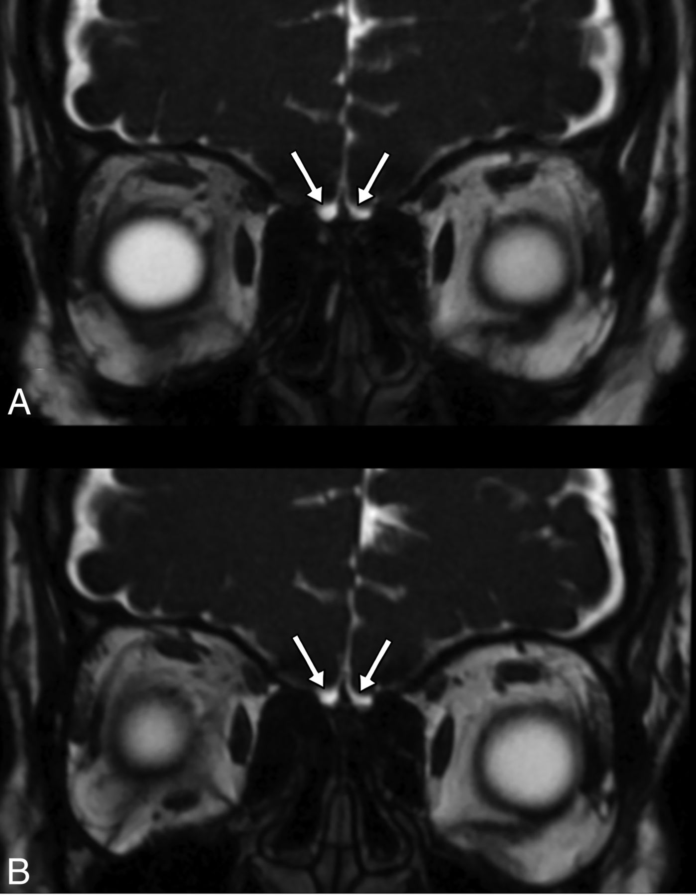
- FIG 5.
Coronal plane 3D-FIESTA-C MR images of a 33-year-old female patient with loss of smell who was proved to have COVID-19. The olfactory bulbs are seen as atrophic (A). The olfactory bulbs are still atrophic, though the loss of smell has improved, on MR image of the same patient 3 months later (B).
- FIG 6.
Boxplots of the right OBV, left OBV, and total OBV values in both patient and control groups.
- FIG 7.
Boxplots of the right OTL, left OTL, right OSD, and left OSD values in both patient and control groups.
- FIG 8.
Coronal 3D FIESTA-C MR images of a 26-year-old male from control group (A) and a 25-year-old female patient with COVID-19 anosmia (B). Normal and increased signal intensity in bilateral olfactory bulbs (red and green arrows, respectively).
- FIG 9.
Coronal 3D-FIESTA-C MR images of a 28-year-old man from the control group (A) and a 36-year-old female patient (B). Note normal and abnormal olfactory bulbs (red and green arrows, respectively) and olfactory sulci (red and green dashed lines, respectively).
- FIG 10.
Sagittal multiplanar reconstruction of 3D-FIESTA-C MR images of a 42-year-old woman from the control group (A) and a 36-year-old female patient (B) showing normal and abnormal olfactory tracts (red and green arrows, respectively).


Tables
Features No. (%) Persistent headache 19 (53%) Taste disturbance 8 (22%) Vertigo/dizziness 6 (17%) Pulmonary involvement 2 (6%) Sinonasal symptoms 0 (0%) - Table 3:
Average, SD, and P values of OBV, OTL, and OSD measurements of patient and control groups
Patient Control Pa R OBV (mm3) 41.57 (SD, 16.96) 66.12 (SD, 16.86) <.001 L OBV (mm3) 40.76 (SD, 15.93) 65.38 (SD, 18.80) <.001 T OBV (mm3) 82.34 (SD, 31.29) 131.50 (SD, 32.27) <.001 R OTL (mm) 11.08 (SD, 2.18) 12.85 (SD, 2.14) <.001 L OTL (mm) 11.24 (SD, 2.57) 12.80 (SD, 2.60) .003 R OSD (mm) 8.33 (SD, 1.65) 9.20 (SD, 1.64) .01 L OSD (mm) 8.64 (SD, 1.40) 9.29 (SD, 1.53) .033 Note:—R indicates right; L, left; T, total.
↵a Independent t test.


