Advertisement

Main menu

  • Home
  • Content
    • Current Issue
    • Accepted Manuscripts
    • Article Preview
    • Past Issue Archive
    • Video Articles
    • AJNR Case Collection
    • Case of the Week Archive
    • Case of the Month Archive
    • Classic Case Archive
  • Special Collections
    • AJNR Awards
    • Low-Field MRI
    • Alzheimer Disease
    • ASNR Foundation Special Collection
    • Photon-Counting CT
    • View All
  • Multimedia
    • AJNR Podcasts
    • AJNR SCANtastic
    • Trainee Corner
    • MRI Safety Corner
    • Imaging Protocols
  • For Authors
    • Submit a Manuscript
    • Submit a Video Article
    • Submit an eLetter to the Editor/Response
    • Manuscript Submission Guidelines
    • Statistical Tips
    • Fast Publishing of Accepted Manuscripts
    • Graphical Abstract Preparation
    • Imaging Protocol Submission
    • Author Policies
  • About Us
    • About AJNR
    • Editorial Board
    • Editorial Board Alumni
  • More
    • Become a Reviewer/Academy of Reviewers
    • Subscribers
    • Permissions
    • Alerts
    • Feedback
    • Advertisers
    • ASNR Home

User menu

  • Alerts
  • Log in

Search

  • Advanced search
American Journal of Neuroradiology

American Journal of Neuroradiology

ASHNR American Society of Functional Neuroradiology ASHNR American Society of Pediatric Neuroradiology ASSR
  • Alerts
  • Log in

Advanced Search

  • Home
  • Content
    • Current Issue
    • Accepted Manuscripts
    • Article Preview
    • Past Issue Archive
    • Video Articles
    • AJNR Case Collection
    • Case of the Week Archive
    • Case of the Month Archive
    • Classic Case Archive
  • Special Collections
    • AJNR Awards
    • Low-Field MRI
    • Alzheimer Disease
    • ASNR Foundation Special Collection
    • Photon-Counting CT
    • View All
  • Multimedia
    • AJNR Podcasts
    • AJNR SCANtastic
    • Trainee Corner
    • MRI Safety Corner
    • Imaging Protocols
  • For Authors
    • Submit a Manuscript
    • Submit a Video Article
    • Submit an eLetter to the Editor/Response
    • Manuscript Submission Guidelines
    • Statistical Tips
    • Fast Publishing of Accepted Manuscripts
    • Graphical Abstract Preparation
    • Imaging Protocol Submission
    • Author Policies
  • About Us
    • About AJNR
    • Editorial Board
    • Editorial Board Alumni
  • More
    • Become a Reviewer/Academy of Reviewers
    • Subscribers
    • Permissions
    • Alerts
    • Feedback
    • Advertisers
    • ASNR Home
  • Follow AJNR on Twitter
  • Visit AJNR on Facebook
  • Follow AJNR on Instagram
  • Join AJNR on LinkedIn
  • RSS Feeds

AJNR Awards, New Junior Editors, and more. Read the latest AJNR updates

Research ArticleSpine Imaging and Spine Image-Guided Interventions

MRI T2-Hyperintense Signal Structures in the Cervical Spinal Cord: Anterior Median Fissure versus Central Canal in Chiari and Control—An Exploratory Pilot Analysis

T.A. Tomsick, L.L. Wang, M. Zuccarello and A.J. Ringer
American Journal of Neuroradiology March 2021, DOI: https://doi.org/10.3174/ajnr.A7046
T.A. Tomsick
aFrom the Department of Radiology (T.A.T., L.L.W.)
  • Find this author on Google Scholar
  • Find this author on PubMed
  • Search for this author on this site
  • ORCID record for T.A. Tomsick
L.L. Wang
aFrom the Department of Radiology (T.A.T., L.L.W.)
  • Find this author on Google Scholar
  • Find this author on PubMed
  • Search for this author on this site
  • ORCID record for L.L. Wang
M. Zuccarello
bNeuroradiology Section and Department of Neurosurgery (M.Z.), University of Cincinnati Medical Center, Cincinnati, Ohio
  • Find this author on Google Scholar
  • Find this author on PubMed
  • Search for this author on this site
  • ORCID record for M. Zuccarello
A.J. Ringer
cMayfield Clinic (A.J.R.), Cincinnati, Ohio
  • Find this author on Google Scholar
  • Find this author on PubMed
  • Search for this author on this site
  • ORCID record for A.J. Ringer
  • Article
  • Figures & Data
  • Supplemental
  • Info & Metrics
  • Responses
  • References
  • PDF
Loading

Article Figures & Data

Figures

  • Tables
  • FIG 1.
    • Download figure
    • Open in new tab
    • Download powerpoint
    FIG 1.

    A patient with Chiari I with 19 HIF up to 3 mm in diameter, 1 AMF, no AMF>HIF, and an SL of various hyperintensity and diameter from C4 through T1, consistent with hydromyelia.
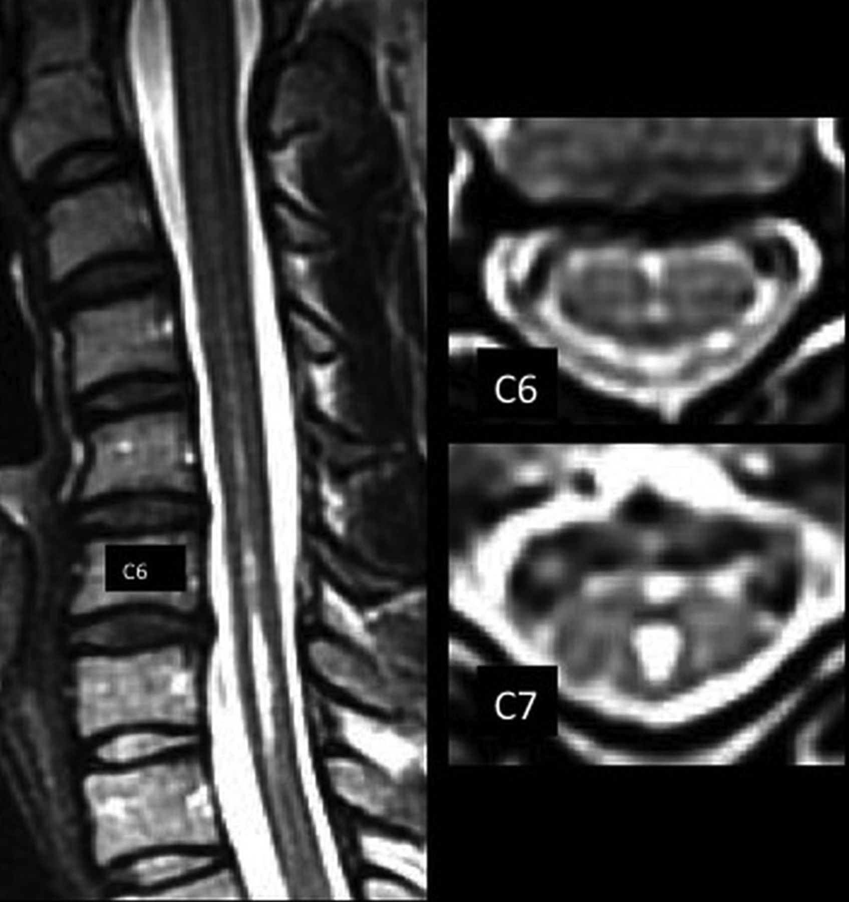
  • FIG 2.
    • Download figure
    • Open in new tab
    • Download powerpoint
    FIG 2.

    Postdecompressive craniectomy patient with Chiari I with 9 HIF, 4 AMFs, 1 AMF>HIF, and sharp and hyperintense SLs at C6–C7 and less hyperintense, sharp, and defined SLs at C2–C6.
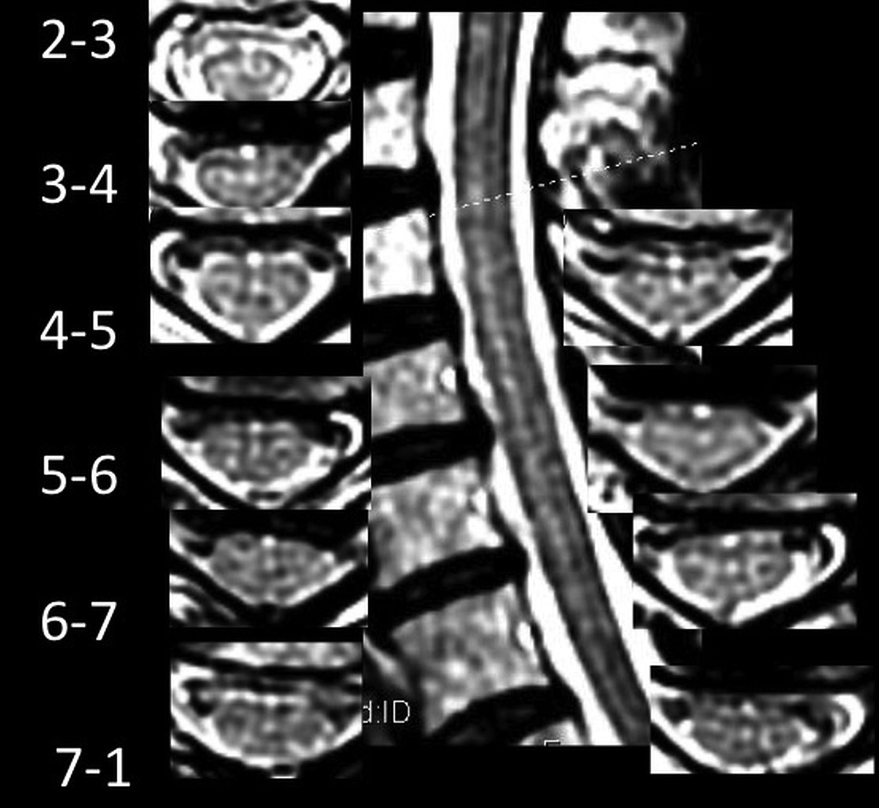
  • FIG 3.
    • Download figure
    • Open in new tab
    • Download powerpoint
    FIG 3.

    Control patient with 15 HIF, 3 AMFs, 3 AMF>HIF, and SL with variably hyperintense and variably sharp dots and dashes from C3–4 to C7. Note hyperintense pixels immediately anterior to the AMF on axial images, despite crescentic flow-related signal loss immediately anterior to the cord on most images.

  • FIG 4.
    • Download figure
    • Open in new tab
    • Download powerpoint
    FIG 4.

    Postoperative patient with Chiari I with 19 HIF, 3 AMFs, and 2 AMF>HIF, with a faintly hyperintense ventral line at C6–T1.

Tables

  • Figures
    • View popup
    Table 1:

    Age, sex, incidence (%), and frequency (No./Pt), for HIF, AMF, SL, and AMF>HIF in the Chiari I and non-Chiari I control groups

    Age (Median)Sex (F) (%)HIF (%)HIF/Pt.AMF (%)AMF/Pt.SL (No.) (%)AMF>HIF (Mean)
    Chiari I (n = 25)4023 (92%)25 (100%)8.5a24 (96%)4.0b14c (56%)2.2
    Control (n = 25)4017 (68%)24 (96%)3.9a22 (88%)2.7b7c (28%)1.0
    • ↵a P = .0004.

    • ↵b P = .025.

    • ↵c P < .02 (χ2).

    • View popup
    Table 2:

    Incidence HIF and AMF/pt., SL incidence, and number of AMFs extending to HIF (AMF>HIF) in Chiari I without an operation versus patients having undergone postdecompressive craniectomy

    HIFAMFSLAMF>HIF
    %HIF/Pt.%AMF/Pt.No. (%)No.
    Chiari I no surgery (n = 15)1008.91004.05 (33%)a1.9b
    Chiari I post-op (n = 10)1008.1904.38 (80%)a2.8b
    • Note:—Post-op indicates postoperative.

    • ↵a P = .009.

    • ↵b P = .07.

    • View popup
    Table 3:

    Number patients, HIF/pt., AMF/pt., HIF to AMF ratio, and AMF>HIF/pt. in HIF or AMF-only, HIF+AMF, and AMF>HIF subgroups for all Chiari I and control groups, with and without SL

     No.HIF/Pt.AMF/Pt.RatioAMF>HIF
    All Chiari I258.64.12.12.2
    All control253.92.71.41
    All Chiari I and controls
     HIF only
      Chiari I17NANA2.3
      Control112NANA1.0
     AMF only0NANANANA
     HIF+AMF
      Chiari I248.64.32.02.1
      Control233.931.31.1
      AMF-HIF
      Chiari I227.94.71.72.5
      Control164.43.31.31.5
     No AMF→HIF
      Chiari I313.71.78.20
      Control931.71.80
     SL
      Chiari I1410.14.62.22.9
      Control773.42.11.3
     HIF+AMF
      Chiari I1410.14.62.22.2
      Control66.241.61.5
     AMF-HIF
      Chiari I128.651.72.9
      Control57.241.81.8
     No SL
      Chiari I116.63.51.91.9
      Control182.72.41.10.8
     HIF+AMF
      Chiari I116.83.91.72.1
      Control172.930.90.9
     AMF-HIF
      Chiari I974.21.72.2
      Control113.131.11.2
    • Note:—NA indicates not applicable.

PreviousNext
Back to top
Advertisement
Print
Download PDF
Email Article

Thank you for your interest in spreading the word on American Journal of Neuroradiology.

NOTE: We only request your email address so that the person you are recommending the page to knows that you wanted them to see it, and that it is not junk mail. We do not capture any email address.

Enter multiple addresses on separate lines or separate them with commas.
MRI T2-Hyperintense Signal Structures in the Cervical Spinal Cord: Anterior Median Fissure versus Central Canal in Chiari and Control—An Exploratory Pilot Analysis
(Your Name) has sent you a message from American Journal of Neuroradiology
(Your Name) thought you would like to see the American Journal of Neuroradiology web site.
CAPTCHA
This question is for testing whether or not you are a human visitor and to prevent automated spam submissions.
Cite this article
T.A. Tomsick, L.L. Wang, M. Zuccarello, A.J. Ringer
MRI T2-Hyperintense Signal Structures in the Cervical Spinal Cord: Anterior Median Fissure versus Central Canal in Chiari and Control—An Exploratory Pilot Analysis
American Journal of Neuroradiology Mar 2021, DOI: 10.3174/ajnr.A7046

Citation Manager Formats

  • BibTeX
  • Bookends
  • EasyBib
  • EndNote (tagged)
  • EndNote 8 (xml)
  • Medlars
  • Mendeley
  • Papers
  • RefWorks Tagged
  • Ref Manager
  • RIS
  • Zotero
0 Responses
Respond to this article
Share
Bookmark this article
MRI T2-Hyperintense Signal Structures in the Cervical Spinal Cord: Anterior Median Fissure versus Central Canal in Chiari and Control—An Exploratory Pilot Analysis
T.A. Tomsick, L.L. Wang, M. Zuccarello, A.J. Ringer
American Journal of Neuroradiology Mar 2021, DOI: 10.3174/ajnr.A7046
del.icio.us logo Twitter logo Facebook logo Mendeley logo
  • Tweet Widget
  • Facebook Like
  • Google Plus One
Purchase

Jump to section

  • Article
    • Abstract
    • ABBREVIATIONS:
    • MATERIALS AND METHODS
    • RESULTS
    • DISCUSSION
    • CONCLUSIONS
    • Footnotes
    • References
  • Figures & Data
  • Supplemental
  • Info & Metrics
  • Responses
  • References
  • PDF

Related Articles

  • PubMed
  • Google Scholar

Cited By...

  • No citing articles found.
  • Crossref
  • Google Scholar

This article has not yet been cited by articles in journals that are participating in Crossref Cited-by Linking.

More in this TOC Section

  • Optimization of Photon Counting CT Myelography
  • Characteristics of SIH Type I Culprit Lesions
  • Management Outcomes For VO Spine Biopsy
Show more Spine Imaging and Spine Image-Guided Interventions

Similar Articles

Advertisement

Indexed Content

  • Current Issue
  • Accepted Manuscripts
  • Article Preview
  • Past Issues
  • Editorials
  • Editor's Choice
  • Fellows' Journal Club
  • Letters to the Editor
  • Video Articles

Cases

  • Case Collection
  • Archive - Case of the Week
  • Archive - Case of the Month
  • Archive - Classic Case

More from AJNR

  • Trainee Corner
  • Imaging Protocols
  • MRI Safety Corner
  • Book Reviews

Multimedia

  • AJNR Podcasts
  • AJNR Scantastics

Resources

  • Turnaround Time
  • Submit a Manuscript
  • Submit a Video Article
  • Submit an eLetter to the Editor/Response
  • Manuscript Submission Guidelines
  • Statistical Tips
  • Fast Publishing of Accepted Manuscripts
  • Graphical Abstract Preparation
  • Imaging Protocol Submission
  • Evidence-Based Medicine Level Guide
  • Publishing Checklists
  • Author Policies
  • Become a Reviewer/Academy of Reviewers
  • News and Updates

About Us

  • About AJNR
  • Editorial Board
  • Editorial Board Alumni
  • Alerts
  • Permissions
  • Not an AJNR Subscriber? Join Now
  • Advertise with Us
  • Librarian Resources
  • Feedback
  • Terms and Conditions
  • AJNR Editorial Board Alumni

American Society of Neuroradiology

  • Not an ASNR Member? Join Now

© 2025 by the American Society of Neuroradiology All rights, including for text and data mining, AI training, and similar technologies, are reserved.
Print ISSN: 0195-6108 Online ISSN: 1936-959X

Powered by HighWire