Advertisement

Main menu

  • Home
  • Content
    • Current Issue
    • Accepted Manuscripts
    • Article Preview
    • Past Issue Archive
    • Video Articles
    • AJNR Case Collection
    • Case of the Week Archive
    • Case of the Month Archive
    • Classic Case Archive
  • Special Collections
    • AJNR Awards
    • Low-Field MRI
    • Alzheimer Disease
    • ASNR Foundation Special Collection
    • Photon-Counting CT
    • View All
  • Multimedia
    • AJNR Podcasts
    • AJNR SCANtastic
    • Trainee Corner
    • MRI Safety Corner
    • Imaging Protocols
  • For Authors
    • Submit a Manuscript
    • Submit a Video Article
    • Submit an eLetter to the Editor/Response
    • Manuscript Submission Guidelines
    • Statistical Tips
    • Fast Publishing of Accepted Manuscripts
    • Graphical Abstract Preparation
    • Imaging Protocol Submission
    • Author Policies
  • About Us
    • About AJNR
    • Editorial Board
    • Editorial Board Alumni
  • More
    • Become a Reviewer/Academy of Reviewers
    • Subscribers
    • Permissions
    • Alerts
    • Feedback
    • Advertisers
    • ASNR Home

User menu

  • Alerts
  • Log in

Search

  • Advanced search
American Journal of Neuroradiology

American Journal of Neuroradiology

ASHNR American Society of Functional Neuroradiology ASHNR American Society of Pediatric Neuroradiology ASSR
  • Alerts
  • Log in

Advanced Search

  • Home
  • Content
    • Current Issue
    • Accepted Manuscripts
    • Article Preview
    • Past Issue Archive
    • Video Articles
    • AJNR Case Collection
    • Case of the Week Archive
    • Case of the Month Archive
    • Classic Case Archive
  • Special Collections
    • AJNR Awards
    • Low-Field MRI
    • Alzheimer Disease
    • ASNR Foundation Special Collection
    • Photon-Counting CT
    • View All
  • Multimedia
    • AJNR Podcasts
    • AJNR SCANtastic
    • Trainee Corner
    • MRI Safety Corner
    • Imaging Protocols
  • For Authors
    • Submit a Manuscript
    • Submit a Video Article
    • Submit an eLetter to the Editor/Response
    • Manuscript Submission Guidelines
    • Statistical Tips
    • Fast Publishing of Accepted Manuscripts
    • Graphical Abstract Preparation
    • Imaging Protocol Submission
    • Author Policies
  • About Us
    • About AJNR
    • Editorial Board
    • Editorial Board Alumni
  • More
    • Become a Reviewer/Academy of Reviewers
    • Subscribers
    • Permissions
    • Alerts
    • Feedback
    • Advertisers
    • ASNR Home
  • Follow AJNR on Twitter
  • Visit AJNR on Facebook
  • Follow AJNR on Instagram
  • Join AJNR on LinkedIn
  • RSS Feeds

AJNR Awards, New Junior Editors, and more. Read the latest AJNR updates

Research ArticlePediatric Neuroimaging

A Diagnostic Algorithm for Posterior Fossa Tumors in Children: A Validation Study

C.A.P.F. Alves, U. Löbel, J.S. Martin-Saavedra, S. Toescu, M.H. Tsunemi, S.R. Teixeira, K. Mankad, D. Hargrave, T.S. Jacques, C. da Costa Leite, F.G. Gonçalves, A. Vossough and F. D’Arco
American Journal of Neuroradiology March 2021, DOI: https://doi.org/10.3174/ajnr.A7057
C.A.P.F. Alves
aFrom the Division of Neuroradiology (C.A.P.F.A., J.S.M.-S., S.R.T., F.G.G., A.V.), Department of Radiology, Children’s Hospital of Philadelphia, Philadelphia, Pennsylvania
  • Find this author on Google Scholar
  • Find this author on PubMed
  • Search for this author on this site
  • ORCID record for C.A.P.F. Alves
U. Löbel
bDepartments of Radiology (U.L., K.M., F.D.)
  • Find this author on Google Scholar
  • Find this author on PubMed
  • Search for this author on this site
  • ORCID record for U. Löbel
J.S. Martin-Saavedra
aFrom the Division of Neuroradiology (C.A.P.F.A., J.S.M.-S., S.R.T., F.G.G., A.V.), Department of Radiology, Children’s Hospital of Philadelphia, Philadelphia, Pennsylvania
  • Find this author on Google Scholar
  • Find this author on PubMed
  • Search for this author on this site
  • ORCID record for J.S. Martin-Saavedra
S. Toescu
cNeurosurgery (S.T.)
  • Find this author on Google Scholar
  • Find this author on PubMed
  • Search for this author on this site
  • ORCID record for S. Toescu
M.H. Tsunemi
eDepartment of Biostatistics (M.H.T.), Instituto de Biociências, São Paulo State University, São Paulo, Brazil
  • Find this author on Google Scholar
  • Find this author on PubMed
  • Search for this author on this site
  • ORCID record for M.H. Tsunemi
S.R. Teixeira
aFrom the Division of Neuroradiology (C.A.P.F.A., J.S.M.-S., S.R.T., F.G.G., A.V.), Department of Radiology, Children’s Hospital of Philadelphia, Philadelphia, Pennsylvania
  • Find this author on Google Scholar
  • Find this author on PubMed
  • Search for this author on this site
  • ORCID record for S.R. Teixeira
K. Mankad
bDepartments of Radiology (U.L., K.M., F.D.)
  • Find this author on Google Scholar
  • Find this author on PubMed
  • Search for this author on this site
  • ORCID record for K. Mankad
D. Hargrave
dPediatric Oncology Unit (D.H.), University College London Great Ormond Street Hospital for Children, London, UK
  • Find this author on Google Scholar
  • Find this author on PubMed
  • Search for this author on this site
  • ORCID record for D. Hargrave
T.S. Jacques
fDevelopmental Biology and Cancer Programme (T.S.J.), University College London Great Ormond Street Institute of Child Health, University College London, London, UK
  • Find this author on Google Scholar
  • Find this author on PubMed
  • Search for this author on this site
  • ORCID record for T.S. Jacques
C. da Costa Leite
gDepartment of Radiology (C.d.C.L.), Hospital das Clinicas, Faculdade de Medicina de Sao Paulo, Sao Paulo, Brazil.
  • Find this author on Google Scholar
  • Find this author on PubMed
  • Search for this author on this site
  • ORCID record for C. da Costa Leite
F.G. Gonçalves
aFrom the Division of Neuroradiology (C.A.P.F.A., J.S.M.-S., S.R.T., F.G.G., A.V.), Department of Radiology, Children’s Hospital of Philadelphia, Philadelphia, Pennsylvania
  • Find this author on Google Scholar
  • Find this author on PubMed
  • Search for this author on this site
  • ORCID record for F.G. Gonçalves
A. Vossough
aFrom the Division of Neuroradiology (C.A.P.F.A., J.S.M.-S., S.R.T., F.G.G., A.V.), Department of Radiology, Children’s Hospital of Philadelphia, Philadelphia, Pennsylvania
  • Find this author on Google Scholar
  • Find this author on PubMed
  • Search for this author on this site
  • ORCID record for A. Vossough
F. D’Arco
bDepartments of Radiology (U.L., K.M., F.D.)
  • Find this author on Google Scholar
  • Find this author on PubMed
  • Search for this author on this site
  • ORCID record for F. D’Arco
  • Article
  • Figures & Data
  • Info & Metrics
  • Responses
  • References
  • PDF
Loading

Article Figures & Data

Figures

  • Tables
  • FIG 1.
    • Download figure
    • Open in new tab
    • Download powerpoint
    FIG 1.

    Predesigned radiologic flow chart created according to the literature before diagnostic accuracy analysis. The asterisk indicates brain stem tumors excluded from the analysis. Double asterisks indicate relative to gray matter. Modified with permission from D’Arco et al.11
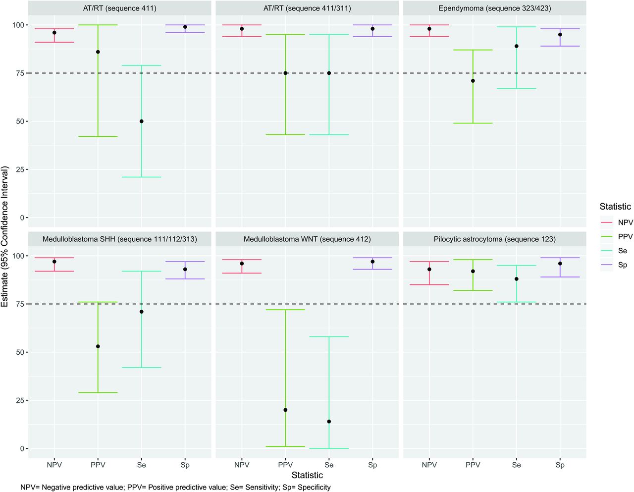
  • FIG 2.
    • Download figure
    • Open in new tab
    • Download powerpoint
    FIG 2.

    Diagnostic accuracy of a predesigned radiologic flow chart to identify different types of cerebellar tumors.
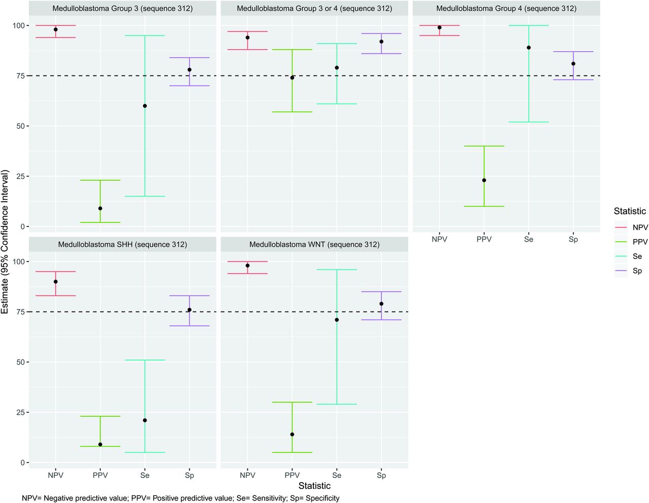
  • FIG 3.
    • Download figure
    • Open in new tab
    • Download powerpoint
    FIG 3.

    Diagnostic accuracy of sequence 312 (all types of medulloblastomas) of the predesigned radiologic flow chart to identify different types of medulloblastomas.

  • FIG 4.
    • Download figure
    • Open in new tab
    • Download powerpoint
    FIG 4.

    Modified radiologic flow chart (flow chart 2) after diagnostic accuracy analysis. The asterisk indicates brain stem tumors excluded from the analysis. Double asterisks indicate relative to gray matter. Triple asterisks indicates low PPV and sensitivity for any particular molecular/histological group of tumor.
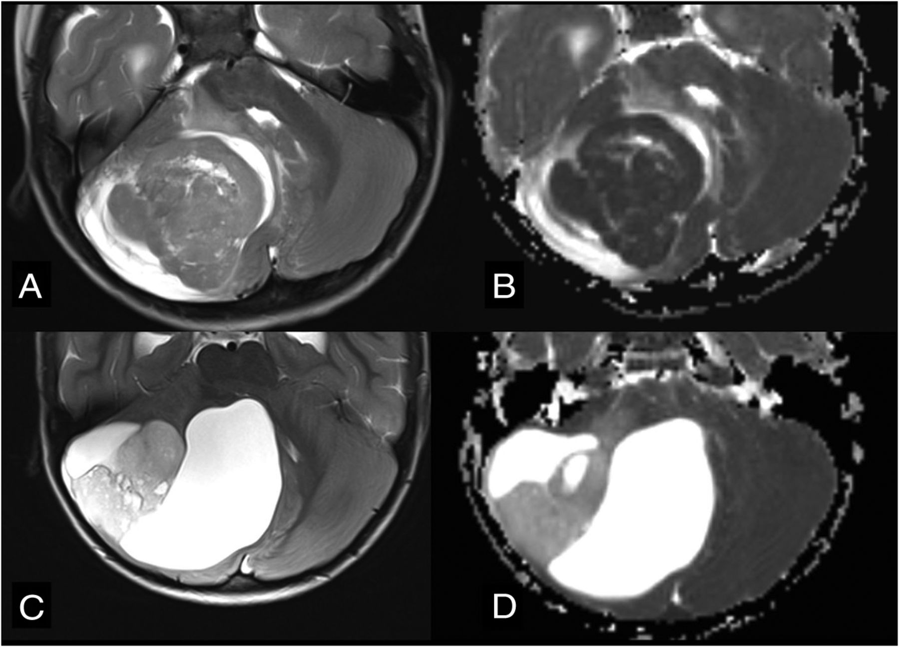
  • FIG 5.
    • Download figure
    • Open in new tab
    • Download powerpoint
    FIG 5.

    Differential diagnoses in cases of posterior fossa tumors originating from the cerebellar hemisphere. Axial T2WI (A) and axial ADC map (B) show SHH medulloblastoma (flow chart 2, number 111) in a typical peripheral location within the cerebellar hemisphere due to its origin from ganglionic cell precursors. Note very low ADC values (ie, diffusion restriction). Axial T2WI (C) and axial ADC map (D) show the typical appearance of a pilocytic astrocytoma (flow chart 2, number 123) originating from the cerebellar hemisphere. Note the typical nodule and appearance of cysts and much higher ADC values in comparison with the medulloblastoma.

  • FIG 6.
    • Download figure
    • Open in new tab
    • Download powerpoint
    FIG 6.

    Differential diagnoses in posterior fossa tumors involving the foramen of Luschka and cerebellopontine angle. Axial T2WI (A) and ADC map (B) in a child with ependymoma (flow chart 2, number 423). Note the presence of internal vessels (arrow) and intermediate ADC values. Axial T2 (C) and ADC maps (D) in a 2-year-old boy with a AT/RT (new flow chart number 411). Note very low values of ADC, suggesting an embryonal tumor and peripheral cysts.

Tables

  • Figures
    • View popup
    Table 1:

    Statistical analysis of the radiologic flow chart to discriminate different types of cerebellar tumors

    Diagnosis EquivalentFlowchart SequenceSensitivity (%)Specificity (%)PPV (%)NPV (%)Accuracy (%)
    Ependymoma323/42389 (67–99)95 (89–98)71 (49–87)98 (94–100)94 (89–97)
    Pilocytic astrocytoma12388 (76–95)96 (89–99)92 (82–98)93 (85–97)93 (87–96)
    AT/RT41150 (21–79)99 (96–100)86 (42–100)96 (91–98)99 (96–100)
    AT/RT411/31175 (43–95)98 (94–100)75 (43–95)98 (94–100)91 (96–98)
    Medulloblastoma SHH111/112/31371 (42–92)93 (88–97)53 (29–76)97 (92–99)72 (64–79)
    Medulloblastoma WNT41214 (0–58)97 (93–99)20 (1–72)96 (91–98)88 (93–97)
    • View popup
    Table 2:

    Likelihood ratio analysis of the radiologic flow chart to discriminate different types of cerebellar tumors

    DiagnosisFlow Chart SequenceLR+ (95% CI)LR– (95% CI)
    AT/RT411/31134 (10.6–109)a0.26 (0.1–0.7)
    Ependymoma323/42316.5 (7.9–35)0.11 (0.03–0.4)a
    Medulloblastoma SHH111/112/31310.6 (5.2–21.7)0.3 (0.1–0.7)
    Medulloblastoma group 3 or 431210.07 (5.3–19.3)0.23 (0.12–0.45)a
    Pilocytic astrocytoma12320 (7.7–52.8)0.13 (0.13–0.26)a
    • ↵a Clinically applicable confidence intervals.

PreviousNext
Back to top
Advertisement
Print
Download PDF
Email Article

Thank you for your interest in spreading the word on American Journal of Neuroradiology.

NOTE: We only request your email address so that the person you are recommending the page to knows that you wanted them to see it, and that it is not junk mail. We do not capture any email address.

Enter multiple addresses on separate lines or separate them with commas.
A Diagnostic Algorithm for Posterior Fossa Tumors in Children: A Validation Study
(Your Name) has sent you a message from American Journal of Neuroradiology
(Your Name) thought you would like to see the American Journal of Neuroradiology web site.
CAPTCHA
This question is for testing whether or not you are a human visitor and to prevent automated spam submissions.
Cite this article
C.A.P.F. Alves, U. Löbel, J.S. Martin-Saavedra, S. Toescu, M.H. Tsunemi, S.R. Teixeira, K. Mankad, D. Hargrave, T.S. Jacques, C. da Costa Leite, F.G. Gonçalves, A. Vossough, F. D’Arco
A Diagnostic Algorithm for Posterior Fossa Tumors in Children: A Validation Study
American Journal of Neuroradiology Mar 2021, DOI: 10.3174/ajnr.A7057

Citation Manager Formats

  • BibTeX
  • Bookends
  • EasyBib
  • EndNote (tagged)
  • EndNote 8 (xml)
  • Medlars
  • Mendeley
  • Papers
  • RefWorks Tagged
  • Ref Manager
  • RIS
  • Zotero
0 Responses
Respond to this article
Share
Bookmark this article
A Diagnostic Algorithm for Posterior Fossa Tumors in Children: A Validation Study
C.A.P.F. Alves, U. Löbel, J.S. Martin-Saavedra, S. Toescu, M.H. Tsunemi, S.R. Teixeira, K. Mankad, D. Hargrave, T.S. Jacques, C. da Costa Leite, F.G. Gonçalves, A. Vossough, F. D’Arco
American Journal of Neuroradiology Mar 2021, DOI: 10.3174/ajnr.A7057
del.icio.us logo Twitter logo Facebook logo Mendeley logo
  • Tweet Widget
  • Facebook Like
  • Google Plus One
Purchase

Jump to section

  • Article
    • Abstract
    • ABBREVIATIONS:
    • MATERIALs and METHODS
    • RESULTS
    • DISCUSSION
    • CONCLUSIONS
    • Footnotes
    • References
  • Figures & Data
  • Info & Metrics
  • Responses
  • References
  • PDF

Related Articles

  • No related articles found.
  • PubMed
  • Google Scholar

Cited By...

  • CNS Embryonal Tumor with PLAGL Amplification, a New Tumor in Children and Adolescents: Insights from a Comprehensive MRI Analysis
  • Crossref
  • Google Scholar

This article has not yet been cited by articles in journals that are participating in Crossref Cited-by Linking.

More in this TOC Section

  • Frontal Paraventricular Cysts
  • Sodium MRI in Pediatric Brain Tumors
  • FRACTURE MR in Congenital Vertebral Anomalies
Show more Pediatric Neuroimaging

Similar Articles

Advertisement

Indexed Content

  • Current Issue
  • Accepted Manuscripts
  • Article Preview
  • Past Issues
  • Editorials
  • Editor's Choice
  • Fellows' Journal Club
  • Letters to the Editor
  • Video Articles

Cases

  • Case Collection
  • Archive - Case of the Week
  • Archive - Case of the Month
  • Archive - Classic Case

More from AJNR

  • Trainee Corner
  • Imaging Protocols
  • MRI Safety Corner
  • Book Reviews

Multimedia

  • AJNR Podcasts
  • AJNR Scantastics

Resources

  • Turnaround Time
  • Submit a Manuscript
  • Submit a Video Article
  • Submit an eLetter to the Editor/Response
  • Manuscript Submission Guidelines
  • Statistical Tips
  • Fast Publishing of Accepted Manuscripts
  • Graphical Abstract Preparation
  • Imaging Protocol Submission
  • Evidence-Based Medicine Level Guide
  • Publishing Checklists
  • Author Policies
  • Become a Reviewer/Academy of Reviewers
  • News and Updates

About Us

  • About AJNR
  • Editorial Board
  • Editorial Board Alumni
  • Alerts
  • Permissions
  • Not an AJNR Subscriber? Join Now
  • Advertise with Us
  • Librarian Resources
  • Feedback
  • Terms and Conditions
  • AJNR Editorial Board Alumni

American Society of Neuroradiology

  • Not an ASNR Member? Join Now

© 2025 by the American Society of Neuroradiology All rights, including for text and data mining, AI training, and similar technologies, are reserved.
Print ISSN: 0195-6108 Online ISSN: 1936-959X

Powered by HighWire