
Article Figures & Data
Figures
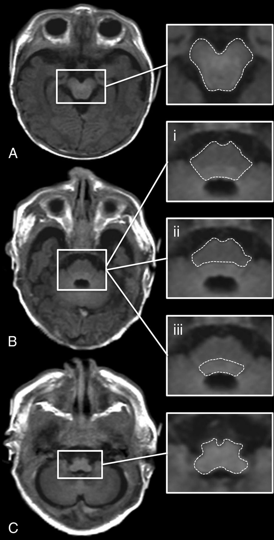
- FIG 1.
ROI placement is shown on an SyMRI-generated T1-weighted MR image (TR/TE = 650/10 ms) of a term-born neonate (GA: 40 + 1 weeks). A, Midbrain. B.i, Pons (basis pontis and tegmentum pontis included). B.ii, Basis pontis (tegmentum pontis excluded). B.iii, Tegmentum pontis (basis pontis excluded). C, Medulla oblongata.
- FIG 2.
Pearson correlation between GA (weeks) at birth (x-axis) and physical MR parameters (y-axis) determined at term-equivalent age by rater 1. Left column: T1R (A–E). Middle column: T2R (F–J). Right column: PD (K–O).
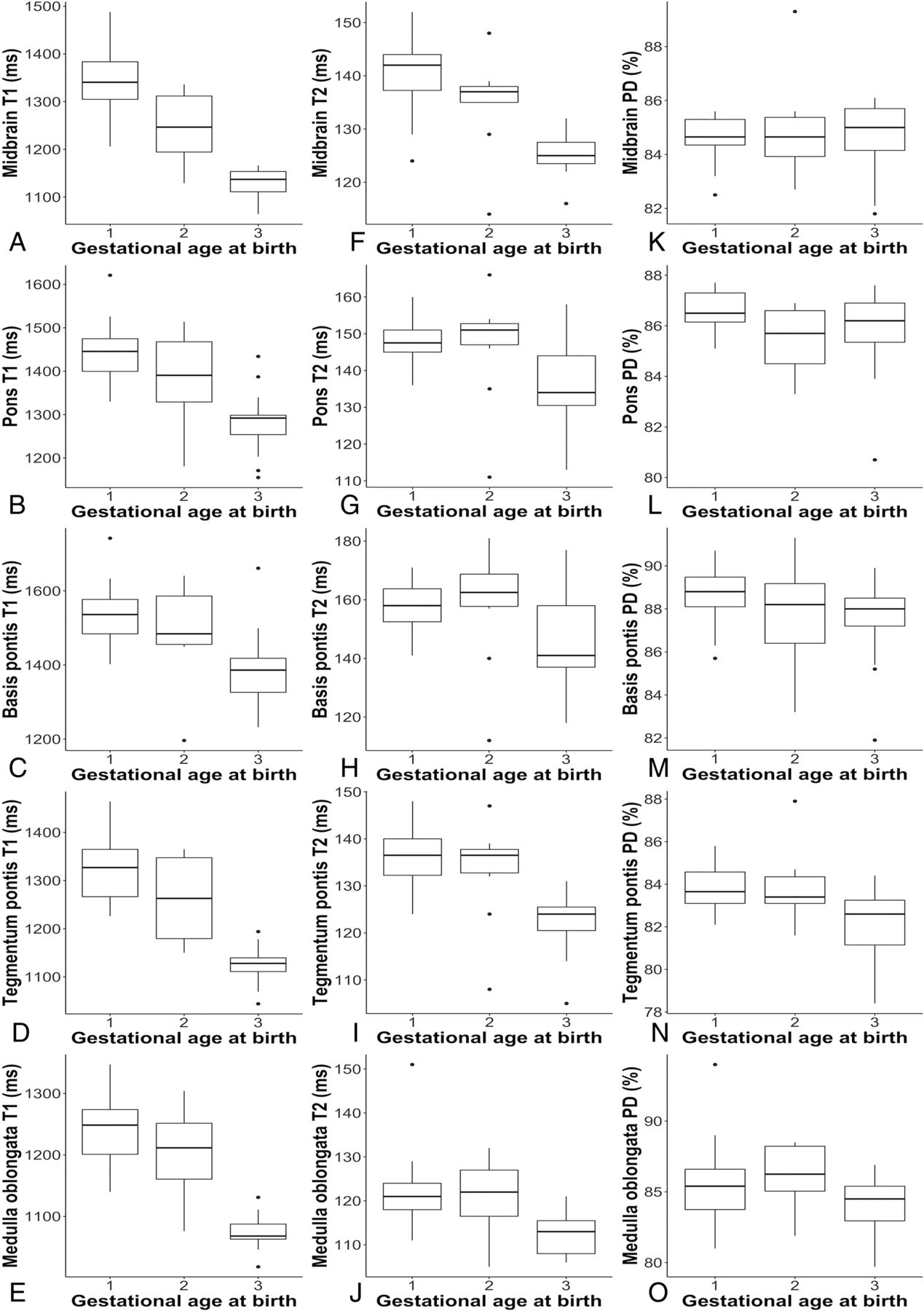
- FIG 3.
The boxplots show descriptive data for quantitative MR parameters (y-axis) of (1) neonates born <28 + 0 weeks GA, (2) neonates born between 28 + 0–36 + 6 weeks GA, and (3) neonates born ≥37 + 0 weeks GA (x-axis) determined at term-equivalent age by rater 1. Left column: T1R (A–E). Middle column: T2R (F–J). Right column: PD (K–O).


Tables
Neonates: n = 55 Extremely Preterm Birth (<28 + 0 weeks GA): n = 30 Preterm Birth (28 + 0–36 + 6 weeks GA): n = 10 Term Born (≥37 + 0 weeks GA): n = 15 Clinical characteristics Male/female 10/20 5/5 6/9 GA at birtha M = 25 + 5, SD = 1 + 4;R = 23 + 0–27 + 6 M = 32 + 4, SD = 3 + 1; R = 28 + 0–36 + 3 M = 39 + 5, SD = 0 + 6; R = 37 + 6–40 + 5 GA at MR imaginga M = 38 + 2, SD = 2 + 0;R = 35 + 1–43 + 4 M = 39 + 0, SD = 6 + 1; R = 33 + 4–54 + 5 M = 41 + 5, SD = 2 + 1; R = 39 + 0–45 + 3 Clinical diagnosis Without pathologic findingsb n = 15 n = 4 n = 5 Hemorrhageb,c n = 10d n = 3e n = 3f Cystic PVLb n = 2 Expired infarctionb n = 1 n = 1 n = 1 Blake pouchb n = 1 Venous vessel malformationb n = 1 n = 1 Intraventricular arachnoid cystb n = 1 HIEb n = 4 Cephalohematomab n = 1 Hygromab n = 1 Note:—HIE indicates hypoxic-ischemic encephalopathy; PVL, periventricular leukomalacia.
↵a Data represented as mean (M), standard deviation (SD), and range (R).
↵b Data represented as total number.
↵c Including hyperacute, acute, subacute, and chronic intraventricular, cortical, subcortical, parenchymal, subarachnoid, and subdural hemorrhage.
↵d Grade III/IV intracranial hemorrhage (according to Deeg et al43) in 1 of 10.
↵e Grade III/IV intracranial hemorrhage (according to Deeg et al43) in 2 of 3.
↵f Grade III/IV intracranial hemorrhage (according to Deeg et al43) in 1 of 3.
ROI Group vs Group Significance Differences: Mean vs Meana Absoluteb Relative (%)c Midbrain T1 relaxation time (ms) Extremely preterm Preterm P < .001 84.4 6.3 Term born P < .001 182.1 13.7 Preterm Term born P < .001 97.7 7.8 Pons T1 relaxation time (ms) Extremely preterm Preterm P = .241 43.7 3 Term born P < .001 124.1 8.7 Preterm Term born P = .02 80.4 5.8 Basis pontis T1 relaxation time (ms) Extremely preterm Preterm P = 1 27.9 1.8 Term born P = .004 97.9 6.5 Preterm Term born P = .147 70 4.7 Tegmentum pontis T1 relaxation time (ms) Extremely preterm Preterm P = .051 54.7 4.2 Term born P < .001 181.4 13.8 Preterm Term born P < .001 126.7 10.1 Medulla oblongata T1 relaxation time (ms) Extremely preterm Preterm P = .334 24.5 2 Term born P < .001 130.9 10.6 Preterm Term born P < .001 106.4 8.8 Midbrain T2 relaxation time (ms) Extremely preterm Preterm P = .02 4.3 3.1 Term born P < .001 10.2 7.3 Preterm Term born P = .006 5.9 4.4 Tegmentum pontis T2 relaxation time (ms) Extremely preterm Preterm P = 1 1.1 0.8 Term born P < .001 8.4 6.3 Preterm Term born P = .009 7.3 5.5 Tegmentum pontis PD (%) Extremely preterm Preterm P = 1 0.1 0.1 Term born P = .004 1 1.2 Preterm Term born P = .026 0.9 1


Jump to section
Related Articles
Cited By...
- Synthetic MRI and MR Fingerprinting-Derived Relaxometry of Antenatal Human Brainstem Myelination: A Postmortem-Based Quantitative Imaging Study
- Synthetic MR Imaging-Based WM Signal Suppression Identifies Neonatal Brainstem Pathways in Vivo
- Different from the Beginning: WM Maturity of Female and Male Extremely Preterm Neonates--A Quantitative MRI Study
- Mapping Human Fetal Brain Maturation In Vivo Using Quantitative MRI