
Article Figures & Data
Figures
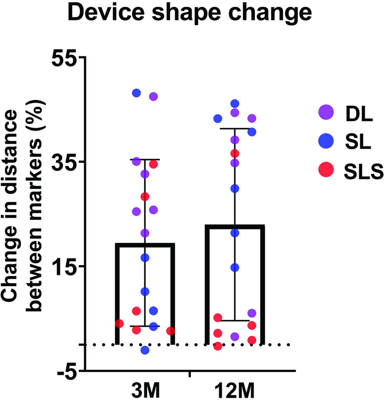
- FIG 1.
Range of WEB device shape change by time and device configuration. The change in distance between the distal and proximal markers of the device at follow-up compared with that immediately posttreatment. Positive values denote device shortening; negative values represent device elongation. Device elongation is seen in only 2 cases, both of which have device elongation of <2%. 3M indicates 3 months; 12M, 12 months.
- FIG 2.
WEB shape changes at 3 months following device implantation. A, Anteroposterior DSA image before device implantation shows an aneurysm cavity (arrow). B, The DSA image immediately after SL device deployment shows complete aneurysm occlusion (arrow). C, Unsubtracted image of B shows proximal and distal markers (arrows) of the WEB device. D, DSA image at 3-month follow-up shows a residual neck (arrow). E, Unsubtracted image of D shows device compression. Note that the distance between the proximal and distal markers (arrows) is reduced compared with that in C, indicating a change in shape.
- FIG 3.
A, Anteroposterior DSA before aneurysm treatment (arrow). B, A DSA image immediately after DL device deployment shows residual aneurysm (arrow). C, A DSA image at 12 months shows complete occlusion (arrow) with substantial shortening of the device. D, Photomicrograph of a section (hematoxylin-eosin, original magnification ×12.5) demonstrates an aneurysm sac filled with loose connective tissue, except for a small neck remnant. A neointimal layer completely traverses the neck interface near the proximal device markers (arrows). E, Photomicrograph of a section shows moderate collagen deposition throughout the aneurysm cavity (Masson trichrome stain, original magnification ×2.3). Relatively high collagen content was noted near the proximal marker (arrow). F, Photomicrograph of a section shows the presence of smooth muscles throughout the aneurysm dome, as well as in the neointimal lining bridging the neck (arrows) (SMA immunostain, original magnification ×2.0). Tx indicates treatment.
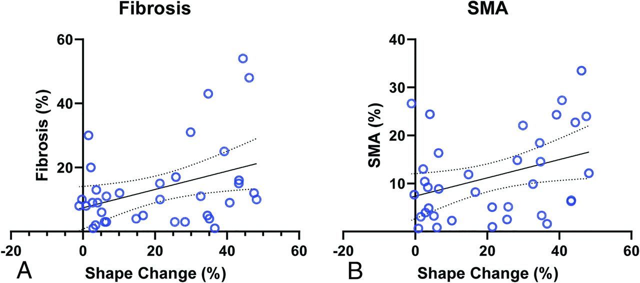
- FIG 4.
Correlation of the degree of device shape change with histologic features. Linear regression shows a significant positive correlation between the percentage of device length change and fibrosis (A) and SMA (B). Straight lines indicate regression; bowed lines indicate 95% CI.


Tables
Groupa DL, No. (%) SL, No. (%) SLS, No. (%) Total, No. (%) 3-Month group Grade I 1 (16.7) 1 (16.7) 3 (50.0) 5 (27.8) Grade II 3 (50.0) 0 (0) 1 (16.7) 4 (22.2) Grade III 1 (16.7) 2 (33.3) 0 (0) 3 (16.7) Grade IV 1 (16.7) 3 (50.0) 2 (33.3) 6 (33.3) Total 6 6 6 18 12-Month group Grade I 3 (50.0) 1 (16.7) 1 (16.7) 5 (27.8) Grade II 1 (16.7) 0 (0) 1 (16.7) 2 (11.1) Grade III 0 (0) 2 (33.3) 1 (16.7) 3 (16.7) Grade IV 2 (33.3) 3 (50.0) 3 (50.0) 8 (44.4) Total 6 6 6 18 ↵a Grade I represents complete occlusion; grade II, complete occlusion with recess filling; grade III, residual neck; grade IV, residual aneurysm.
Follow-Up, Occlusion Groupa DL (n = 12) SL (n = 12) SLS (n = 12) Total (n = 36) With WSM, No. (%) Without WSM, No. (%) With WSM, No. (%) Without WSM, No. (%) With WSM, No. (%) Without WSM, No. (%) With WSM, No. (%) Without WSM, No. (%) 3 Months Grade I or II 4 (33.3) 0 0 1 (8.3) 1 (8.3) 3 (25.0) 5 (13.9) 4 (11.1) Grade III or IV 2 (16.7) 0 3 (25.0) 2 (16.7) 1 (8.3) 1 (8.3) 6 (16.7) 3 (8.3) 12 Months Grade I or II 3 (25.0) 1 (8.3) 1 (8.3) 0 0 2 (16.7) 4 (11.1) 3 (8.3) Grade III or IV 1 (8.3) 1 (8.3) 5 (41.7) 0 1 (8.3) 3 (25.0) 7 (19.4) 4 (11.1) Total 10 (83.3) 2 (16.7) 9 (75.0) 3 (25.0) 3 (25.0) 9 (33.3) 22 (61.1) 14 (38.9) ↵a Grade I represents complete occlusion; grade II, complete occlusion with recess filling; grade III, residual neck; grade IV, residual aneurysm.


Jump to section
Related Articles
Cited By...
- Neck apposition is a key factor for aneurysm occlusion after Woven EndoBridge device embolization
- A novel intrasaccular aneurysm device with high complete occlusion rate: initial results in a rabbit model
- WEB shape modifications: angiography-histopathology correlations in rabbits
- WEB shape modifications: angiography-histopathology correlations in rabbits
- Determinants of cerebral aneurysm occlusion after embolization with the WEB device: a single-institution series of 215 cases with angiographic follow-up
- Determinants of cerebral aneurysm occlusion after embolization with the WEB device: a single-institution series of 215 cases with angiographic follow-up
- Impact of A1 Asymmetry on the Woven EndoBridge Device in Anterior Communicating Artery Aneurysms