
Article Figures & Data
Figures
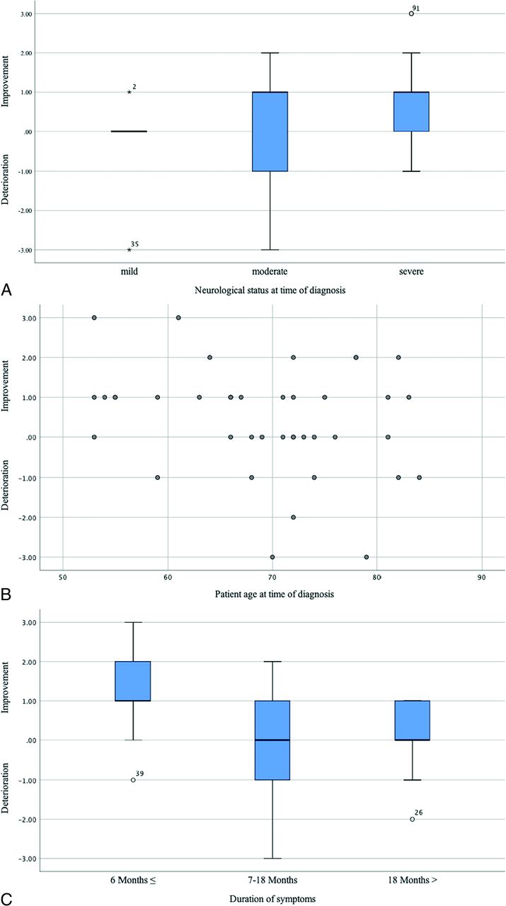
- Fig 1.
A, Boxplot demonstrates the correlation between long-term outcome and neurologic status at diagnosis evaluated by the AL-score and dichotomized into mild: AL-score 0–1; moderate: AL-score 2–3; and severe: AL-score: 4–5. B, Scatterplot demonstrates the relationship between long-term outcome and patient age at diagnosis. C, Boxplot demonstrates the correlation between long-term outcome and the duration of symptoms at diagnosis. The x-axis indicates the initial and follow-up AL-scores.
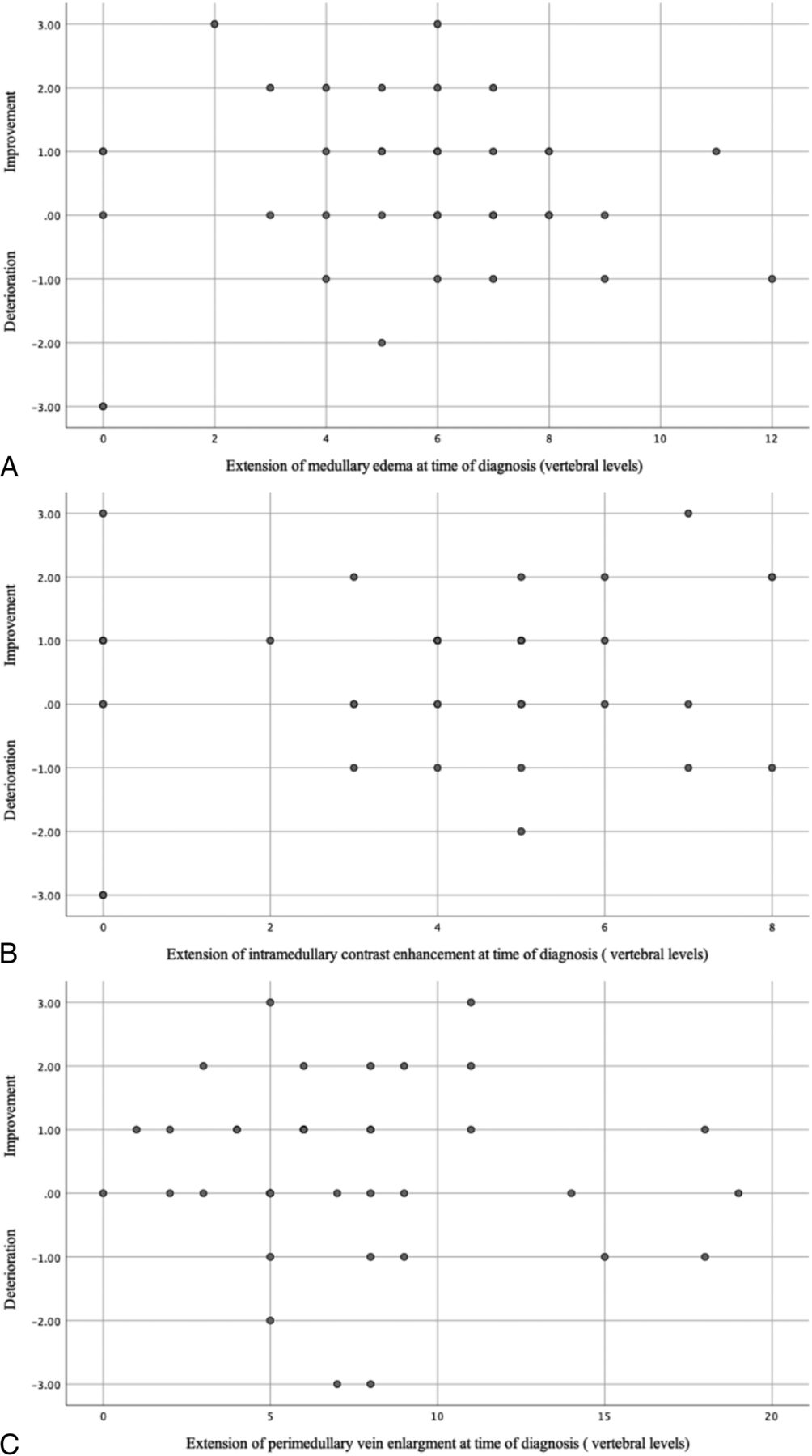
- Fig 2.
Scatterplot demonstrates the relationship between long-term outcome and extension of medullary edema (A), intramedullary contrast enhancement (B), and perimedullary vein enlargement at diagnosis (C). The x-axis indicates the initial and follow-up AL-scores.
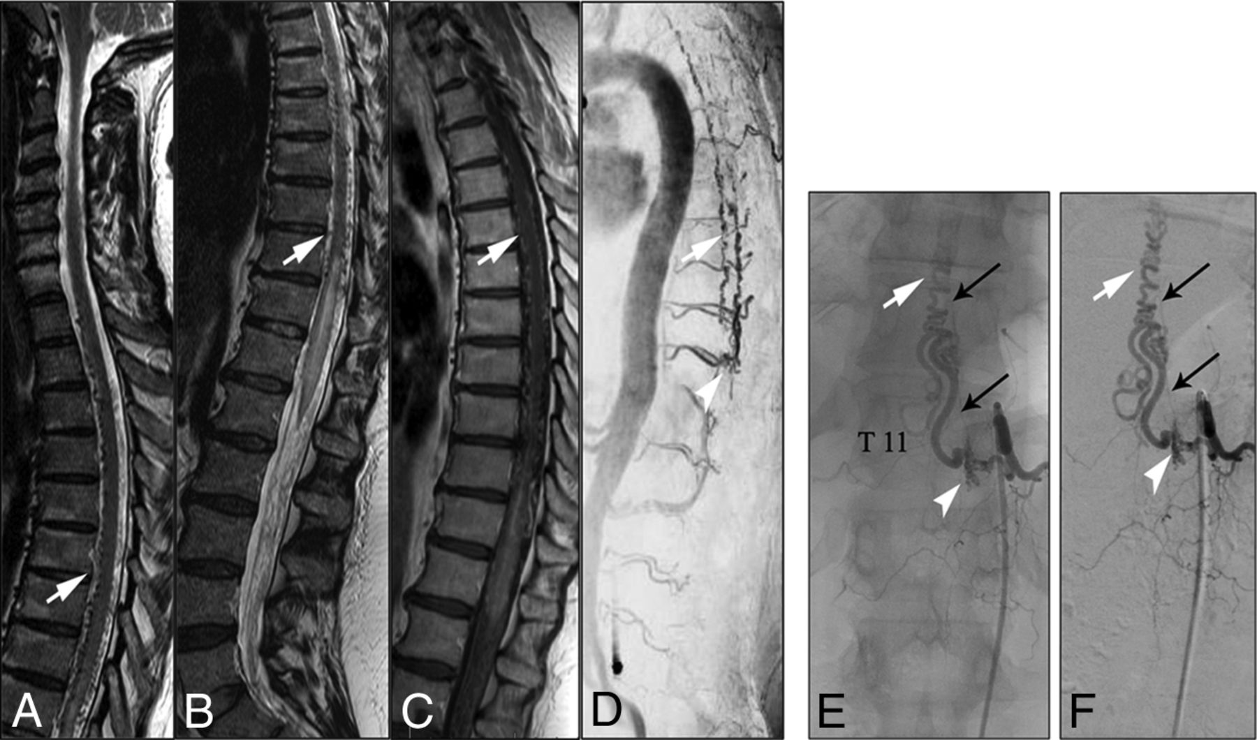
- Fig 3.
A–C, T2- and T1-weighted MR images demonstrate elongated perimedullary veins (white arrows) associated with medullary edema and centro-medullary contrast enhancement in the lower thoracic region. D, CE-MRA image shows the arterialized perimedullary veins in the thoracic region (white arrow) and depicts the shunt zone at the T11 vertebral level (white arrowhead). E and F, Spinal DSA examination (posterior-anterior projection) of the left T11 segmental artery shows the hypervascularized fistula zone (white arrowhead) with a dilated and elongated intradural drainage vein. Note the origin of the Adamkiewicz artery from the same fistula side (small black arrows).
- Fig 4.
A, Intraoperative images show the fistula zone (black arrowhead), a narrow arterial feeder (small black arrow), and the elongated radicular drainage vein (white arrow), B, Indocyanine green images confirm the pathologic arterialization of the elongated radicular vein. C and D, The radicular drainage vein is disconnected via clip ligation, and indocyanine green videoangiography confirms the interruption of the pathologic arterialization.
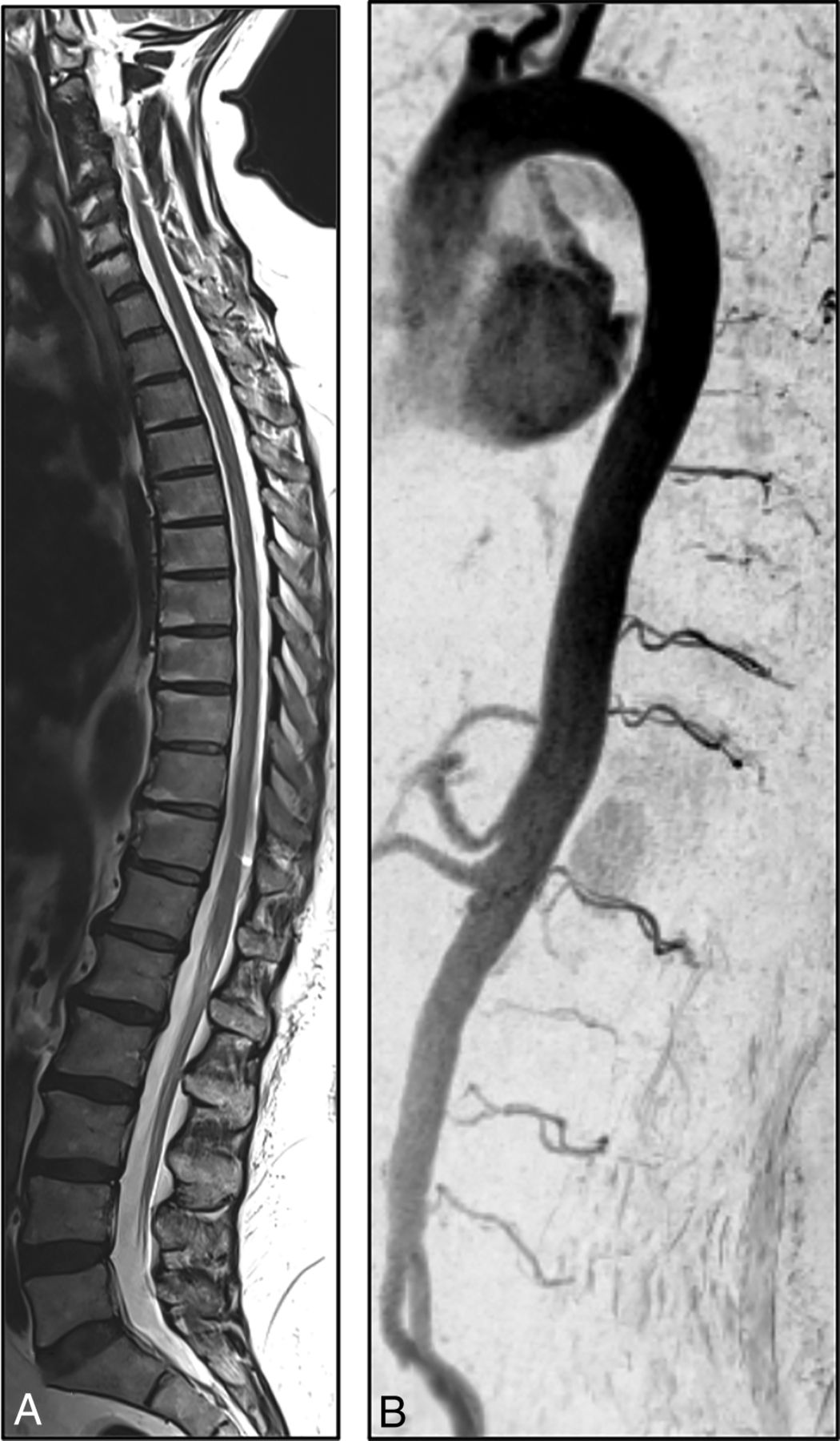
- Fig 5.
Follow-up T2-weighted MR image and CE-MRA 3 years after treatment show a complete regression of medullary edema and alteration of perimedullary veins.


Tables
Aminoff-Logue disability score for gait
Grade of Gait Disturbances Characteristics 0 Normal 1 Leg weakness, abnormal walk or stance, but no restriction of activity 2 Restricted activity 3 Requiring 1 cane for walking 4 Requiring 2 canes, crutches, or walker 5 Confined to wheelchair


Jump to section
Related Articles
Cited By...
- Application of Spinal Subtraction and Bone Background Fusion CTA in the Accurate Diagnosis and Evaluation of Spinal Vascular Malformations
- Multiple synchronous spinal dural arteriovenous fistulas: A systematic literature review
- Long-term outcomes and prognostic factors in patients with treated spinal dural arteriovenous fistulas: a prospective cohort study