
Article Figures & Data
Figures
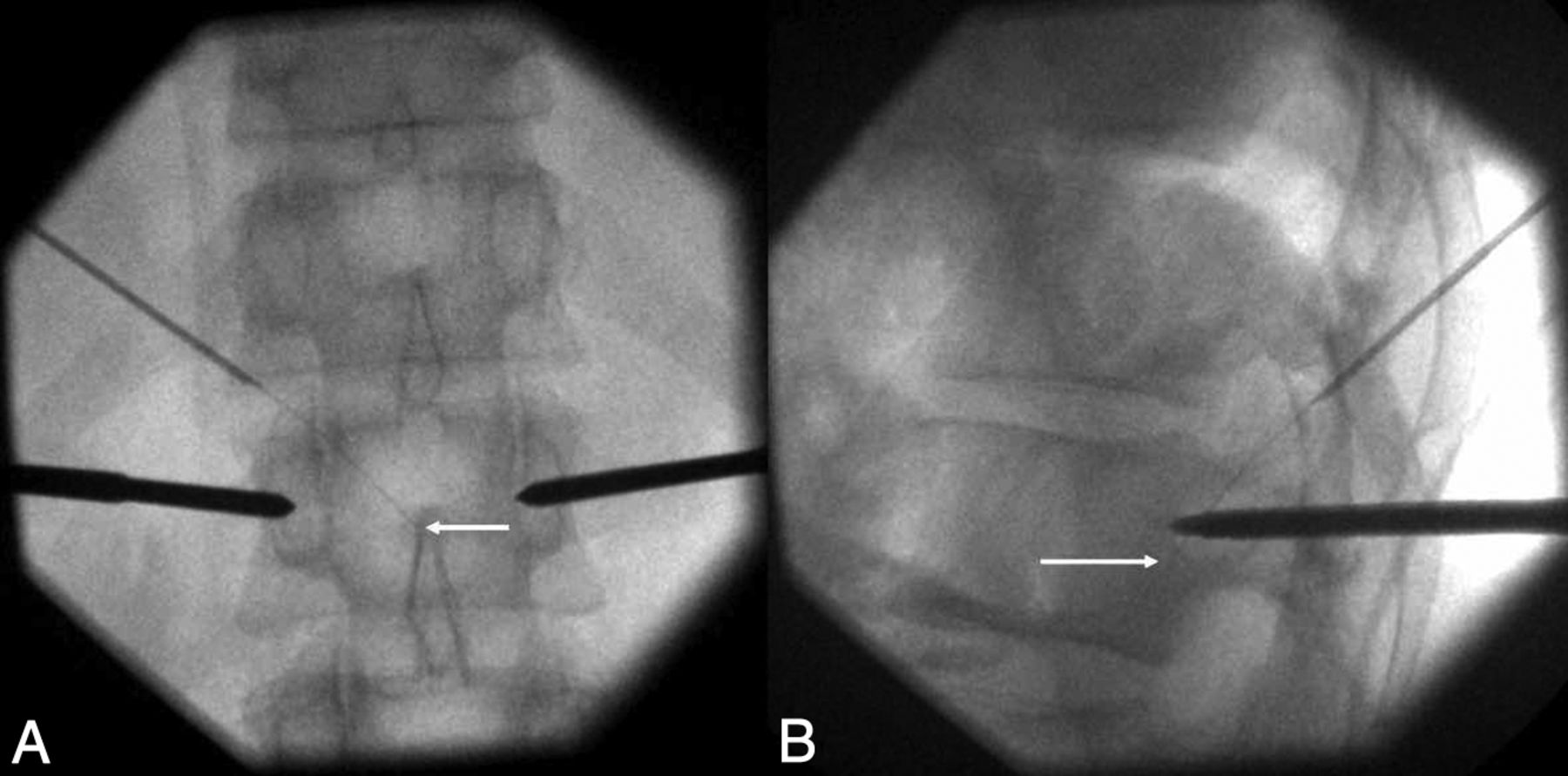
- Fig 1.
Double-oblique transforaminal approach on fluoroscopy (patient 12). Anterior-posterior (A) and lateral (B) fluoroscopic projections demonstrate the tip of the thermosensor (white arrow) just posterior to the vertebral body, at its mid portion (B), and in the midline (A), thanks to an oblique approach in both anterior-posterior and lateral views.
- Fig 2.
Representation and principle of the double-oblique approach. A, Drawing from a sagittal perspective. The craniocaudal approach in the sagittal plane enables going through the posterior and inferior parts of the foramen, away from the radicular nerve and vessels. B, Drawing from an oblique axial view (in the axis of the sagittal angulation). The lateromedial approach allows slipping along the facet joint with the 18-ga needle (arrow), away from the nerve and vessels (arrowhead). The 28-ga thermosensor (dotted arrows) can then be advanced into the anterior epidural space toward the posterior wall of the vertebral body.
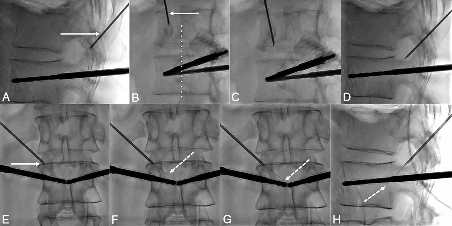
- Fig 3.
Fluoroscopic details of the technique of the transforaminal approach. A, The craniocaudal angulation in the sagittal plane is estimated on the lateral projection to point the 18-ga needle (white arrow) used as a landmark on the skin of the patient toward the posterior and inferior parts of the foramen. B, The 35° oblique view (from the anterior-posterior view) is then used to define the distance of the entry point laterally. The 18-ga spinal needle (arrow) should be pointed toward the lateral part of the facet joint (dotted line). C, The needle is advanced in the oblique view toward the foramen. D, Once in the vicinity of the foramen, the lateral view confirms that the needle enters it at its posterior and inferior parts. E, Satisfactory localization of the needle tip (arrow) inside the foramen is confirmed on anterior-posterior projection. F, The 28-ga thermosensor (dotted arrow) is gently advanced into the canal until it reaches the midline (G), where resistance is felt. H, At this point, the tip of the thermometer (dotted arrow) should be located at the middle portion of the vertebral body on the lateral view.
- Fig 4.
Thermal monitoring combined with hydrodissection. A, Lateral fluoroscopic view demonstrates the 18-ga needle in the foramen (arrow) and the thermosensor in contact with the posterior wall (dotted arrow). B, Conebeam CT acquisition with reconstruction in the axis of the needle and thermometer confirms the findings of fluoroscopy with the 18-ga needle (arrow) and the thermosensor (dotted arrow). C, Lateral view after injection of dextrose mixed with contrast shows satisfactory diffusion of the fluid into the anterior epidural space (white asterisks) separating the dural sac from the vertebral body. D, This is again outlined on the conebeam CT acquisition, which demonstrates the hydrodissection (black asterisk) between the posterior wall and the dural sac.


Tables
Patient Age (yr) Primary Cancer Lesion No. Level Posterior Cortex Disruption Epidural Involvement RFA Type (Device, Company) 1 55 Colon 1 L1 No No Bipolar RFA (OsteoCool; Medtronic)a 2 78 Bladder 1 L2 No No Bipolar RFA (OsteoCool; Medtronic) 3 46 Breast 1 L1 No No Monopolar RFA (Cool-tip; Medtronic) 4 66 Kidney 1 L2 No Yes Bipolar RFA (OsteoCool; Medtronic) 5 52 Breast 1 L3 No No Bipolar RFA (OsteoCool; Medtronic) 2 L4 No No Bipolar RFA (OsteoCool; Medtronic) 6 73 Lung 1 L1 No No Bipolar RFA (STAR; Merrit Medical)b 7 70 Melanoma 1 L1 No Yes Bipolar RFA (OsteoCool; Medtronic) 8 76 Lung 1 L2 Yes Yes Bipolar RFA (OsteoCool; Medtronic) 9 64 Breast 1 L1 No No Bipolar RFA (OsteoCool; Medtronic) 10 63 Lung 1 L4 No No Bipolar RFA (OsteoCool; Medtronic) 11 67 Rectum 1 L3 No No Bipolar RFA (OsteoCool; Medtronic) 12 41 Lung 1 L1 No No Bipolar RFA (OsteoCool; Medtronic) 13 40 Breast 1 L3 Yes Yes Bipolar RFA (OsteoCool; Medtronic) Patient Foramen Guidance Time (min) Obliquity in the Sagittal Plane Technical Successa Hydrodissection Maximal Temperature (°C) 1 T12–L1 Fluoro 11 40° Yes Yes, effective 39 2 L1–L2 Fluoro 11 40° Yes Yes, effective 41 3 T12–L1 CT & fluoro 9 36° Yes Yes, effective 45 4 L1–L2 Fluoro 12 35° Yes No 45 5 L2–L3 Fluoro 10 43° Yes Yes, effective 43 L3–L4 Fluoro 6 38° Yes No 44 6 T12–L1 CT & fluoro 7 42° Yes Yes, effective 41 7 T12–L1 Fluoro 8 44° Yes Yes, ineffective 44 8 L1–L2 Fluoro & CBCT 38 33° No (4 mm too cranial and 5 mm too lateral) Yes, ineffective 39 9 T12–L1 Fluoro 5 36° Yes Yes, effective 43 10 L3–L4 Fluoro 8 47° Yes Yes, effective 38 11 L2–L3 CT & fluoro 9 37° Yes Yes, effective 39 12 T12–L1 CT & fluoro 7 47° Yes Yes, effective 45 13 L2–L3 CT & fluoro 7 44° Yes No 42 Note:—Fluoro indicates fluoroscopy; CBCT, conbeam CT.
a Technical success was defined by a position of the thermosensor in the midline on anteroposterior view and at the midportion of the posterior wall on lateral view.


Jump to section
Related Articles
Cited By...
- No citing articles found.