Advertisement

Main menu

  • Home
  • Content
    • Current Issue
    • Accepted Manuscripts
    • Article Preview
    • Past Issue Archive
    • Video Articles
    • AJNR Case Collection
    • Case of the Week Archive
    • Case of the Month Archive
    • Classic Case Archive
  • Special Collections
    • AJNR Awards
    • Low-Field MRI
    • Alzheimer Disease
    • ASNR Foundation Special Collection
    • Photon-Counting CT
    • View All
  • Multimedia
    • AJNR Podcasts
    • AJNR SCANtastic
    • Trainee Corner
    • MRI Safety Corner
    • Imaging Protocols
  • For Authors
    • Submit a Manuscript
    • Submit a Video Article
    • Submit an eLetter to the Editor/Response
    • Manuscript Submission Guidelines
    • Statistical Tips
    • Fast Publishing of Accepted Manuscripts
    • Graphical Abstract Preparation
    • Imaging Protocol Submission
    • Author Policies
  • About Us
    • About AJNR
    • Editorial Board
    • Editorial Board Alumni
  • More
    • Become a Reviewer/Academy of Reviewers
    • Subscribers
    • Permissions
    • Alerts
    • Feedback
    • Advertisers
    • ASNR Home

User menu

  • Alerts
  • Log in

Search

  • Advanced search
American Journal of Neuroradiology

American Journal of Neuroradiology

ASHNR American Society of Functional Neuroradiology ASHNR American Society of Pediatric Neuroradiology ASSR
  • Alerts
  • Log in

Advanced Search

  • Home
  • Content
    • Current Issue
    • Accepted Manuscripts
    • Article Preview
    • Past Issue Archive
    • Video Articles
    • AJNR Case Collection
    • Case of the Week Archive
    • Case of the Month Archive
    • Classic Case Archive
  • Special Collections
    • AJNR Awards
    • Low-Field MRI
    • Alzheimer Disease
    • ASNR Foundation Special Collection
    • Photon-Counting CT
    • View All
  • Multimedia
    • AJNR Podcasts
    • AJNR SCANtastic
    • Trainee Corner
    • MRI Safety Corner
    • Imaging Protocols
  • For Authors
    • Submit a Manuscript
    • Submit a Video Article
    • Submit an eLetter to the Editor/Response
    • Manuscript Submission Guidelines
    • Statistical Tips
    • Fast Publishing of Accepted Manuscripts
    • Graphical Abstract Preparation
    • Imaging Protocol Submission
    • Author Policies
  • About Us
    • About AJNR
    • Editorial Board
    • Editorial Board Alumni
  • More
    • Become a Reviewer/Academy of Reviewers
    • Subscribers
    • Permissions
    • Alerts
    • Feedback
    • Advertisers
    • ASNR Home
  • Follow AJNR on Twitter
  • Visit AJNR on Facebook
  • Follow AJNR on Instagram
  • Join AJNR on LinkedIn
  • RSS Feeds

AJNR Awards, New Junior Editors, and more. Read the latest AJNR updates

Getting new auth cookie, if you see this message a lot, tell someone!
Research ArticleHead and Neck Imaging

Contrast-Enhanced CISS Imaging for Evaluation of Neurovascular Compression in Trigeminal Neuralgia: Improved Correlation with Symptoms and Prediction of Surgical Outcomes

A.M. Blitz, B. Northcutt, J. Shin, N. Aygun, D.A. Herzka, D. Theodros, C.R. Goodwin, M. Lim and D.P. Seeburg
American Journal of Neuroradiology August 2018, DOI: https://doi.org/10.3174/ajnr.A5743
A.M. Blitz
aFrom the Departments of Radiology and Radiological Science (A.M.B., B.N., J.S., N.A., D.P.S.)
  • Find this author on Google Scholar
  • Find this author on PubMed
  • Search for this author on this site
  • ORCID record for A.M. Blitz
B. Northcutt
aFrom the Departments of Radiology and Radiological Science (A.M.B., B.N., J.S., N.A., D.P.S.)
  • Find this author on Google Scholar
  • Find this author on PubMed
  • Search for this author on this site
  • ORCID record for B. Northcutt
J. Shin
aFrom the Departments of Radiology and Radiological Science (A.M.B., B.N., J.S., N.A., D.P.S.)
  • Find this author on Google Scholar
  • Find this author on PubMed
  • Search for this author on this site
  • ORCID record for J. Shin
N. Aygun
aFrom the Departments of Radiology and Radiological Science (A.M.B., B.N., J.S., N.A., D.P.S.)
  • Find this author on Google Scholar
  • Find this author on PubMed
  • Search for this author on this site
  • ORCID record for N. Aygun
D.A. Herzka
bBiomedical Engineering (D.A.H.)
  • Find this author on Google Scholar
  • Find this author on PubMed
  • Search for this author on this site
  • ORCID record for D.A. Herzka
D. Theodros
cNeurosurgery (D.T., C.R.G., M.L.), Johns Hopkins University School of Medicine, Baltimore, Maryland.
  • Find this author on Google Scholar
  • Find this author on PubMed
  • Search for this author on this site
  • ORCID record for D. Theodros
C.R. Goodwin
cNeurosurgery (D.T., C.R.G., M.L.), Johns Hopkins University School of Medicine, Baltimore, Maryland.
  • Find this author on Google Scholar
  • Find this author on PubMed
  • Search for this author on this site
  • ORCID record for C.R. Goodwin
M. Lim
cNeurosurgery (D.T., C.R.G., M.L.), Johns Hopkins University School of Medicine, Baltimore, Maryland.
  • Find this author on Google Scholar
  • Find this author on PubMed
  • Search for this author on this site
  • ORCID record for M. Lim
D.P. Seeburg
aFrom the Departments of Radiology and Radiological Science (A.M.B., B.N., J.S., N.A., D.P.S.)
  • Find this author on Google Scholar
  • Find this author on PubMed
  • Search for this author on this site
  • ORCID record for D.P. Seeburg
  • Article
  • Figures & Data
  • Supplemental
  • Info & Metrics
  • Responses
  • References
  • PDF
Loading

Article Figures & Data

Figures

  • Tables
  • Fig 1.
    • Download figure
    • Open in new tab
    • Download powerpoint
    Fig 1.

    Grades 0 and 1 neurovascular conflict. Coronal NE-CISS (A) and CE-CISS (B) images show grade 1 (simple contact) on the patient's right side with a branch from the superior cerebellar artery (white solid arrow) contacting the cisternal segment of the trigeminal nerve root (dashed black arrow) from above. Note enhancement of the artery on the CE-CISS image. On the patient's left, the cisternal trigeminal nerve root (dashed black arrow) has no neurovascular conflict (grade 0).

  • Fig 2.
    • Download figure
    • Open in new tab
    • Download powerpoint
    Fig 2.

    Grade 2 neurovascular conflict. Coronal NE-CISS (A) and CE-CISS (B) and sagittal NE-CISS (C) and CE-CISS (D) images show neurovascular conflict of the cisternal segment of the patient's right trigeminal nerve with a branch of the superior cerebellar artery from above (solid white arrow) and the superior petrosal vein from below (dashed white arrow), resulting in flattening of the nerve near the porus trigeminus. On the NE-CISS images (A and C), the nerve is not well-delineated from the adjacent vascular structures. On the CE-CISS images (B and D), the vessels enhance, outlining the compressed nerve between them. Zoomed-in images of the site of neurovascular conflict in the coronal plane (E and F) illustrate the poor contrast between vessels and nerve on the NE-CISS image (E) and the improved contrast after administration of gadolinium contrast material (F), allowing more confident delineation of the compressed nerve from the adjacent vessels (G). Both NE-CISS and CE-CISS images were interpreted as grade 2 compression. On the patient's left, grade 0 was given for both the NE-CISS and CE-CISS images.
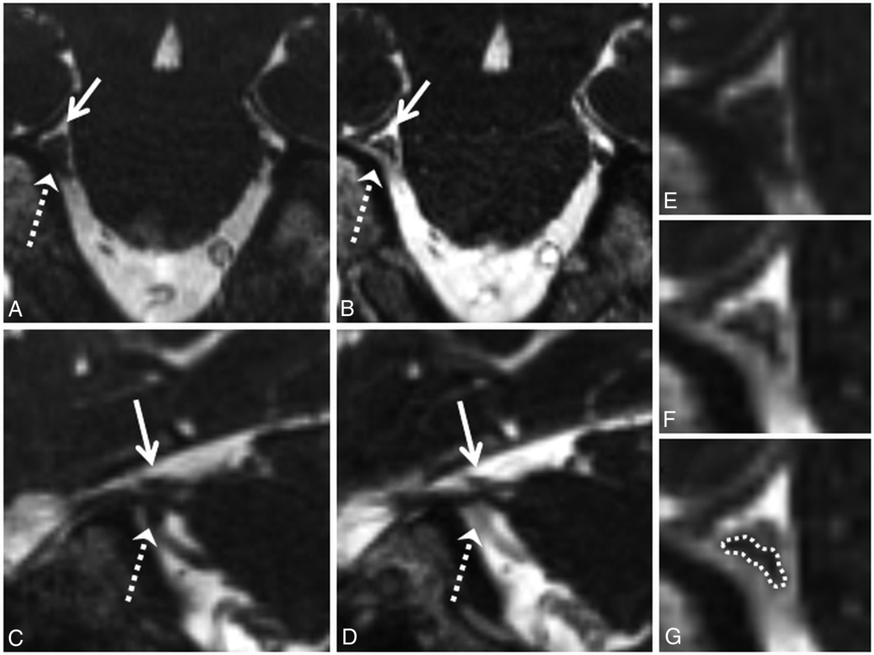
  • Fig 3.
    • Download figure
    • Open in new tab
    • Download powerpoint
    Fig 3.

    Grade 3 neurovascular conflict. Coronal NE-CISS (A) and CE-CISS (B) images show compression of the cisternal segment of the patient's right trigeminal nerve (black dashed arrow) by branches of the superior cerebellar artery from above (solid white arrow). On the unenhanced image (A), the nerve root is not well-distinguished from the compressing arterial branches. After contrast administration (B), at least 2 arterial branches enhance, allowing more confident delineation of the markedly compressed nerve. Zoomed-in images of the site of neurovascular conflict in the coronal plane (C–E) illustrate the poor contrast between vessels and nerve on the NE-CISS image (C) and the improved contrast after administration of gadolinium contrast material (D), allowing more confident delineation of the compressed nerve from the adjacent vessels (E). In this patient, grade 3 was given for both the NE-CISS and CE-CISS images, but the measured CSA was lower and the degree of flattening was more pronounced on the CE-CISS images. On the patient's left, grade 0 was given for both the NE-CISS and CE-CISS images.

  • Fig 4.
    • Download figure
    • Open in new tab
    • Download powerpoint
    Fig 4.

    High-grade neurovascular conflict. Coronal NE-CISS (A) and CE-CISS (B) images show compression of the distal cisternal segment of the patient's left trigeminal nerve (gray dashed arrow) by a branch of the superior petrosal venous complex from below (solid white arrow). On the unenhanced image (A), the nerve root is not well-distinguished from the compressing venous branch. After contrast administration (B), the venous branch fills with contrast, showing a severely compressed unenhanced adjacent trigeminal nerve root at the level of the porus trigeminus. Zoomed-in images of the site of neurovascular conflict in the coronal plane (C–E) illustrate the poor contrast between vessels and nerve on the NE-CISS image (C) and the improved contrast after administration of gadolinium contrast material (D), allowing more confident delineation of the compressed nerve from the adjacent vessel (E). In this patient, grade 2 was given on the NE-CISS and grade 3 was given on CE-CISS images. On the patient's right, grade 1 was given on both the NE-CISS and CE-CISS images.

  • Fig 5.
    • Download figure
    • Open in new tab
    • Download powerpoint
    Fig 5.

    Correlation of the metrics of neurovascular conflict with postsurgical outcomes after MVD. Grades of neurovascular conflict (A), CSA (B), and DOF (C) were correlated with different postsurgical outcomes after MVD, as described in the individual graphs. Non-con indicates non-contrast; Con, contrast-enhanced.

  • Fig 6.
    • Download figure
    • Open in new tab
    • Download powerpoint
    Fig 6.

    Receiver operating curves assessing the performance of the grade of neurovascular conflict (A, D, and G), CSA (B, E, and H), and DOF (C, F, and I) in predicting complete relief without further need for analgesic medication (type 1 outcome) after MVD (A–C), complete relief with or without further need for analgesic medication (type 1 or 2 outcome) after MVD (D–F), and symptom side in patients with TN (G–I) for both NE-CISS and CE-CISS images, as delineated in the graphs. The area under the curve and P values comparing the NE-CISS and CE-CISS curves are provided in the individual graphs.

Tables

  • Figures
    • View popup
    Table 1:

    Prevalence of various grades of neurovascular conflict and types of vascular involvement in patients with TN and in controls

    Contrast EnhancementSymptomatic Side (No.) (%) of 81 CasesAsymptomatic Side (No.) (%) of 81 CasesControl (No.) (%) of 30 Cases
    Grades of neurovascular conflict
        0Contrast−10 (12.3)27 (33.3)12 (40.0)
    Contrast+10 (12.3)27 (33.3)12 (40.0)
        1Contrast−33 (40.7)48 (59.3)18 (60.0)
    Contrast+29 (35.8)48 (59.3)18 (60.0)
        2Contrast−26 (32.1)6 (7.4)0 (0.0)
    Contrast+15 (18.5)6 (7.4)0 (0.0)
        3Contrast−12 (14.8)0 (0)0 (0.0)
    Contrast+27 (33.3)0 (0)0 (0.0)
        1, 2, or 3Contrast−71 (87.7)54 (66.7)18 (60.0)
    Contrast+71 (87.7)54 (66.7)18 (60.0)
        2 and 3Contrast−38 (46.9)6 (7.4)0 (0.0)
    Contrast+42 (51.9)6 (7.4)0 (0.0)
    Artery alone27 (33.3)17 (21.0)4 (13.3)
    Artery involved49 (60.5)30 (37.0)5 (16.7)
    Vein alone22 (27.2)24 (29.6)12 (40.0)
    Vein involved44 (54.3)37 (45.7)13 (43.3)
    Mixed22 (27.2)13 (16.0)1 (3.3)
    • Note:—Contrast+ indicates contrast enhancement; Contrast−, no contrast enhancement.

    • View popup
    Table 2:

    Statistical analysis

    Grades of Neurovascular ConflictContrast EnhancementP ValuesContrast vs NoncontrastSensitivitySpecificity
    Sym vs AsymSym vs CntrlAsym vs CntrlSymAsym
    0Contrast−.002.002.511112.3%66.7%
    Contrast+.002.003.5112.3%66.7%
    1Contrast−.03.0910.63140.7%40.5%
    Contrast+.004.03135.8%40.5%
    2Contrast−<.001<.001.190.07132.1%94.6%
    Contrast+.06.01.1918.5%94.6%
    3Contrast−<.001.0310.001114.8%100.0%
    Contrast+<.001<.001133.3%100.0%
    1, 2, or 3Contrast−.002.003.511187.7%35.1%
    Contrast+.002.002.5187.7%35.1%
    2 and 3Contrast−<.001<.001.190.64146.9%94.6%
    Contrast+<.001<.001.1951.9%94.6%
    • Note:—Sym indicates symptomatic; Asym, asymptomatic; Cntrl, control; Contrast +, contrast enhancement; Contrast −, no contrast enhancement.

    • View popup
    Table 3:

    Characteristics of neurovascular involvement

    Involvement of Neurovascular ConflictP Values
    Sym vs AsymSym vs CntrlAsym vs CntrlContrast vsNoncontrast
    Artery alone0.110.060.4333.3%81.1%
    Artery involved.005<.001.0660.5%68.5%
    Vein alone0.860.250.3627.2%67.6%
    Vein involved0.350.39154.3%55.0%
    Mixed0.130.0070.1127.2%87.4%
    • View popup
    Table 4:

    Cutoff values for grade of neurovascular conflict, CSA, and DOF to obtain the highest sum of sensitivity and specificity in predicting complete relief of pain after MVD (type 1 outcome), complete relief of pain with or without need for analgesic medication (type 1 or 2 outcome), or in predicting the side of symptoms in patients with TN

    OutcomeCutoffContrastSensitivitySpecificity
    Type 1
        Grade≥2−59.690.7
    ≥2+67.390.7
        CSA≤5.1−63.567.6
    ≤4.15+55.879.9
        DOF≥2.0−69.273.4
    ≥2.5+59.687.1
    Type 2
        Grade≥2−52.491.4
    ≥2+58.791.4
        CSA≤4.7−57.172.7
    ≤4.15+5482
        DOF≥2.0−63.574.3
    ≥2.5+5488.3
    Predicting side of sym in patients with TN
        Grade≥2−46.994.6
    ≥2+51.994.6
        CSA≤4.7−55.675.7
    ≤4.15+50.685.6
        DOF≥2.2−53.184.7
    ≥2.5+49.491
    • Note:—sym indicates symptoms; −, absent; +, present.

Next
Back to top
Advertisement
Print
Download PDF
Email Article

Thank you for your interest in spreading the word on American Journal of Neuroradiology.

NOTE: We only request your email address so that the person you are recommending the page to knows that you wanted them to see it, and that it is not junk mail. We do not capture any email address.

Enter multiple addresses on separate lines or separate them with commas.
Contrast-Enhanced CISS Imaging for Evaluation of Neurovascular Compression in Trigeminal Neuralgia: Improved Correlation with Symptoms and Prediction of Surgical Outcomes
(Your Name) has sent you a message from American Journal of Neuroradiology
(Your Name) thought you would like to see the American Journal of Neuroradiology web site.
CAPTCHA
This question is for testing whether or not you are a human visitor and to prevent automated spam submissions.
Cite this article
A.M. Blitz, B. Northcutt, J. Shin, N. Aygun, D.A. Herzka, D. Theodros, C.R. Goodwin, M. Lim, D.P. Seeburg
Contrast-Enhanced CISS Imaging for Evaluation of Neurovascular Compression in Trigeminal Neuralgia: Improved Correlation with Symptoms and Prediction of Surgical Outcomes
American Journal of Neuroradiology Aug 2018, DOI: 10.3174/ajnr.A5743

Citation Manager Formats

  • BibTeX
  • Bookends
  • EasyBib
  • EndNote (tagged)
  • EndNote 8 (xml)
  • Medlars
  • Mendeley
  • Papers
  • RefWorks Tagged
  • Ref Manager
  • RIS
  • Zotero
0 Responses
Respond to this article
Share
Bookmark this article
Contrast-Enhanced CISS Imaging for Evaluation of Neurovascular Compression in Trigeminal Neuralgia: Improved Correlation with Symptoms and Prediction of Surgical Outcomes
A.M. Blitz, B. Northcutt, J. Shin, N. Aygun, D.A. Herzka, D. Theodros, C.R. Goodwin, M. Lim, D.P. Seeburg
American Journal of Neuroradiology Aug 2018, DOI: 10.3174/ajnr.A5743
del.icio.us logo Twitter logo Facebook logo Mendeley logo
  • Tweet Widget
  • Facebook Like
  • Google Plus One
Purchase

Jump to section

  • Article
    • Abstract
    • ABBREVIATIONS:
    • Materials and Methods
    • Results
    • Discussion
    • Conclusions
    • Footnotes
    • References
  • Figures & Data
  • Supplemental
  • Info & Metrics
  • Responses
  • References
  • PDF

Related Articles

  • PubMed
  • Google Scholar

Cited By...

  • Contrast-Enhanced CISS/FIESTA Imaging for Increased Conspicuity of Pituitary Microadenomas in Cushing Disease
  • Crossref
  • Google Scholar

This article has not yet been cited by articles in journals that are participating in Crossref Cited-by Linking.

More in this TOC Section

  • MRI of the Parasellar Ligaments
  • ASL Sensitivity for Head and Neck Paraganglioma
  • Post SRS Peritumoral Hyperintense Signal of VSs
Show more Head and Neck Imaging

Similar Articles

Advertisement

Indexed Content

  • Current Issue
  • Accepted Manuscripts
  • Article Preview
  • Past Issues
  • Editorials
  • Editor's Choice
  • Fellows' Journal Club
  • Letters to the Editor
  • Video Articles

Cases

  • Case Collection
  • Archive - Case of the Week
  • Archive - Case of the Month
  • Archive - Classic Case

More from AJNR

  • Trainee Corner
  • Imaging Protocols
  • MRI Safety Corner
  • Book Reviews

Multimedia

  • AJNR Podcasts
  • AJNR Scantastics

Resources

  • Turnaround Time
  • Submit a Manuscript
  • Submit a Video Article
  • Submit an eLetter to the Editor/Response
  • Manuscript Submission Guidelines
  • Statistical Tips
  • Fast Publishing of Accepted Manuscripts
  • Graphical Abstract Preparation
  • Imaging Protocol Submission
  • Evidence-Based Medicine Level Guide
  • Publishing Checklists
  • Author Policies
  • Become a Reviewer/Academy of Reviewers
  • News and Updates

About Us

  • About AJNR
  • Editorial Board
  • Editorial Board Alumni
  • Alerts
  • Permissions
  • Not an AJNR Subscriber? Join Now
  • Advertise with Us
  • Librarian Resources
  • Feedback
  • Terms and Conditions
  • AJNR Editorial Board Alumni

American Society of Neuroradiology

  • Not an ASNR Member? Join Now

© 2025 by the American Society of Neuroradiology All rights, including for text and data mining, AI training, and similar technologies, are reserved.
Print ISSN: 0195-6108 Online ISSN: 1936-959X

Powered by HighWire