Advertisement

Main menu

  • Home
  • Content
    • Current Issue
    • Accepted Manuscripts
    • Article Preview
    • Past Issue Archive
    • Video Articles
    • AJNR Case Collection
    • Case of the Week Archive
    • Case of the Month Archive
    • Classic Case Archive
  • Special Collections
    • AJNR Awards
    • Low-Field MRI
    • Alzheimer Disease
    • ASNR Foundation Special Collection
    • Photon-Counting CT
    • View All
  • Multimedia
    • AJNR Podcasts
    • AJNR SCANtastic
    • Trainee Corner
    • MRI Safety Corner
    • Imaging Protocols
  • For Authors
    • Submit a Manuscript
    • Submit a Video Article
    • Submit an eLetter to the Editor/Response
    • Manuscript Submission Guidelines
    • Statistical Tips
    • Fast Publishing of Accepted Manuscripts
    • Graphical Abstract Preparation
    • Imaging Protocol Submission
    • Author Policies
  • About Us
    • About AJNR
    • Editorial Board
    • Editorial Board Alumni
  • More
    • Become a Reviewer/Academy of Reviewers
    • Subscribers
    • Permissions
    • Alerts
    • Feedback
    • Advertisers
    • ASNR Home

User menu

  • Alerts
  • Log in

Search

  • Advanced search
American Journal of Neuroradiology

American Journal of Neuroradiology

ASHNR American Society of Functional Neuroradiology ASHNR American Society of Pediatric Neuroradiology ASSR
  • Alerts
  • Log in

Advanced Search

  • Home
  • Content
    • Current Issue
    • Accepted Manuscripts
    • Article Preview
    • Past Issue Archive
    • Video Articles
    • AJNR Case Collection
    • Case of the Week Archive
    • Case of the Month Archive
    • Classic Case Archive
  • Special Collections
    • AJNR Awards
    • Low-Field MRI
    • Alzheimer Disease
    • ASNR Foundation Special Collection
    • Photon-Counting CT
    • View All
  • Multimedia
    • AJNR Podcasts
    • AJNR SCANtastic
    • Trainee Corner
    • MRI Safety Corner
    • Imaging Protocols
  • For Authors
    • Submit a Manuscript
    • Submit a Video Article
    • Submit an eLetter to the Editor/Response
    • Manuscript Submission Guidelines
    • Statistical Tips
    • Fast Publishing of Accepted Manuscripts
    • Graphical Abstract Preparation
    • Imaging Protocol Submission
    • Author Policies
  • About Us
    • About AJNR
    • Editorial Board
    • Editorial Board Alumni
  • More
    • Become a Reviewer/Academy of Reviewers
    • Subscribers
    • Permissions
    • Alerts
    • Feedback
    • Advertisers
    • ASNR Home
  • Follow AJNR on Twitter
  • Visit AJNR on Facebook
  • Follow AJNR on Instagram
  • Join AJNR on LinkedIn
  • RSS Feeds

AJNR Awards, New Junior Editors, and more. Read the latest AJNR updates

Research ArticleSpine Imaging and Spine Image-Guided Interventions

Simultaneous Bipedicular Radiofrequency Ablation Combined with Vertebral Augmentation for Local Tumor Control of Spinal Metastases

A. Tomasian, T.J. Hillen, R.O. Chang and J.W. Jennings
American Journal of Neuroradiology August 2018, DOI: https://doi.org/10.3174/ajnr.A5752
A. Tomasian
aFrom the Department of Radiology (A.T.), University of Southern California, Los Angeles, California
  • Find this author on Google Scholar
  • Find this author on PubMed
  • Search for this author on this site
  • ORCID record for A. Tomasian
T.J. Hillen
bthe Mallinckrodt Institute of Radiology (T.J.H., R.O.C., J.W.J.), St. Louis, Missouri.
  • Find this author on Google Scholar
  • Find this author on PubMed
  • Search for this author on this site
  • ORCID record for T.J. Hillen
R.O. Chang
bthe Mallinckrodt Institute of Radiology (T.J.H., R.O.C., J.W.J.), St. Louis, Missouri.
  • Find this author on Google Scholar
  • Find this author on PubMed
  • Search for this author on this site
  • ORCID record for R.O. Chang
J.W. Jennings
bthe Mallinckrodt Institute of Radiology (T.J.H., R.O.C., J.W.J.), St. Louis, Missouri.
  • Find this author on Google Scholar
  • Find this author on PubMed
  • Search for this author on this site
  • ORCID record for J.W. Jennings
  • Article
  • Figures & Data
  • Info & Metrics
  • Responses
  • References
  • PDF
Loading

Article Figures & Data

Figures

  • Fig 1.
    • Download figure
    • Open in new tab
    • Download powerpoint
    Fig 1.

    A 63-year-old man with chest wall melanoma and painful L5 metastasis. Axial FDG-PET/CT (A) and axial T1-weighted fat-saturated contrast-enhanced MR imaging (B) show hypermetabolic bone marrow replacing lesion in the L5 vertebral body extending to the right pedicle (A and B, arrows). Axial (C) and sagittal (D) stereotactic body radiation therapy planning CT images show stereotactic body radiation therapy contours with clinical target volume including the entire vertebral body and pedicles.
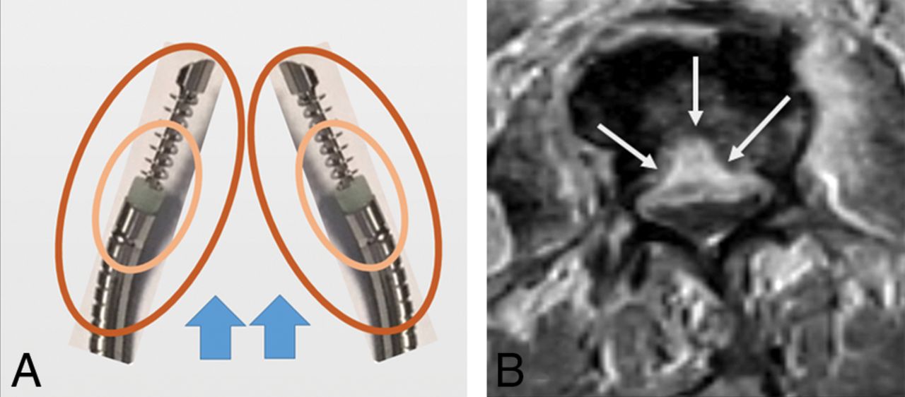
  • Fig 2.
    • Download figure
    • Open in new tab
    • Download powerpoint
    Fig 2.

    Illustration of simultaneous bipedicular RF ablation (A) depicts individual zones of resistive and conductive heating (central and peripheral ovoids, respectively) around 2 adjacent RF electrodes, resulting in a diminished convective cooling effect (ie, heat sink due to blood and CSF flow) (A, arrows). Adjacent areas of thermal spread result in reduction in the power required to conduct heat in tissue, decreased risk of thermal injury, and impedance-related issues. Axial T1-weighted fat-saturated contrast-enhanced MR imaging (B) following bilateral RF ablation using 2 straight electrodes shows ablation failure along the posterior third vertebral body centrally due to lack of confluent ablation zones (B, arrows).
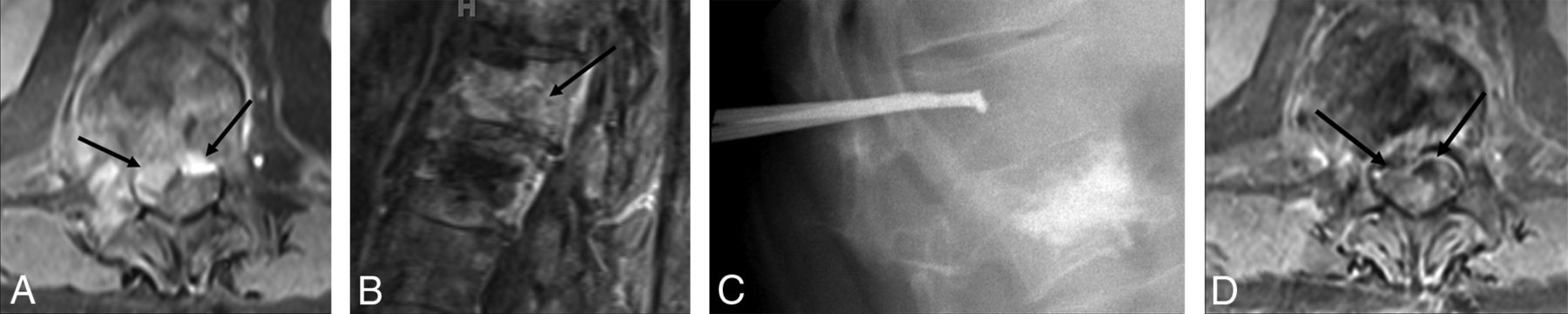
  • Fig 3.
    • Download figure
    • Open in new tab
    • Download powerpoint
    Fig 3.

    An 86-year-old man with metastatic melanoma and a painful L1 lesion. Axial contrast-enhanced CT (A) shows a destructive osteolytic mass within the vertebral body with partial disruption of the posterior wall and a small component extending to the anterior central canal (A, arrow). An anteroposterior fluoroscopic image during simultaneous bipedicular RF ablation (B) shows medial articulation of electrode tips, which are 5–10 mm apart (the width of the spinous process as a landmark). Lateral fluoroscopic images (C–E) show ablation of the anterior vertebral body first (C), followed by ablation of the posterior vertebral body and pedicles (D), and vertebral augmentation (E). Axial T1-weighted fat-saturated contrast-enhanced MR images obtained 2 weeks (F) and 52 weeks (G) following treatment show local tumor control with granulation tissues along the periphery of ablation zone (F and G, arrows).

  • Fig 4.
    • Download figure
    • Open in new tab
    • Download powerpoint
    Fig 4.

    A 70-year-old man with thigh metastatic undifferentiated pleomorphic sarcoma and a painful T12 lesion. Axial and sagittal T1-weighted fat-saturated contrast-enhanced MR images (A and B, respectively) show bone marrow replacing lesion in the T12 vertebral body with posterior wall destruction, epidural extension of tumor, and thecal sac compression (A and B, arrows). Note the previously treated L1 lesion (B). Lateral fluoroscopic image during simultaneous bipedicular RF ablation (C) shows aggressive ablation of the posterior vertebral body and pedicles. Axial T1-weighted fat-saturated contrast-enhanced MR image (D) obtained 30 weeks following treatment shows local tumor control with no evidence of recurrence and retraction of epidural component (D, arrows).

Next
Back to top
Advertisement
Print
Download PDF
Email Article

Thank you for your interest in spreading the word on American Journal of Neuroradiology.

NOTE: We only request your email address so that the person you are recommending the page to knows that you wanted them to see it, and that it is not junk mail. We do not capture any email address.

Enter multiple addresses on separate lines or separate them with commas.
Simultaneous Bipedicular Radiofrequency Ablation Combined with Vertebral Augmentation for Local Tumor Control of Spinal Metastases
(Your Name) has sent you a message from American Journal of Neuroradiology
(Your Name) thought you would like to see the American Journal of Neuroradiology web site.
CAPTCHA
This question is for testing whether or not you are a human visitor and to prevent automated spam submissions.
Cite this article
A. Tomasian, T.J. Hillen, R.O. Chang, J.W. Jennings
Simultaneous Bipedicular Radiofrequency Ablation Combined with Vertebral Augmentation for Local Tumor Control of Spinal Metastases
American Journal of Neuroradiology Aug 2018, DOI: 10.3174/ajnr.A5752

Citation Manager Formats

  • BibTeX
  • Bookends
  • EasyBib
  • EndNote (tagged)
  • EndNote 8 (xml)
  • Medlars
  • Mendeley
  • Papers
  • RefWorks Tagged
  • Ref Manager
  • RIS
  • Zotero
0 Responses
Respond to this article
Share
Bookmark this article
Simultaneous Bipedicular Radiofrequency Ablation Combined with Vertebral Augmentation for Local Tumor Control of Spinal Metastases
A. Tomasian, T.J. Hillen, R.O. Chang, J.W. Jennings
American Journal of Neuroradiology Aug 2018, DOI: 10.3174/ajnr.A5752
del.icio.us logo Twitter logo Facebook logo Mendeley logo
  • Tweet Widget
  • Facebook Like
  • Google Plus One
Purchase

Jump to section

  • Article
    • Abstract
    • ABBREVIATIONS:
    • Materials and Methods
    • Results
    • Discussion
    • Conclusions
    • Footnotes
    • References
  • Figures & Data
  • Info & Metrics
  • Responses
  • References
  • PDF

Related Articles

  • No related articles found.
  • PubMed
  • Google Scholar

Cited By...

  • Spine Microwave Ablation: Safety and Efficacy for Treatment of Vertebral Metastases
  • Reply:
  • Percutaneous CT-Guided Microwave Ablation Combined with Vertebral Augmentation for Treatment of Painful Spinal Metastases
  • Crossref
  • Google Scholar

This article has not yet been cited by articles in journals that are participating in Crossref Cited-by Linking.

More in this TOC Section

Spine Imaging and Spine Image-Guided Interventions

  • Optimization of Photon Counting CT Myelography
  • Characteristics of SIH Type I Culprit Lesions
  • Management Outcomes For VO Spine Biopsy
Show more Spine Imaging and Spine Image-Guided Interventions

Neurointervention

  • Rescue Reentry in Carotid Near-Occlusion
  • Contour Neurovascular System: Five Year Follow Up
  • Effect of SARS-CoV2 on Endovascular Thrombectomy
Show more Neurointervention

Similar Articles

Advertisement

Indexed Content

  • Current Issue
  • Accepted Manuscripts
  • Article Preview
  • Past Issues
  • Editorials
  • Editor's Choice
  • Fellows' Journal Club
  • Letters to the Editor
  • Video Articles

Cases

  • Case Collection
  • Archive - Case of the Week
  • Archive - Case of the Month
  • Archive - Classic Case

More from AJNR

  • Trainee Corner
  • Imaging Protocols
  • MRI Safety Corner
  • Book Reviews

Multimedia

  • AJNR Podcasts
  • AJNR Scantastics

Resources

  • Turnaround Time
  • Submit a Manuscript
  • Submit a Video Article
  • Submit an eLetter to the Editor/Response
  • Manuscript Submission Guidelines
  • Statistical Tips
  • Fast Publishing of Accepted Manuscripts
  • Graphical Abstract Preparation
  • Imaging Protocol Submission
  • Evidence-Based Medicine Level Guide
  • Publishing Checklists
  • Author Policies
  • Become a Reviewer/Academy of Reviewers
  • News and Updates

About Us

  • About AJNR
  • Editorial Board
  • Editorial Board Alumni
  • Alerts
  • Permissions
  • Not an AJNR Subscriber? Join Now
  • Advertise with Us
  • Librarian Resources
  • Feedback
  • Terms and Conditions
  • AJNR Editorial Board Alumni

American Society of Neuroradiology

  • Not an ASNR Member? Join Now

© 2025 by the American Society of Neuroradiology All rights, including for text and data mining, AI training, and similar technologies, are reserved.
Print ISSN: 0195-6108 Online ISSN: 1936-959X

Powered by HighWire