Advertisement

Main menu

  • Home
  • Content
    • Current Issue
    • Accepted Manuscripts
    • Article Preview
    • Past Issue Archive
    • Video Articles
    • AJNR Case Collection
    • Case of the Week Archive
    • Case of the Month Archive
    • Classic Case Archive
  • Special Collections
    • AJNR Awards
    • Low-Field MRI
    • Alzheimer Disease
    • ASNR Foundation Special Collection
    • Photon-Counting CT
    • View All
  • Multimedia
    • AJNR Podcasts
    • AJNR SCANtastic
    • Trainee Corner
    • MRI Safety Corner
    • Imaging Protocols
  • For Authors
    • Submit a Manuscript
    • Submit a Video Article
    • Submit an eLetter to the Editor/Response
    • Manuscript Submission Guidelines
    • Statistical Tips
    • Fast Publishing of Accepted Manuscripts
    • Graphical Abstract Preparation
    • Imaging Protocol Submission
    • Author Policies
  • About Us
    • About AJNR
    • Editorial Board
    • Editorial Board Alumni
  • More
    • Become a Reviewer/Academy of Reviewers
    • Subscribers
    • Permissions
    • Alerts
    • Feedback
    • Advertisers
    • ASNR Home

User menu

  • Alerts
  • Log in

Search

  • Advanced search
American Journal of Neuroradiology

American Journal of Neuroradiology

ASHNR American Society of Functional Neuroradiology ASHNR American Society of Pediatric Neuroradiology ASSR
  • Alerts
  • Log in

Advanced Search

  • Home
  • Content
    • Current Issue
    • Accepted Manuscripts
    • Article Preview
    • Past Issue Archive
    • Video Articles
    • AJNR Case Collection
    • Case of the Week Archive
    • Case of the Month Archive
    • Classic Case Archive
  • Special Collections
    • AJNR Awards
    • Low-Field MRI
    • Alzheimer Disease
    • ASNR Foundation Special Collection
    • Photon-Counting CT
    • View All
  • Multimedia
    • AJNR Podcasts
    • AJNR SCANtastic
    • Trainee Corner
    • MRI Safety Corner
    • Imaging Protocols
  • For Authors
    • Submit a Manuscript
    • Submit a Video Article
    • Submit an eLetter to the Editor/Response
    • Manuscript Submission Guidelines
    • Statistical Tips
    • Fast Publishing of Accepted Manuscripts
    • Graphical Abstract Preparation
    • Imaging Protocol Submission
    • Author Policies
  • About Us
    • About AJNR
    • Editorial Board
    • Editorial Board Alumni
  • More
    • Become a Reviewer/Academy of Reviewers
    • Subscribers
    • Permissions
    • Alerts
    • Feedback
    • Advertisers
    • ASNR Home
  • Follow AJNR on Twitter
  • Visit AJNR on Facebook
  • Follow AJNR on Instagram
  • Join AJNR on LinkedIn
  • RSS Feeds

AJNR Awards, New Junior Editors, and more. Read the latest AJNR updates

Research ArticlePediatric Neuroimaging
Open Access

Altered White Matter Microstructure in the Corpus Callosum and Its Cerebral Interhemispheric Tracts in Adolescent Idiopathic Scoliosis: Diffusion Tensor Imaging Analysis

C. Xue, L. Shi, S.C.N. Hui, D. Wang, T.P. Lam, C.-B. Ip, B.K.W. Ng, J.C.Y. Cheng and W.C.W. Chu
American Journal of Neuroradiology April 2018, DOI: https://doi.org/10.3174/ajnr.A5634
C. Xue
aFrom the Departments of Imaging and Interventional Radiology (C.X., L.S., S.C.N.H., D.W., C.-B.I., W.C.W.C.)
  • Find this author on Google Scholar
  • Find this author on PubMed
  • Search for this author on this site
  • ORCID record for C. Xue
L. Shi
aFrom the Departments of Imaging and Interventional Radiology (C.X., L.S., S.C.N.H., D.W., C.-B.I., W.C.W.C.)
  • Find this author on Google Scholar
  • Find this author on PubMed
  • Search for this author on this site
  • ORCID record for L. Shi
S.C.N. Hui
aFrom the Departments of Imaging and Interventional Radiology (C.X., L.S., S.C.N.H., D.W., C.-B.I., W.C.W.C.)
  • Find this author on Google Scholar
  • Find this author on PubMed
  • Search for this author on this site
  • ORCID record for S.C.N. Hui
D. Wang
aFrom the Departments of Imaging and Interventional Radiology (C.X., L.S., S.C.N.H., D.W., C.-B.I., W.C.W.C.)
  • Find this author on Google Scholar
  • Find this author on PubMed
  • Search for this author on this site
  • ORCID record for D. Wang
T.P. Lam
bOrthopedics and Traumatology (T.P.L., B.K.W.N., J.C.Y.C.), Prince of Wales Hospital, The Chinese University of Hong Kong, Shatin, New Territories, Hong Kong.
  • Find this author on Google Scholar
  • Find this author on PubMed
  • Search for this author on this site
  • ORCID record for T.P. Lam
C.-B. Ip
aFrom the Departments of Imaging and Interventional Radiology (C.X., L.S., S.C.N.H., D.W., C.-B.I., W.C.W.C.)
  • Find this author on Google Scholar
  • Find this author on PubMed
  • Search for this author on this site
  • ORCID record for C.-B. Ip
B.K.W. Ng
bOrthopedics and Traumatology (T.P.L., B.K.W.N., J.C.Y.C.), Prince of Wales Hospital, The Chinese University of Hong Kong, Shatin, New Territories, Hong Kong.
  • Find this author on Google Scholar
  • Find this author on PubMed
  • Search for this author on this site
  • ORCID record for B.K.W. Ng
J.C.Y. Cheng
bOrthopedics and Traumatology (T.P.L., B.K.W.N., J.C.Y.C.), Prince of Wales Hospital, The Chinese University of Hong Kong, Shatin, New Territories, Hong Kong.
  • Find this author on Google Scholar
  • Find this author on PubMed
  • Search for this author on this site
  • ORCID record for J.C.Y. Cheng
W.C.W. Chu
aFrom the Departments of Imaging and Interventional Radiology (C.X., L.S., S.C.N.H., D.W., C.-B.I., W.C.W.C.)
  • Find this author on Google Scholar
  • Find this author on PubMed
  • Search for this author on this site
  • ORCID record for W.C.W. Chu
  • Article
  • Figures & Data
  • Info & Metrics
  • Responses
  • References
  • PDF
Loading

Article Figures & Data

Figures

  • Tables
  • Fig 1.
    • Download figure
    • Open in new tab
    • Download powerpoint
    Fig 1.

    Subdivision of the CC based on the Witelson template19 in the midsagittal view in 2D segmentation (A), 3D segmentation (B), and fiber tractography (C).
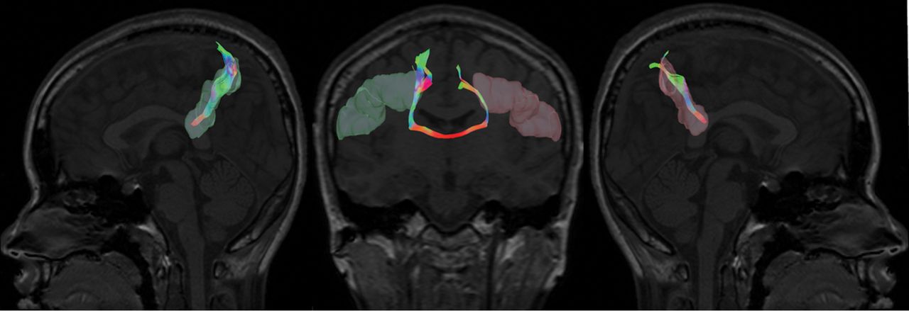
  • Fig 2.
    • Download figure
    • Open in new tab
    • Download powerpoint
    Fig 2.

    Illustration of the interhemispheric tract with the ROIs (left and right Brodmann 2) selected from the Brodmann atlas.

  • Fig 3.
    • Download figure
    • Open in new tab
    • Download powerpoint
    Fig 3.

    Results of voxelwise statistical analysis using the TBSS pipeline. The red lines show the lower FA value in the voxels in the subject with AIS compared with control subjects. S indicates superior; I, inferior; R, right; L, left; P, posterior; A, anterior.

  • Fig 4.
    • Download figure
    • Open in new tab
    • Download powerpoint
    Fig 4.

    Summary of FA values of each subdivision of the corpus callosum divided by the Witelson template19 in the 2D segmentation ROI analysis. Blue represents controls, and red represents patients with AIS.

  • Fig 5.
    • Download figure
    • Open in new tab
    • Download powerpoint
    Fig 5.

    Summary of the mean FA values on the right and left of 3D segmentation of the CC for each subdivision according to the Witelson template19 in controls and patients with AIS.

  • Fig 6.
    • Download figure
    • Open in new tab
    • Download powerpoint
    Fig 6.

    The laterality index of the CC for both patients with AIS (red) and controls (blue). The laterality index was calculated by the following formula: LI = (Mean FALHS − Mean FARHS) / (Mean FALHS + Mean FARHS).

Tables

  • Figures
    • View popup
    Table 1:

    Demographics of subjects with AIS with different curve severities and healthy controls

    AISControls
    MildModerateSevereOverall
    Cobb angle13°–19°20°–39°40°–79°13°–79°–
    Age (range) (mean) (yr)12.2–16.4 (13.3)11.3–16.7 (14.6)12.7–16.4 (14.9)11.3–16.7 (14.5)12.6–16.4 (14.6)
    No. of subjects9 AIS (9 RT)43 AIS (40 RT, 3 RTL)17 AIS (16 RT, 1 RTL)69 AIS (65 RT, 4 RTL)40
    • Note:—RT indicates right thoracic; RTL, right thoracolumbar.

    • View popup
    Table 2:

    Descriptive statistics of the FA comparison between controls and patients with AIS in 2D segmentationa

    ControlsAISP Value (Uncorrected)P Value (Corrected)b
    Rostrum0.7528 ± 0.027350.7455 ± 0.04039.31.62
    Genu0.6997 ± 0.025520.6870 ± 0.02955.025.08
    Anterior midbody0.6607 ± 0.043300.6647 ± 0.0366.61.73
    Posterior midbody0.6445 ± 0.051310.6516 ± 0.04586.46.68
    Isthmus0.7630 ± 0.035500.7631 ± 0.03340.99.99
    Splenium0.7845 ± 0.032280.7630 ± 0.03855.004.02
    • ↵a Data are means unless otherwise indicated.

    • ↵b P value after multiple comparison correction.

    • View popup
    Table 3:

    Descriptive statistics of the FA comparison between controls and patients with AIS in 3D segmentationa

    ControlsAISP Value (Uncorrected)P Value (Corrected)b
    LHRHLHRHLHRHLHRH
    Rostrum0.7592 ± 0.021060.7664 ± 0.022600.7548 ± 0.025220.7645 ± 0.02396.33.691.83
    Genu0.7349 ± 0.017350.7180 ± 0.01750.7246 ± 0.018430.7073 ± 0.02316.005c.47.03c.94
    Anterior midbody0.7144 ± 0.027820.6985 ± 0.020270.7150 ± 0.021420.7004 ± 0.02379.91.67.91.90
    Posterior midbody0.6921 ± 0.039980.6764 ± 0.032160.6973 ± 0.031410.6780 ± 0.03358.48.81.82.88
    Isthmus0.7655 ± 0.019780.7574 ± 0.021340.7612 ± 0.025470.7548 ± 0.02395.36.56.87.85
    Splenium0.7842 ± 0.021280.7883 ± 0.019900.7701 ± 0.021540.7841 ± 0.02404.001c.34.012c1
    • Note:—LH indicates left-hand side; RH, right-hand side.

    • ↵a Data are means unless otherwise indicated.

    • ↵b P value after multiple comparison correction.

    • ↵c Statistically significant (P < .05).

    • View popup
    Table 4:

    Descriptive statistics of the laterality index comparison between controls and patients with AISa

    ControlsAISP Value (Uncorrected)P Value (Corrected)b
    Rostrum−0.0047 ± 0.00965−0.0064 ± 0.01120.41.61
    Genu0.0116 ± 0.007500.0068 ± 0.01001.01c.03c
    Anterior midbody0.0111 ± 0.012200.0104 ± 0.01156.76.76
    Posterior midbody0.0112 ± 0.013310.0142 ± 0.01521.31.61
    Isthmus0.00053 ± 0.010010.0042 ± 0.01101.60.72
    Splenium−0.00026 ± 0.00848−0.009 ± 0.01108.002c.012c
    • ↵a Data are means unless otherwise indicated.

    • ↵b P value after multiple comparison correction.

    • ↵c Statistically significant (P < .05).

Next
Back to top
Advertisement
Print
Download PDF
Email Article

Thank you for your interest in spreading the word on American Journal of Neuroradiology.

NOTE: We only request your email address so that the person you are recommending the page to knows that you wanted them to see it, and that it is not junk mail. We do not capture any email address.

Enter multiple addresses on separate lines or separate them with commas.
Altered White Matter Microstructure in the Corpus Callosum and Its Cerebral Interhemispheric Tracts in Adolescent Idiopathic Scoliosis: Diffusion Tensor Imaging Analysis
(Your Name) has sent you a message from American Journal of Neuroradiology
(Your Name) thought you would like to see the American Journal of Neuroradiology web site.
CAPTCHA
This question is for testing whether or not you are a human visitor and to prevent automated spam submissions.
Cite this article
C. Xue, L. Shi, S.C.N. Hui, D. Wang, T.P. Lam, C.-B. Ip, B.K.W. Ng, J.C.Y. Cheng, W.C.W. Chu
Altered White Matter Microstructure in the Corpus Callosum and Its Cerebral Interhemispheric Tracts in Adolescent Idiopathic Scoliosis: Diffusion Tensor Imaging Analysis
American Journal of Neuroradiology Apr 2018, DOI: 10.3174/ajnr.A5634

Citation Manager Formats

  • BibTeX
  • Bookends
  • EasyBib
  • EndNote (tagged)
  • EndNote 8 (xml)
  • Medlars
  • Mendeley
  • Papers
  • RefWorks Tagged
  • Ref Manager
  • RIS
  • Zotero
0 Responses
Respond to this article
Share
Bookmark this article
Altered White Matter Microstructure in the Corpus Callosum and Its Cerebral Interhemispheric Tracts in Adolescent Idiopathic Scoliosis: Diffusion Tensor Imaging Analysis
C. Xue, L. Shi, S.C.N. Hui, D. Wang, T.P. Lam, C.-B. Ip, B.K.W. Ng, J.C.Y. Cheng, W.C.W. Chu
American Journal of Neuroradiology Apr 2018, DOI: 10.3174/ajnr.A5634
del.icio.us logo Twitter logo Facebook logo Mendeley logo
  • Tweet Widget
  • Facebook Like
  • Google Plus One
Purchase

Jump to section

  • Article
    • Abstract
    • ABBREVIATIONS:
    • Materials and Methods
    • Results
    • Discussion
    • Conclusions
    • Footnotes
    • References
  • Figures & Data
  • Info & Metrics
  • Responses
  • References
  • PDF

Related Articles

  • No related articles found.
  • PubMed
  • Google Scholar

Cited By...

  • Quantification of Structural Brain Connectivity via a Conductance Model
  • Crossref
  • Google Scholar

This article has not yet been cited by articles in journals that are participating in Crossref Cited-by Linking.

More in this TOC Section

  • Frontal Paraventricular Cysts
  • Sodium MRI in Pediatric Brain Tumors
  • FRACTURE MR in Congenital Vertebral Anomalies
Show more Pediatric Neuroimaging

Similar Articles

Advertisement

Indexed Content

  • Current Issue
  • Accepted Manuscripts
  • Article Preview
  • Past Issues
  • Editorials
  • Editor's Choice
  • Fellows' Journal Club
  • Letters to the Editor
  • Video Articles

Cases

  • Case Collection
  • Archive - Case of the Week
  • Archive - Case of the Month
  • Archive - Classic Case

More from AJNR

  • Trainee Corner
  • Imaging Protocols
  • MRI Safety Corner
  • Book Reviews

Multimedia

  • AJNR Podcasts
  • AJNR Scantastics

Resources

  • Turnaround Time
  • Submit a Manuscript
  • Submit a Video Article
  • Submit an eLetter to the Editor/Response
  • Manuscript Submission Guidelines
  • Statistical Tips
  • Fast Publishing of Accepted Manuscripts
  • Graphical Abstract Preparation
  • Imaging Protocol Submission
  • Evidence-Based Medicine Level Guide
  • Publishing Checklists
  • Author Policies
  • Become a Reviewer/Academy of Reviewers
  • News and Updates

About Us

  • About AJNR
  • Editorial Board
  • Editorial Board Alumni
  • Alerts
  • Permissions
  • Not an AJNR Subscriber? Join Now
  • Advertise with Us
  • Librarian Resources
  • Feedback
  • Terms and Conditions
  • AJNR Editorial Board Alumni

American Society of Neuroradiology

  • Not an ASNR Member? Join Now

© 2025 by the American Society of Neuroradiology All rights, including for text and data mining, AI training, and similar technologies, are reserved.
Print ISSN: 0195-6108 Online ISSN: 1936-959X

Powered by HighWire