Advertisement

Main menu

  • Home
  • Content
    • Current Issue
    • Accepted Manuscripts
    • Article Preview
    • Past Issue Archive
    • Video Articles
    • AJNR Case Collection
    • Case of the Week Archive
    • Case of the Month Archive
    • Classic Case Archive
  • Special Collections
    • AJNR Awards
    • Low-Field MRI
    • Alzheimer Disease
    • ASNR Foundation Special Collection
    • Photon-Counting CT
    • View All
  • Multimedia
    • AJNR Podcasts
    • AJNR SCANtastic
    • Trainee Corner
    • MRI Safety Corner
    • Imaging Protocols
  • For Authors
    • Submit a Manuscript
    • Submit a Video Article
    • Submit an eLetter to the Editor/Response
    • Manuscript Submission Guidelines
    • Statistical Tips
    • Fast Publishing of Accepted Manuscripts
    • Graphical Abstract Preparation
    • Imaging Protocol Submission
    • Author Policies
  • About Us
    • About AJNR
    • Editorial Board
    • Editorial Board Alumni
  • More
    • Become a Reviewer/Academy of Reviewers
    • Subscribers
    • Permissions
    • Alerts
    • Feedback
    • Advertisers
    • ASNR Home

User menu

  • Alerts
  • Log in

Search

  • Advanced search
American Journal of Neuroradiology

American Journal of Neuroradiology

ASHNR American Society of Functional Neuroradiology ASHNR American Society of Pediatric Neuroradiology ASSR
  • Alerts
  • Log in

Advanced Search

  • Home
  • Content
    • Current Issue
    • Accepted Manuscripts
    • Article Preview
    • Past Issue Archive
    • Video Articles
    • AJNR Case Collection
    • Case of the Week Archive
    • Case of the Month Archive
    • Classic Case Archive
  • Special Collections
    • AJNR Awards
    • Low-Field MRI
    • Alzheimer Disease
    • ASNR Foundation Special Collection
    • Photon-Counting CT
    • View All
  • Multimedia
    • AJNR Podcasts
    • AJNR SCANtastic
    • Trainee Corner
    • MRI Safety Corner
    • Imaging Protocols
  • For Authors
    • Submit a Manuscript
    • Submit a Video Article
    • Submit an eLetter to the Editor/Response
    • Manuscript Submission Guidelines
    • Statistical Tips
    • Fast Publishing of Accepted Manuscripts
    • Graphical Abstract Preparation
    • Imaging Protocol Submission
    • Author Policies
  • About Us
    • About AJNR
    • Editorial Board
    • Editorial Board Alumni
  • More
    • Become a Reviewer/Academy of Reviewers
    • Subscribers
    • Permissions
    • Alerts
    • Feedback
    • Advertisers
    • ASNR Home
  • Follow AJNR on Twitter
  • Visit AJNR on Facebook
  • Follow AJNR on Instagram
  • Join AJNR on LinkedIn
  • RSS Feeds

AJNR Awards, New Junior Editors, and more. Read the latest AJNR updates

Research ArticleHead and Neck Imaging

Effect of an Arm Traction Device on Image Quality and Radiation Exposure during Neck CT: A Prospective Study

Y.J. Choi, J.H. Lee, D.H. Yoon, H.J. Kim, K.J. Seo, K.-H. Do and J.H. Baek
American Journal of Neuroradiology November 2017, DOI: https://doi.org/10.3174/ajnr.A5418
Y.J. Choi
aFrom the Departments of Radiology and Research Institute of Radiology (Y.J.C., J.H.L., K.J.S., K.-H.D., J.H.B.)
  • Find this author on Google Scholar
  • Find this author on PubMed
  • Search for this author on this site
  • ORCID record for Y.J. Choi
J.H. Lee
aFrom the Departments of Radiology and Research Institute of Radiology (Y.J.C., J.H.L., K.J.S., K.-H.D., J.H.B.)
  • Find this author on Google Scholar
  • Find this author on PubMed
  • Search for this author on this site
  • ORCID record for J.H. Lee
D.H. Yoon
bOncology (D.H.Y.)
  • Find this author on Google Scholar
  • Find this author on PubMed
  • Search for this author on this site
  • ORCID record for D.H. Yoon
H.J. Kim
cClinical Epidemiology and Biostatistics (H.J.K.), University of Ulsan College of Medicine, Asan Medical Center, Seoul, Korea.
  • Find this author on Google Scholar
  • Find this author on PubMed
  • Search for this author on this site
  • ORCID record for H.J. Kim
K.J. Seo
aFrom the Departments of Radiology and Research Institute of Radiology (Y.J.C., J.H.L., K.J.S., K.-H.D., J.H.B.)
  • Find this author on Google Scholar
  • Find this author on PubMed
  • Search for this author on this site
  • ORCID record for K.J. Seo
K.-H. Do
aFrom the Departments of Radiology and Research Institute of Radiology (Y.J.C., J.H.L., K.J.S., K.-H.D., J.H.B.)
  • Find this author on Google Scholar
  • Find this author on PubMed
  • Search for this author on this site
  • ORCID record for K.-H. Do
J.H. Baek
aFrom the Departments of Radiology and Research Institute of Radiology (Y.J.C., J.H.L., K.J.S., K.-H.D., J.H.B.)
  • Find this author on Google Scholar
  • Find this author on PubMed
  • Search for this author on this site
  • ORCID record for J.H. Baek
  • Article
  • Figures & Data
  • Info & Metrics
  • Responses
  • References
  • PDF
Loading

Article Figures & Data

Figures

  • Tables
  • Fig 1.
    • Download figure
    • Open in new tab
    • Download powerpoint
    Fig 1.

    Patient position for neck CT imaging. Patients in the intervention CT protocol were examined with the arm traction device (A). Patients in the standard CT protocol were examined in a relaxed supine position (B). Reprinted with permission from Choi et al.9
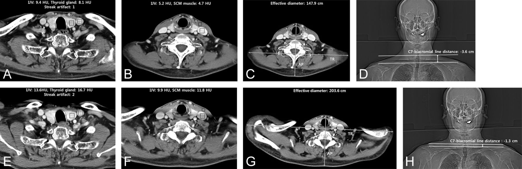
  • Fig 2.
    • Download figure
    • Open in new tab
    • Download powerpoint
    Fig 2.

    Representative neck CT images obtained with the intervention (A–D) and standard (E–H) protocols. The images demonstrate the measurements for image noise at the lower neck (first costovertebral joint level, A and E) and midneck (cricoid cartilage level, B and F), and the effective diameter measurement at the midneck (C and G). The C7-biacromial line distance was determined as illustrated on images D and H. The white line indicates the biacromial line; arrow, the C7-biacromial line distance; SCM, sternocleidomastoid; IJV, internal jugular vein; HU, Hounsfield units.
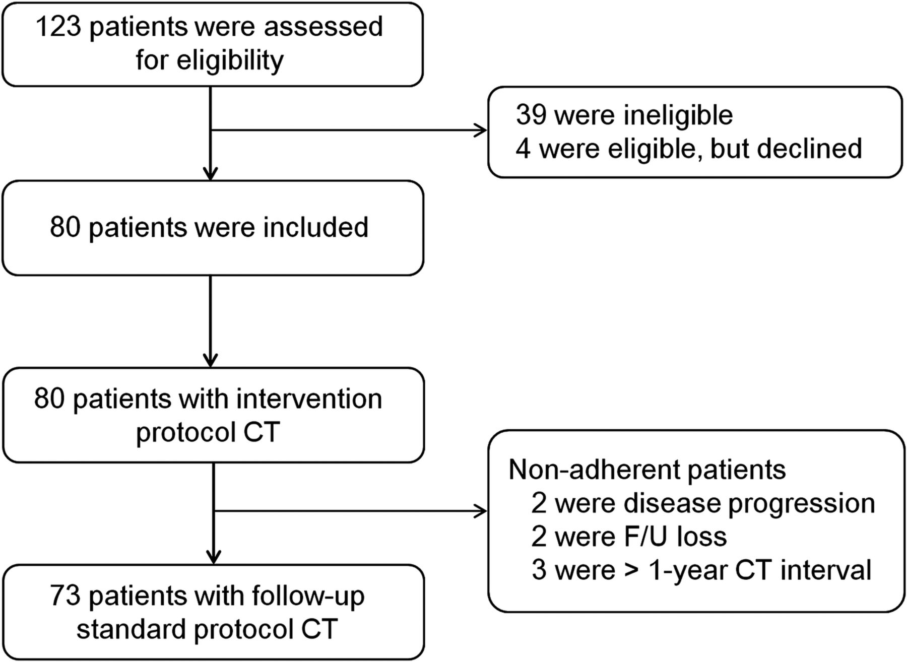
  • Fig 3.
    • Download figure
    • Open in new tab
    • Download powerpoint
    Fig 3.

    Study flow diagram.

Tables

  • Figures
    • View popup
    Table 1:

    Baseline characteristics of the patients enrolled in the study

    ParameterValue
    Age (yr)
        Mean50.6
        Range19–76
    Sex (No.) (%)
        Men39 (53.4)
        Women34 (46.6)
    Body weight (kg)
        Mean64.7
        Range35.7–98.1
    Interval of CT scans (days)
        Mean155
        Range58–355
    • View popup
    Table 2:

    Clinical outcomes in the intervention and standard CT protocolsa

    OutcomeIntervention CT Protocol (n = 73)Standard CT Protocol (n = 73)P Value
    Primary end points
        Image noise, lower neckb
            Thyroid gland12.3 ± 5.117.2 ± 5.5<.001
            Internal jugular vein10.7 ± 4.414.3 ± 5.1<.001
        DLP (mGy × cm)397.6 ± 52.8413.9 ± 56.3<.001
    Secondary end points
        Streak artifactsc1.6 ± 0.51.9 ± 0.4<.001
            126 (35.6%)11 (15.1%)
            246 (63.0%)59 (80.8%)
            31 (1.4%)3 (4.1%)
        Image noise, midneckb
            Internal jugular vein7.1 ± 3.79.1 ± 5.8.009
            SCM muscle5.6 ± 2.47.3 ± 4.0<.001
    CTDIvol (mGy)13.4 ± 0.913.9 ± 0.9<.001
    Effective diameter (cm)d16.2 ± 4.419.6 ± 4.9<.001
    C7-biacromial line distance (cm)e−2.4 ± 1.1−0.3 ± 1.1<.001
    • Note:—SCM indicates sternocleidomastoid.

    • ↵a Data are expressed as mean ± SD.

    • ↵b Based on the evaluation of image noise, by measurement of the SD of the CT values in Hounsfield units.

    • ↵c Streak artifacts at the supraclavicular fossa (1, none or minimal; 2, mild; and 3, severe).

    • ↵d Effective diameter of the neck at the level of the lower margin of the cricoid cartilage.

    • ↵e The C7-biacromial line distance was defined as the distance from the intersection of a line connecting the acromion processes to the upper endplate of C7. Negative values indicate that the biacromial line was located caudal to the upper margin of C7, and positive values, that it was located cranial of the upper margin of C7.

Next
Back to top
Advertisement
Print
Download PDF
Email Article

Thank you for your interest in spreading the word on American Journal of Neuroradiology.

NOTE: We only request your email address so that the person you are recommending the page to knows that you wanted them to see it, and that it is not junk mail. We do not capture any email address.

Enter multiple addresses on separate lines or separate them with commas.
Effect of an Arm Traction Device on Image Quality and Radiation Exposure during Neck CT: A Prospective Study
(Your Name) has sent you a message from American Journal of Neuroradiology
(Your Name) thought you would like to see the American Journal of Neuroradiology web site.
CAPTCHA
This question is for testing whether or not you are a human visitor and to prevent automated spam submissions.
Cite this article
Y.J. Choi, J.H. Lee, D.H. Yoon, H.J. Kim, K.J. Seo, K.-H. Do, J.H. Baek
Effect of an Arm Traction Device on Image Quality and Radiation Exposure during Neck CT: A Prospective Study
American Journal of Neuroradiology Nov 2017, DOI: 10.3174/ajnr.A5418

Citation Manager Formats

  • BibTeX
  • Bookends
  • EasyBib
  • EndNote (tagged)
  • EndNote 8 (xml)
  • Medlars
  • Mendeley
  • Papers
  • RefWorks Tagged
  • Ref Manager
  • RIS
  • Zotero
0 Responses
Respond to this article
Share
Bookmark this article
Effect of an Arm Traction Device on Image Quality and Radiation Exposure during Neck CT: A Prospective Study
Y.J. Choi, J.H. Lee, D.H. Yoon, H.J. Kim, K.J. Seo, K.-H. Do, J.H. Baek
American Journal of Neuroradiology Nov 2017, DOI: 10.3174/ajnr.A5418
del.icio.us logo Twitter logo Facebook logo Mendeley logo
  • Tweet Widget
  • Facebook Like
  • Google Plus One
Purchase

Jump to section

  • Article
    • Abstract
    • ABBREVIATIONS:
    • Materials and Methods
    • Results
    • Discussion
    • Conclusions
    • Footnotes
    • References
  • Figures & Data
  • Info & Metrics
  • Responses
  • References
  • PDF

Related Articles

  • No related articles found.
  • PubMed
  • Google Scholar

Cited By...

  • No citing articles found.
  • Crossref
  • Google Scholar

This article has not yet been cited by articles in journals that are participating in Crossref Cited-by Linking.

More in this TOC Section

  • MRI of the Parasellar Ligaments
  • ASL Sensitivity for Head and Neck Paraganglioma
  • Post SRS Peritumoral Hyperintense Signal of VSs
Show more Head and Neck Imaging

Similar Articles

Advertisement

Indexed Content

  • Current Issue
  • Accepted Manuscripts
  • Article Preview
  • Past Issues
  • Editorials
  • Editor's Choice
  • Fellows' Journal Club
  • Letters to the Editor
  • Video Articles

Cases

  • Case Collection
  • Archive - Case of the Week
  • Archive - Case of the Month
  • Archive - Classic Case

More from AJNR

  • Trainee Corner
  • Imaging Protocols
  • MRI Safety Corner
  • Book Reviews

Multimedia

  • AJNR Podcasts
  • AJNR Scantastics

Resources

  • Turnaround Time
  • Submit a Manuscript
  • Submit a Video Article
  • Submit an eLetter to the Editor/Response
  • Manuscript Submission Guidelines
  • Statistical Tips
  • Fast Publishing of Accepted Manuscripts
  • Graphical Abstract Preparation
  • Imaging Protocol Submission
  • Evidence-Based Medicine Level Guide
  • Publishing Checklists
  • Author Policies
  • Become a Reviewer/Academy of Reviewers
  • News and Updates

About Us

  • About AJNR
  • Editorial Board
  • Editorial Board Alumni
  • Alerts
  • Permissions
  • Not an AJNR Subscriber? Join Now
  • Advertise with Us
  • Librarian Resources
  • Feedback
  • Terms and Conditions
  • AJNR Editorial Board Alumni

American Society of Neuroradiology

  • Not an ASNR Member? Join Now

© 2025 by the American Society of Neuroradiology All rights, including for text and data mining, AI training, and similar technologies, are reserved.
Print ISSN: 0195-6108 Online ISSN: 1936-959X

Powered by HighWire