
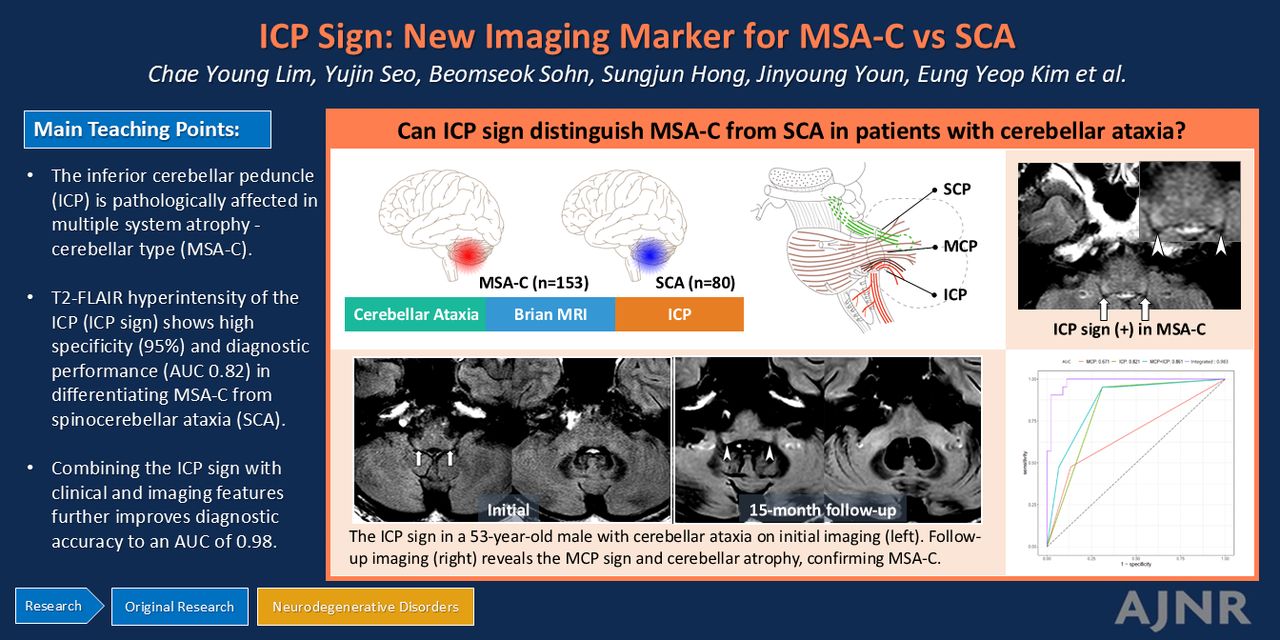
Article Figures & Data
Figures
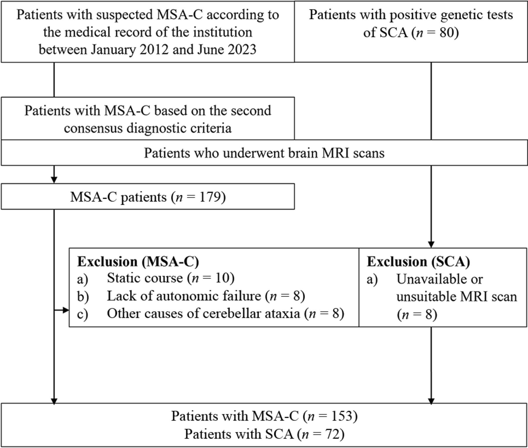
- FIG 1.
Study flow chart.
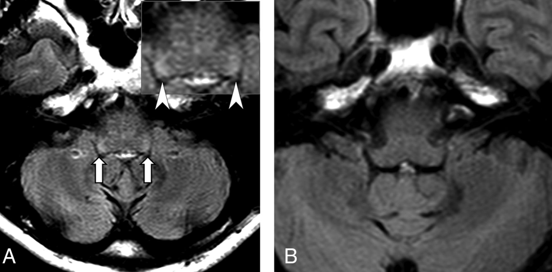
- FIG 2.
Representative cases of positive/negative ICP signs. A, A 45-year-old woman presented with cerebellar ataxia. A FLAIR image reveals increased signal intensity in the bilateral inferior cerebellar peduncles (arrows; arrowheads in an inset) compared with the medulla. The patient was diagnosed with MSA-C. B, In the T2 FLAIR image of a 48-year-old man diagnosed with spinocerebellar ataxia, no ICP sign is seen.
- FIG 3.
FLAIR imaging in a 53-year-old man who presented with cerebellar ataxia. The initial imaging (left) showed high signal intensity in both inferior cerebellar peduncles (arrows), defined as the ICP sign. However, the MCP sign was not evident. On 15-month follow-up MRI (right), the ICP sign was again noted (arrowheads), and both the HCB and MCP signs appeared. The patient was diagnosed as having MSA-C.
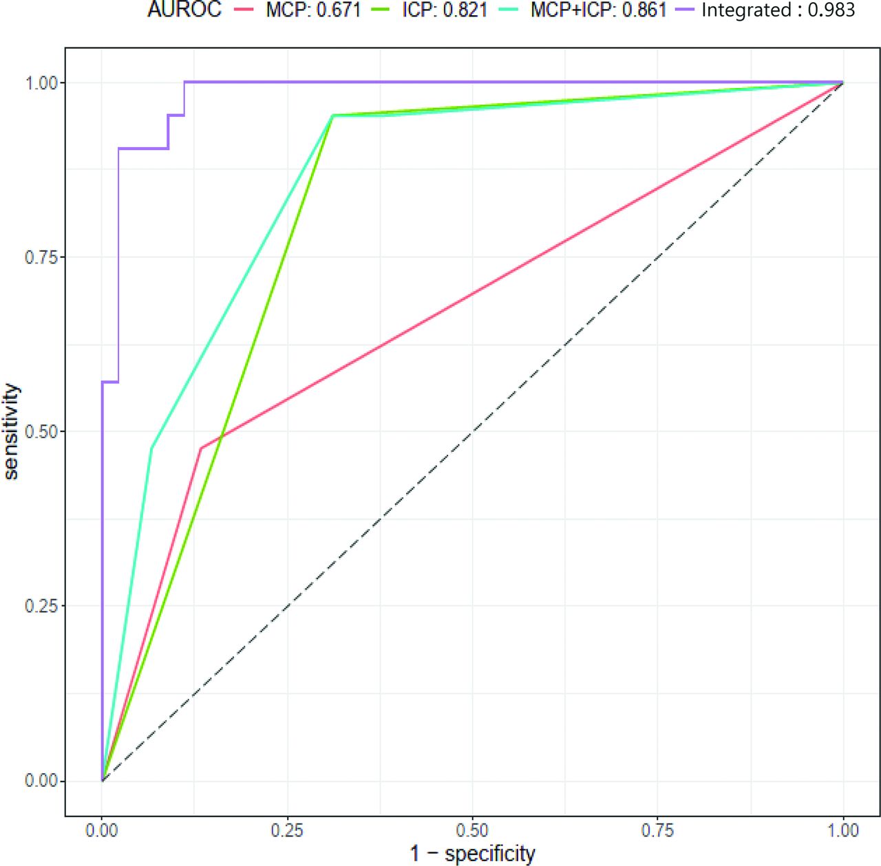
- FIG 4.
Receiver operating curves of ICP, MCP, combined ICP and MCP signs and the integrated clinicoradiologic model for predicting MSA-C. The AUCs of the ICP sign, MCP sign, and combined ICP and MCP signs are 0.82, 0.67, and 0.86, respectively. The integrated model shows the highest diagnostic performance (AUC = 0.98, 95% CI, 0.96–1.00).
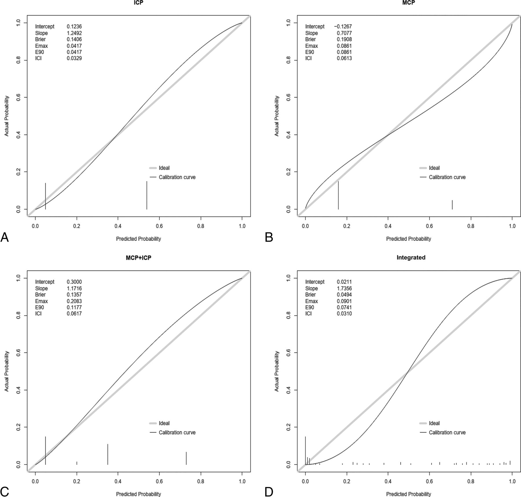
- FIG 5.
Calibration plots with ICIs of the ICP, MCP, combined ICP and MCP hyperintensity signs, and the integrated clinicoradiologic model for predicting MSA-C. Calibration plots comparing the ICP sign (intercept = 0.12, slope = 1.25, ICI = 0.03), MCP sign (intercept = –0.13, slope = 0.71, ICI = 0.61), combined signs (intercept = 0.30, slope = 1.17, ICI = 0.62), and integrated model (intercept= 0.02, slope = 1.74, ICI = 0.03). The ICP sign demonstrates reliable calibration overall. Bars in the calibration plot denote observed outcome frequencies within each bin, reflecting the calibration level of the prediction model. The diagonal line indicates perfect calibration. Brier indicates the Brier score (average squared difference in predicted and actual probabilities); Emax/E90/ICI, the maximum/90th quantile/weighted average of absolute difference in predicted and actual calibrated probabilities.


Tables
Variables Total (n = 225) MSA-C (n = 153) SCA (n = 72) P Values Clinical features Agea 59 (53–65) 60 (56–67) 53 (44–61) <.001 Disease duration (yr)a 3 (1–5) 2 (1–3.3) 5 (2–11) <.001 Sex Male 124 (55)b 79 (52) 45 (63) .204 Female 101 (45) 74 (48) 27 (38) Imaging features Scanner .053 A 182 (81) 119 (78) 63 (88) B 6 (2.7) 4 (2.6) 2 (2.8) C 36 (16) 30 (20) 6 (8.3) D 1 (0.4) 0 (0.0) 1 (1.4) ICP sign 105 (47) 100 (65) 5 (6.9) <.001 HCB sign 129 (57) 110 (72) 19 (26) <.001 MCP sign 164 (73) 134 (88) 30 (42) <.001 Cerebellar atrophy 191 (85) 135 (88) 56 (78) .065 Vertical line 180 (80) 132 (86) 48 (67) .011 - Table 2:
Univariable and multivariable logistic regression analyses for predicting the MSA with predominant cerebellar ataxia group
Variables Univariable Analysis Multivariable Analysis OR 95% CI P Value OR 95% CI P Value Age 1.10 1.07–1.14 <.001 1.16 1.09–1.28 <.001 Disease duration (yr) 0.77 0.70–0.85 <.001 0.63 0.53–0.75 <.001 Sex (male) 1.58 0.89–2.81 .117 Scanner (A)a 2.02 0.91–4.47 .084 ICP sign (absence) 25.7 9.78–67.9 <.001 32.7 5.74–186 <.001 HCB sign (absence) 7.31 3.88–13.8 <.001 MCP sign (absence) 10.4 5.28–20.6 <.001 16.8 5.08–55.8 <.001 Cerebellar atrophy (absence) 2.27 1.07–4.81 .032 Vertical line (absence) 3.30 1.67–6.54 .001 ↵a Scanner A (the most frequently used scanner type) is the reference, with the OR calculated for other scanner types combined. The reference category for each variable is presented in parentheses.
- Table 3:
Diagnostic performance of imaging and clinical features for predicting the MSA with the predominant cerebellar ataxia group
Parameters Sensitivitya Specificitya F1 Score AUCb P Valuec ICP 0.69 [31/45] 0.95 [20/21] 0.73 0.82 (0.74–0.90) – MCP 0.87 [39/45] 0.48 [10/21] 0.69 0.67 (0.55–0.79) .029 HCB 0.69 [31/45] 0.71 [15/21] 0.64 0.70 (0.58,0.82) .052 ICP + MCP 0.62 [28/45] 0.95 [20/21] 0.70 0.86 (0.78–0.98) .114 Integrated modeld 0.87 [39/45] 1.00 [21/21] 0.88 0.98 (0.96–1.00) <.001 Note:—The en dash indicates not applicable.
↵a Data in brackets indicate the number of corresponding patients for each of the metric.
↵b Data in parentheses are 95% CIs with the DeLong test.
↵c P values of AUCs compared with ICP as a reference.
↵d Integrated model includes the ICP, MCP sign, age, and disease duration as variables.


Jump to section
Related Articles
Cited By...
- No citing articles found.