
Article Figures & Data
Figures
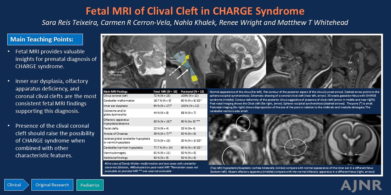
- FIG 1.
A, Parasagittal T2-weighted half-Fourier-acquired single-shot turbo spin echo (HASTE) image of a 35 weeks’ gestation fetus demonstrates normal appearance of the clivus. Note the flat contour of the posterior aspect of the clivus (curved arrow). The dashed arrow anteriorly points to a T2 hyperintense line that corresponds to the spheno-occipital synchondrosis. B, Schematic drawing of a coronal clival cleft (arrow). Case c008, a 35-week gestation fetus with CHARGE syndrome (C through E). C, Parasagittal T2 HASTE and D, T1-weighted images. C, There is contour deformity of the posterior clivus suggestive of presence of clival cleft, better seen on D (arrow). E, Postnatal sagittal T1-weighted image of the fetus shown in C and D. Clival cleft (arrows in D and E). Spheno-occipital synchondrosis (dashed arrows in A through E). The pons (* in C) is small in AP dimension and height, and measurements are below the 10th percentile for reported gestational age. This is better seen on postnatal imaging (E) with a disproportion of the size of the pons (*) in relation to the midbrain and medulla oblongata. The cerebellar vermis is also small in craniocaudal and AP dimension, both on fetal and postnatal MRI.
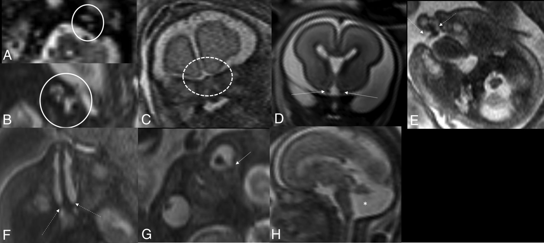
- FIG 2.
Prenatal MR neuroimaging findings in fetuses with CHARGE syndrome. A, Prenatal axial T2-weighted image of a 25.6 weeks’ gestation fetus. Hypoplastic/dysplastic cochlea bilaterally (circles). The labyrinth, including semicircular canals, is not visible (not shown). Compare with normal appearance of the inner ear in a different fetus at 33 weeks’ gestation (B). C, 30.6 weeks’ gestation fetus. Absent olfactory apparatus on coronal T2-weighted image (dashed circle). D, For comparison, coronal T2-weighted image of a 24 weeks’ gestation fetus shows T2 hypointense structures beneath the frontal lobes bilaterally consistent with the normal olfactory apparatus (arrows). E, 30.6 weeks’ gestation fetus, same fetus as shown in C. Prenatal axial T2-weighted image at the level of the maxilla shows bilateral cleft palate. F, 34.1 weeks’ gestation fetus, prenatal axial T2-weighted image shows atresia of choanae bilaterally (dashed arrows). G, 34 weeks’ gestation fetus. Prenatal axial T2-weighted image at the level of the orbits shows bilateral eye dysmorphism with left eye coloboma (arrow) and persistent hyaloid vascular structures. Midline sagittal T2-weighted image of a 22.9 weeks’ gestation fetus (H) shows hypoplastic vermis with enlarged posterior fossa consistent with Dandy-Walker malformation (*).
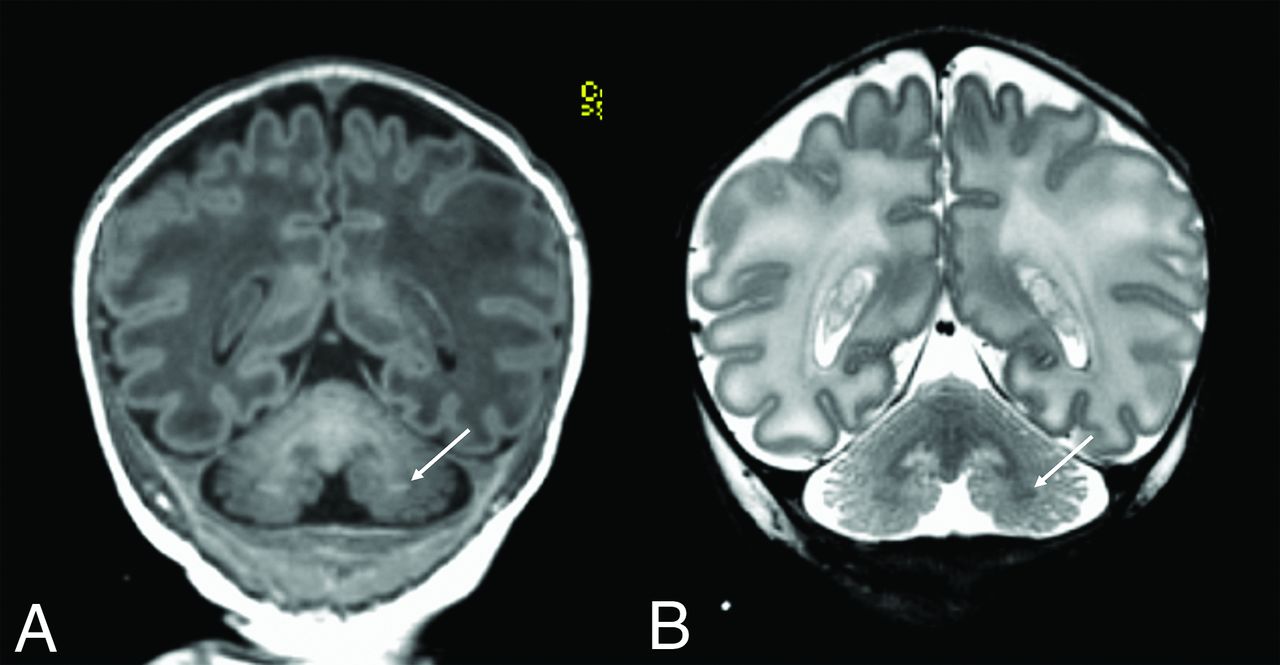
- FIG 3.
Two-day-old male, c008. Coronal T1 (A) and T2-weighted (B) images show symmetric, T1 hyperintense, and T2 hypointense foci of signal abnormality in the deep cerebellar white matter (arrows) consistent with cerebellar gray matter heterotopia.


Jump to section
Related Articles
Cited By...
- No citing articles found.