Advertisement

Main menu

  • Home
  • Content
    • Current Issue
    • Accepted Manuscripts
    • Article Preview
    • Past Issue Archive
    • Video Articles
    • AJNR Case Collection
    • Case of the Week Archive
    • Case of the Month Archive
    • Classic Case Archive
  • Special Collections
    • AJNR Awards
    • Low-Field MRI
    • Alzheimer Disease
    • ASNR Foundation Special Collection
    • Photon-Counting CT
    • View All
  • Multimedia
    • AJNR Podcasts
    • AJNR SCANtastic
    • Trainee Corner
    • MRI Safety Corner
    • Imaging Protocols
  • For Authors
    • Submit a Manuscript
    • Submit a Video Article
    • Submit an eLetter to the Editor/Response
    • Manuscript Submission Guidelines
    • Statistical Tips
    • Fast Publishing of Accepted Manuscripts
    • Graphical Abstract Preparation
    • Imaging Protocol Submission
    • Author Policies
  • About Us
    • About AJNR
    • Editorial Board
    • Editorial Board Alumni
  • More
    • Become a Reviewer/Academy of Reviewers
    • Subscribers
    • Permissions
    • Alerts
    • Feedback
    • Advertisers
    • ASNR Home

User menu

  • Alerts
  • Log in

Search

  • Advanced search
American Journal of Neuroradiology

American Journal of Neuroradiology

ASHNR American Society of Functional Neuroradiology ASHNR American Society of Pediatric Neuroradiology ASSR
  • Alerts
  • Log in

Advanced Search

  • Home
  • Content
    • Current Issue
    • Accepted Manuscripts
    • Article Preview
    • Past Issue Archive
    • Video Articles
    • AJNR Case Collection
    • Case of the Week Archive
    • Case of the Month Archive
    • Classic Case Archive
  • Special Collections
    • AJNR Awards
    • Low-Field MRI
    • Alzheimer Disease
    • ASNR Foundation Special Collection
    • Photon-Counting CT
    • View All
  • Multimedia
    • AJNR Podcasts
    • AJNR SCANtastic
    • Trainee Corner
    • MRI Safety Corner
    • Imaging Protocols
  • For Authors
    • Submit a Manuscript
    • Submit a Video Article
    • Submit an eLetter to the Editor/Response
    • Manuscript Submission Guidelines
    • Statistical Tips
    • Fast Publishing of Accepted Manuscripts
    • Graphical Abstract Preparation
    • Imaging Protocol Submission
    • Author Policies
  • About Us
    • About AJNR
    • Editorial Board
    • Editorial Board Alumni
  • More
    • Become a Reviewer/Academy of Reviewers
    • Subscribers
    • Permissions
    • Alerts
    • Feedback
    • Advertisers
    • ASNR Home
  • Follow AJNR on Twitter
  • Visit AJNR on Facebook
  • Follow AJNR on Instagram
  • Join AJNR on LinkedIn
  • RSS Feeds

AJNR Awards, New Junior Editors, and more. Read the latest AJNR updates

Review ArticleBrain Tumor Imaging

Meningioma: Molecular Updates from the 2021 World Health Organization Classification of CNS Tumors and Imaging Correlates

Neetu Soni, Manish Ora, Girish Bathla, Denes Szekeres, Amit Desai, Jay J. Pillai and Amit Agarwal
American Journal of Neuroradiology February 2025, 46 (2) 240-250; DOI: https://doi.org/10.3174/ajnr.A8368
Neetu Soni
aFrom the Department of Radiology (N.S., J.J.P., A.D., A.A.), Mayo Clinic, Jacksonville, Florida
  • Find this author on Google Scholar
  • Find this author on PubMed
  • Search for this author on this site
  • ORCID record for Neetu Soni
Manish Ora
bDepartment of Nuclear Medicine (M.O.), Sanjay Gandhi Post-Graduate Institute of Medical Sciences, Lucknow, India
  • Find this author on Google Scholar
  • Find this author on PubMed
  • Search for this author on this site
  • ORCID record for Manish Ora
Girish Bathla
cDepartment of Radiology (G.B., J.P.), Mayo Clinic, Rochester, Minnesota
  • Find this author on Google Scholar
  • Find this author on PubMed
  • Search for this author on this site
  • ORCID record for Girish Bathla
Denes Szekeres
dUniversity of Rochester School of Medicine and Dentistry (D.S.), Rochester, New York
  • Find this author on Google Scholar
  • Find this author on PubMed
  • Search for this author on this site
Amit Desai
aFrom the Department of Radiology (N.S., J.J.P., A.D., A.A.), Mayo Clinic, Jacksonville, Florida
  • Find this author on Google Scholar
  • Find this author on PubMed
  • Search for this author on this site
  • ORCID record for Amit Desai
Jay J. Pillai
cDepartment of Radiology (G.B., J.P.), Mayo Clinic, Rochester, Minnesota
  • Find this author on Google Scholar
  • Find this author on PubMed
  • Search for this author on this site
  • ORCID record for Jay J. Pillai
Amit Agarwal
aFrom the Department of Radiology (N.S., J.J.P., A.D., A.A.), Mayo Clinic, Jacksonville, Florida
  • Find this author on Google Scholar
  • Find this author on PubMed
  • Search for this author on this site
  • ORCID record for Amit Agarwal
  • Article
  • Figures & Data
  • Supplemental
  • Info & Metrics
  • Responses
  • References
  • PDF
Loading

Article Figures & Data

Figures

  • Tables
  • FIG 1.
    • Download figure
    • Open in new tab
    • Download powerpoint
    FIG 1.

    Illustration depicting the common location of meningiomas and associated genetic/cytogenetic alteration and grades. NF2 and SMARCB1 mutations are more frequently seen in convexity meningiomas. Grade 2 and 3 meningiomas are more common along the convexity than the skull base. AKT1, KLF4, TRAF7, and POLR2A genetic changes are more frequently seen in skull base meningiomas. Grade 1 is more common in the posterior fossa. Grade 2 (atypical) meningiomas are more frequently seen along the brain convexity and spine and can have a loss of a copy of chromosomes 1, 10, or 14.
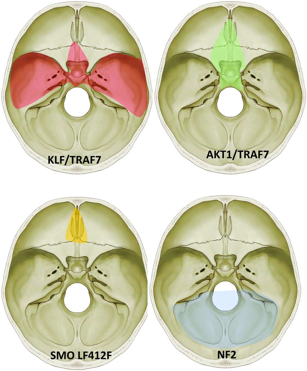
  • FIG 2.
    • Download figure
    • Open in new tab
    • Download powerpoint
    FIG 2.

    “Mutation map” of skull base meningiomas with a regional propensity of meningiomas dependent on specific mutations. Meningiomas arising from the sphenoid wing have KLF4/TRAF7 mutations, midline tumors have AKT1/TRAF7 mutations, and tumors originating from the olfactory groove tend to have SMO mutations. Meningiomas along the posterior skull base commonly have a loss of chromosome 22-loss (NF2). Adapted from Baranoski J. Smarcb1-Mutant Intracranial Meningiomas: A Distinct Subtype of Nf2-Mutant Tumors, 2015. Yale Medicine Thesis Digital Library. 1947.

  • FIG 3.
    • Download figure
    • Open in new tab
    • Download powerpoint
    FIG 3.

    “Mutation map” of convexity meningiomas with the regional propensity of meningiomas dependent on a specific mutation. Meningiomas along the falx and midline parasagittal region tend to have the SMARCB1 mutation. Meningiomas along the posterior cerebral convexity tend to have NF2/chromosomal 22 with higher chances of TRAF mutation along the anterior cerebral convexity. Adapted from Baranoski J, Smarcb1-Mutant Intracranial Meningiomas: A Distinct Subtype of Nf2-Mutant Tumors, 2015. Yale Medicine Thesis Digital Library. 1947.

  • FIG 4.
    • Download figure
    • Open in new tab
    • Download powerpoint
    FIG 4.

    Chart illustration of the grading of meningiomas according to the 2016 WHO classification scheme and the changes made in the 2021 CNS5 classification, with the CNS5 addition of new molecular entities for grading and removal of a few grade 3 histologic subtypes.

  • FIG 5.
    • Download figure
    • Open in new tab
    • Download powerpoint
    FIG 5.

    Transcalvarial frontal atypical, grade 2 meningioma in a 54-year-old man. Sagittal T1-weighted (A) and contrast-enhanced (B) images reveal a large frontal convexity meningioma with intraosseous components and transcalvarial extension into the scalp (B, arrow). Mild hyperostosis of the adjacent calvaria along with a “hair-on-end” periosteal reaction along the outer table is seen on CT (C, arrow). Tumor shows meningothelial morphology, with, however, other features such as focal spontaneous necrosis (D, asterisk) and peripheral nests of tumor cells (D, arrows). A high Ki-67 rate in hot spot areas and areas of tumor that are devoid of progesterone receptors is also noted, supportive of the diagnosis of atypical meningioma (WHO grade 2). Mitotic count of 4 mitoses per 10 high-power fields is noted, on its own fulfilling the criteria for a diagnosis of an atypical meningioma.

  • FIG 6.
    • Download figure
    • Open in new tab
    • Download powerpoint
    FIG 6.

    An anaplastic (malignant) grade 3 meningioma along the right middle cranial fossa floor. Coronal T2-weighted (A) and coronal and axial contrast-enhanced (B and C) images depict the meningioma with strongly suggestive imaging features of parenchymal invasion, including poor interface between the tumor and cortex (A, arrow) and nodules as foci of cortical enhancement (B and C, white arrows) along with linear leptomeningeal enhancement (B, black arrow). Histopathology shows frank anaplasia cells with numerous pleomorphic cells giving this tumor a sarcomatoid appearance (D, arrows) with a grade 3 classification. Mitotic count of 22 mitoses per 10 high-power fields was noted, on its own fulfilling the criteria for the diagnosis of an anaplastic meningioma.

  • FIG 7.
    • Download figure
    • Open in new tab
    • Download powerpoint
    FIG 7.

    Right intraorbital CNS WHO grade 3 meningioma with activation of the TERT promoter. A meningioma is initially classified as grade 2 (atypical) based on histopathologic features; however, it was upgraded to grade 3 on the basis of detection of TERT activation on chromosomal microarray. Preoperative contrast-enhanced MRI at presentation (A) and at 6 months’ follow-up (B) reveals rapid enlargement of the intraorbital meningioma (arrows). The patient had multiple postoperative recurrences during the next 18 months. Preoperative contrast-enhanced MRI (C and D) performed at 3-month intervals shows rapid enlargement of recurrent enhancing tumor along the orbital roof (C and D, arrows).
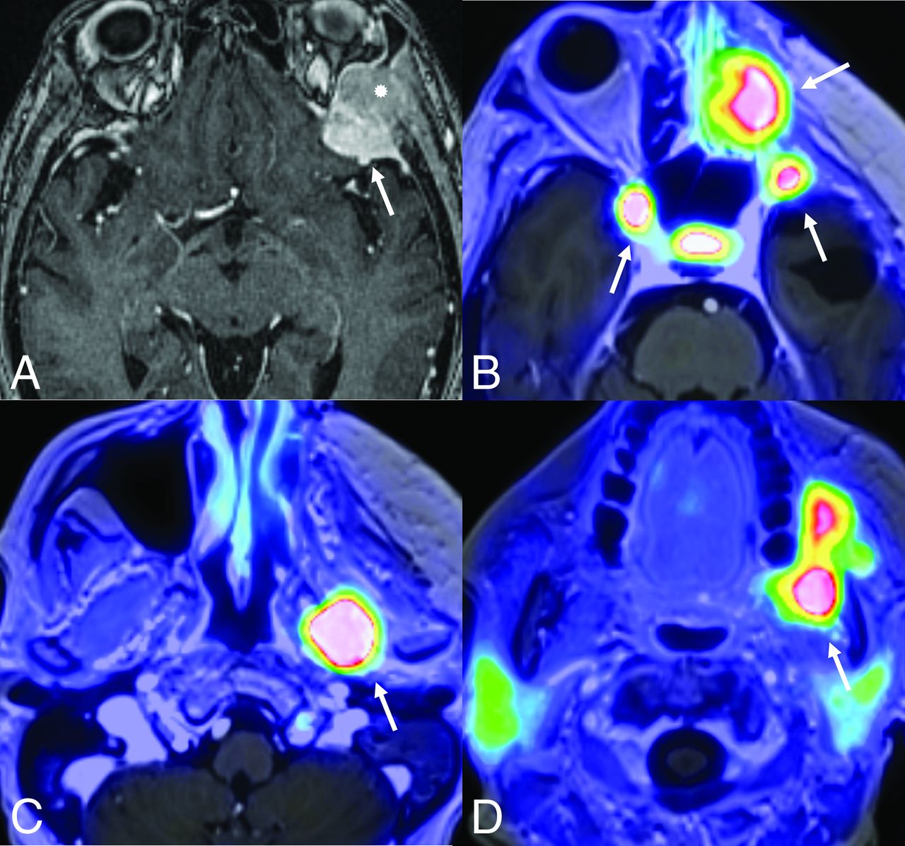
  • FIG 8.
    • Download figure
    • Open in new tab
    • Download powerpoint
    FIG 8.

    Histologic grade 1 skull base meningiomas in 2 different patients (patients A and B) with upgrading to grade 3 (anaplastic) in patient B based on CDKN2 deletion. Axial (A) contrast-enhanced MRI in patient A reveals a left middle cranial fossa meningioma (arrow) with no recurrence and minimal residual activity on the 1-year postresection follow-up DOTATATE scan (B, arrow). A right sphenoid wing meningioma with intraorbital extension is seen on axial contrast-enhanced MRI (C, arrow) in patient B. Tumor recurrence was noted at the 1-year postresection follow-up DOTATATE scan (D, arrows), consistent with high-grade morphology. Despite histologic grade 1, the meningioma was upgraded to grade 3 (anaplastic) on the basis of identification of a CDKN2 deletion.
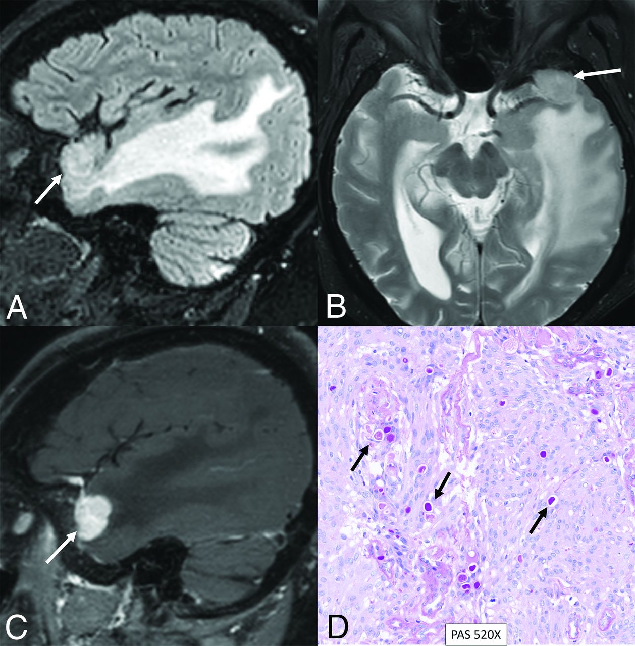
  • FIG 9.
    • Download figure
    • Open in new tab
    • Download powerpoint
    FIG 9.

    Secretory meningioma with a TRAF7 mutation in a 71-year-old woman. Sagittal FLAIR (A), axial T2-weighted (B), and sagittal contrast-enhanced (C) images reveal a small meningioma along the left greater wing of the sphenoid bone (arrows). Extensive parenchymal edema is noted in the right temporal lobe, disproportionate to the size of the meningioma. Histopathology revealed a secretory histologic subtype, with clusters of eosinophilic globules highlighted by pseudopsammoma bodies (D, arrows) with low mitotic activity (<1 mitosis in 10 high-power fields) and no atypical features, supporting a CNS WHO grade 1 designation. Next-generation sequencing studies demonstrated a pathogenic variant in TRAF7 (c.1136-1G>A).

  • FIG 10.
    • Download figure
    • Open in new tab
    • Download powerpoint
    FIG 10.

    Rapid multifocal recurrence of CDKN2A/B-deleted CNS WHO grade 3 meningioma along the left sphenoid wing. The meningioma was initially classified as grade 2 (atypical) on the basis of histopathologic features; however, it was upgraded to grade 3 on the basis of detection of a homozygous deletion of CDKN2A/B on next-generation sequencing. Contrast-enhanced axial MRI depicts the left sphenoid wing meningioma (A, arrow) with intraosseous extension (asterisk). Rapid multifocal recurrence is noted on PET (DOTATATE) MRI (B–D, arrows) within a few months postresection.

Tables

  • Figures
  • Commonly identified germline and somatic mutations in meningiomas with corresponding WHO grade, location, and clinical significance

    Genetic Mutation NameWHO GradeTypical LocationClinical Significance
    NF21−3Parafalcine, posterior fossa–falcotentorialMost common70% of fibroblastic and transitional meningiomasSporadic mutations present in 40%−60% of meningiomas50%−75% of patients with germline mutations develop meningiomasAssociated with larger, more aggressive course
    TRAF71−3Central and lateral skull base2nd most commonSecretory subtypeHigh likelihood of hyperostosisMeningiomas tend to be benign, chromosome-stable
    TERT3Any locationCommonly seen in higher grade, particularly grade 3Associated with shorter time to progression, shorter overall survival, and higher recurrence
    CDKN2A/B3Any locationMutations associated with shorter time to recurrenceClassification criteria for WHO grade 3 meningiomas
    SMO/SUFU1Olfactory groove meningiomas, anterior skull baseHigher recurrence rates among olfactory groove meningiomasLarger tumor volume among anterior skull base meningiomasLinked to development of isolated familial and multiple meningiomas
    KLF41Central and lateral skull baseSecretory subtypeLarger peritumoral brain edemaResults in up-regulation of HIF-1a pathwayMay respond to mTOR inhibition
    POLR2A1Parasellar/tuberculum sellaeFound almost exclusively in WHO grade 1 meningiomas (meningothelial)
    AKT1Anterior and middle skull base, posterior fossaMeningothelialMutations occur with higher frequency among skull base meningiomasAssociated with shorter time to recurrence
    PIK3CA1–3Anterior and middle skull baseLow recurrence riskProgesterone and cyproterone antiandrogen therapy show higher PIK3CA mutation rates in skull base meningiomas
    HedgehogMidline anterior skull baseLow-grade and less aggressive
    SMARCB1SMARCE11–3Parafalcine and lateral skull baseClear cell typeSMARCB1 has been linked to multiple meningiomasSMARCE1 mutations linked with familial multiple spinal meningiomasHigher recurrence risk, faster growth
    BAP1PBRM13Rhabdoid and papillary subtypesAggressive clinical behavior (consistent with CNS5 WHO grade 3)
    • Note:— HIF-1-α indicates hypoxia-inducible factor 1.

PreviousNext
Back to top

In this issue

American Journal of Neuroradiology: 46 (2)
American Journal of Neuroradiology
Vol. 46, Issue 2
1 Feb 2025
  • Table of Contents
  • Index by author
  • Complete Issue (PDF)
Advertisement
Print
Download PDF
Email Article

Thank you for your interest in spreading the word on American Journal of Neuroradiology.

NOTE: We only request your email address so that the person you are recommending the page to knows that you wanted them to see it, and that it is not junk mail. We do not capture any email address.

Enter multiple addresses on separate lines or separate them with commas.
Meningioma: Molecular Updates from the 2021 World Health Organization Classification of CNS Tumors and Imaging Correlates
(Your Name) has sent you a message from American Journal of Neuroradiology
(Your Name) thought you would like to see the American Journal of Neuroradiology web site.
CAPTCHA
This question is for testing whether or not you are a human visitor and to prevent automated spam submissions.
Cite this article
Neetu Soni, Manish Ora, Girish Bathla, Denes Szekeres, Amit Desai, Jay J. Pillai, Amit Agarwal
Meningioma: Molecular Updates from the 2021 World Health Organization Classification of CNS Tumors and Imaging Correlates
American Journal of Neuroradiology Feb 2025, 46 (2) 240-250; DOI: 10.3174/ajnr.A8368

Citation Manager Formats

  • BibTeX
  • Bookends
  • EasyBib
  • EndNote (tagged)
  • EndNote 8 (xml)
  • Medlars
  • Mendeley
  • Papers
  • RefWorks Tagged
  • Ref Manager
  • RIS
  • Zotero
2 Responses
Respond to this article
Share
Bookmark this article
Meningioma Updates from 2021 WHO Classification
Neetu Soni, Manish Ora, Girish Bathla, Denes Szekeres, Amit Desai, Jay J. Pillai, Amit Agarwal
American Journal of Neuroradiology Feb 2025, 46 (2) 240-250; DOI: 10.3174/ajnr.A8368
del.icio.us logo Twitter logo Facebook logo Mendeley logo
  • Tweet Widget
  • Facebook Like
  • Google Plus One
Purchase

Jump to section

  • Article
    • Abstract
    • ABBREVIATIONS:
    • DISCUSSION
    • CONCLUSIONS
    • Footnotes
    • References
  • Figures & Data
  • Supplemental
  • Info & Metrics
  • Responses
  • References
  • PDF

Related Articles

  • PubMed
  • Google Scholar

Cited By...

  • No citing articles found.
  • Crossref
  • Google Scholar

This article has not yet been cited by articles in journals that are participating in Crossref Cited-by Linking.

More in this TOC Section

  • Brain Tumor Subtyping Image-Based search via MRI
  • Neuroimaging of Erdheim-Chester Disease
  • CE MRI for Brain Metastasis Detection
Show more Brain Tumor Imaging

Similar Articles

Advertisement

Indexed Content

  • Current Issue
  • Accepted Manuscripts
  • Article Preview
  • Past Issues
  • Editorials
  • Editor's Choice
  • Fellows' Journal Club
  • Letters to the Editor
  • Video Articles

Cases

  • Case Collection
  • Archive - Case of the Week
  • Archive - Case of the Month
  • Archive - Classic Case

More from AJNR

  • Trainee Corner
  • Imaging Protocols
  • MRI Safety Corner
  • Book Reviews

Multimedia

  • AJNR Podcasts
  • AJNR Scantastics

Resources

  • Turnaround Time
  • Submit a Manuscript
  • Submit a Video Article
  • Submit an eLetter to the Editor/Response
  • Manuscript Submission Guidelines
  • Statistical Tips
  • Fast Publishing of Accepted Manuscripts
  • Graphical Abstract Preparation
  • Imaging Protocol Submission
  • Evidence-Based Medicine Level Guide
  • Publishing Checklists
  • Author Policies
  • Become a Reviewer/Academy of Reviewers
  • News and Updates

About Us

  • About AJNR
  • Editorial Board
  • Editorial Board Alumni
  • Alerts
  • Permissions
  • Not an AJNR Subscriber? Join Now
  • Advertise with Us
  • Librarian Resources
  • Feedback
  • Terms and Conditions
  • AJNR Editorial Board Alumni

American Society of Neuroradiology

  • Not an ASNR Member? Join Now

© 2025 by the American Society of Neuroradiology All rights, including for text and data mining, AI training, and similar technologies, are reserved.
Print ISSN: 0195-6108 Online ISSN: 1936-959X

Powered by HighWire