Advertisement

Main menu

  • Home
  • Content
    • Current Issue
    • Accepted Manuscripts
    • Article Preview
    • Past Issue Archive
    • Video Articles
    • AJNR Case Collection
    • Case of the Week Archive
    • Case of the Month Archive
    • Classic Case Archive
  • Special Collections
    • AJNR Awards
    • Low-Field MRI
    • Alzheimer Disease
    • ASNR Foundation Special Collection
    • Photon-Counting CT
    • View All
  • Multimedia
    • AJNR Podcasts
    • AJNR SCANtastic
    • Trainee Corner
    • MRI Safety Corner
    • Imaging Protocols
  • For Authors
    • Submit a Manuscript
    • Submit a Video Article
    • Submit an eLetter to the Editor/Response
    • Manuscript Submission Guidelines
    • Statistical Tips
    • Fast Publishing of Accepted Manuscripts
    • Graphical Abstract Preparation
    • Imaging Protocol Submission
    • Author Policies
  • About Us
    • About AJNR
    • Editorial Board
    • Editorial Board Alumni
  • More
    • Become a Reviewer/Academy of Reviewers
    • Subscribers
    • Permissions
    • Alerts
    • Feedback
    • Advertisers
    • ASNR Home

User menu

  • Alerts
  • Log in

Search

  • Advanced search
American Journal of Neuroradiology

American Journal of Neuroradiology

ASHNR American Society of Functional Neuroradiology ASHNR American Society of Pediatric Neuroradiology ASSR
  • Alerts
  • Log in

Advanced Search

  • Home
  • Content
    • Current Issue
    • Accepted Manuscripts
    • Article Preview
    • Past Issue Archive
    • Video Articles
    • AJNR Case Collection
    • Case of the Week Archive
    • Case of the Month Archive
    • Classic Case Archive
  • Special Collections
    • AJNR Awards
    • Low-Field MRI
    • Alzheimer Disease
    • ASNR Foundation Special Collection
    • Photon-Counting CT
    • View All
  • Multimedia
    • AJNR Podcasts
    • AJNR SCANtastic
    • Trainee Corner
    • MRI Safety Corner
    • Imaging Protocols
  • For Authors
    • Submit a Manuscript
    • Submit a Video Article
    • Submit an eLetter to the Editor/Response
    • Manuscript Submission Guidelines
    • Statistical Tips
    • Fast Publishing of Accepted Manuscripts
    • Graphical Abstract Preparation
    • Imaging Protocol Submission
    • Author Policies
  • About Us
    • About AJNR
    • Editorial Board
    • Editorial Board Alumni
  • More
    • Become a Reviewer/Academy of Reviewers
    • Subscribers
    • Permissions
    • Alerts
    • Feedback
    • Advertisers
    • ASNR Home
  • Follow AJNR on Twitter
  • Visit AJNR on Facebook
  • Follow AJNR on Instagram
  • Join AJNR on LinkedIn
  • RSS Feeds

AJNR Awards, New Junior Editors, and more. Read the latest AJNR updates

Research ArticleBrain Tumor Imaging
Open Access

Gadolinium-Enhanced T2 FLAIR Is an Imaging Biomarker of Radiation Necrosis and Tumor Progression in Patients with Brain Metastases

Chris Heyn, Jonathan Bishop, Alan R. Moody, Tony Kang, Erin Wong, Peter Howard, Pejman Maralani, Sean Symons, Bradley J. MacIntosh, Julia Keith, Mary Jane Lim-Fat, James Perry, Sten Myrehaug, Jay Detsky, Chia-Lin Tseng, Hanbo Chen, Arjun Sahgal and Hany Soliman
American Journal of Neuroradiology January 2025, 46 (1) 129-135; DOI: https://doi.org/10.3174/ajnr.A8431
Chris Heyn
aFrom the Department of Medical Imaging (C.H., J.B., A.R.M., T.K., E.W., P.H., P.M., S.S.), Sunnybrook Health Sciences Centre, Toronto, Ontario, Canada
bDepartment of Medical Imaging (C.H., A.R.M., T.K., E.W., P.H., P.M., S.S.), University of Toronto, Toronto, Ontario, Canada
cSunnybrook Research Institute, Sunnybrook Health Sciences Centre (C.H., A.R.M., P.M., S.S., B.J.M.), Toronto, Ontario, Canada
  • Find this author on Google Scholar
  • Find this author on PubMed
  • Search for this author on this site
  • ORCID record for Chris Heyn
Jonathan Bishop
aFrom the Department of Medical Imaging (C.H., J.B., A.R.M., T.K., E.W., P.H., P.M., S.S.), Sunnybrook Health Sciences Centre, Toronto, Ontario, Canada
  • Find this author on Google Scholar
  • Find this author on PubMed
  • Search for this author on this site
  • ORCID record for Jonathan Bishop
Alan R. Moody
aFrom the Department of Medical Imaging (C.H., J.B., A.R.M., T.K., E.W., P.H., P.M., S.S.), Sunnybrook Health Sciences Centre, Toronto, Ontario, Canada
bDepartment of Medical Imaging (C.H., A.R.M., T.K., E.W., P.H., P.M., S.S.), University of Toronto, Toronto, Ontario, Canada
cSunnybrook Research Institute, Sunnybrook Health Sciences Centre (C.H., A.R.M., P.M., S.S., B.J.M.), Toronto, Ontario, Canada
  • Find this author on Google Scholar
  • Find this author on PubMed
  • Search for this author on this site
  • ORCID record for Alan R. Moody
Tony Kang
aFrom the Department of Medical Imaging (C.H., J.B., A.R.M., T.K., E.W., P.H., P.M., S.S.), Sunnybrook Health Sciences Centre, Toronto, Ontario, Canada
bDepartment of Medical Imaging (C.H., A.R.M., T.K., E.W., P.H., P.M., S.S.), University of Toronto, Toronto, Ontario, Canada
  • Find this author on Google Scholar
  • Find this author on PubMed
  • Search for this author on this site
  • ORCID record for Tony Kang
Erin Wong
aFrom the Department of Medical Imaging (C.H., J.B., A.R.M., T.K., E.W., P.H., P.M., S.S.), Sunnybrook Health Sciences Centre, Toronto, Ontario, Canada
bDepartment of Medical Imaging (C.H., A.R.M., T.K., E.W., P.H., P.M., S.S.), University of Toronto, Toronto, Ontario, Canada
  • Find this author on Google Scholar
  • Find this author on PubMed
  • Search for this author on this site
  • ORCID record for Erin Wong
Peter Howard
aFrom the Department of Medical Imaging (C.H., J.B., A.R.M., T.K., E.W., P.H., P.M., S.S.), Sunnybrook Health Sciences Centre, Toronto, Ontario, Canada
bDepartment of Medical Imaging (C.H., A.R.M., T.K., E.W., P.H., P.M., S.S.), University of Toronto, Toronto, Ontario, Canada
  • Find this author on Google Scholar
  • Find this author on PubMed
  • Search for this author on this site
Pejman Maralani
aFrom the Department of Medical Imaging (C.H., J.B., A.R.M., T.K., E.W., P.H., P.M., S.S.), Sunnybrook Health Sciences Centre, Toronto, Ontario, Canada
bDepartment of Medical Imaging (C.H., A.R.M., T.K., E.W., P.H., P.M., S.S.), University of Toronto, Toronto, Ontario, Canada
cSunnybrook Research Institute, Sunnybrook Health Sciences Centre (C.H., A.R.M., P.M., S.S., B.J.M.), Toronto, Ontario, Canada
  • Find this author on Google Scholar
  • Find this author on PubMed
  • Search for this author on this site
  • ORCID record for Pejman Maralani
Sean Symons
aFrom the Department of Medical Imaging (C.H., J.B., A.R.M., T.K., E.W., P.H., P.M., S.S.), Sunnybrook Health Sciences Centre, Toronto, Ontario, Canada
bDepartment of Medical Imaging (C.H., A.R.M., T.K., E.W., P.H., P.M., S.S.), University of Toronto, Toronto, Ontario, Canada
cSunnybrook Research Institute, Sunnybrook Health Sciences Centre (C.H., A.R.M., P.M., S.S., B.J.M.), Toronto, Ontario, Canada
  • Find this author on Google Scholar
  • Find this author on PubMed
  • Search for this author on this site
  • ORCID record for Sean Symons
Bradley J. MacIntosh
cSunnybrook Research Institute, Sunnybrook Health Sciences Centre (C.H., A.R.M., P.M., S.S., B.J.M.), Toronto, Ontario, Canada
  • Find this author on Google Scholar
  • Find this author on PubMed
  • Search for this author on this site
Julia Keith
dDepartment of Anatomy and Pathology (J.K.), Sunnybrook Health Sciences Centre, Toronto Ontario, Canada
  • Find this author on Google Scholar
  • Find this author on PubMed
  • Search for this author on this site
  • ORCID record for Julia Keith
Mary Jane Lim-Fat
eDivision of Neurology (M.J.L-F, J.P.), Department of Medicine, Sunnybrook Health Sciences Centre, Toronto, Ontario, Canada
  • Find this author on Google Scholar
  • Find this author on PubMed
  • Search for this author on this site
  • ORCID record for Mary Jane Lim-Fat
James Perry
eDivision of Neurology (M.J.L-F, J.P.), Department of Medicine, Sunnybrook Health Sciences Centre, Toronto, Ontario, Canada
  • Find this author on Google Scholar
  • Find this author on PubMed
  • Search for this author on this site
  • ORCID record for James Perry
Sten Myrehaug
fDepartment of Radiation Oncology (S.M., J.D., C.-L.T., H.C., A.S., H.S.), Sunnybrook Health Sciences Centre, Toronto, Ontario, Canada
  • Find this author on Google Scholar
  • Find this author on PubMed
  • Search for this author on this site
  • ORCID record for Sten Myrehaug
Jay Detsky
fDepartment of Radiation Oncology (S.M., J.D., C.-L.T., H.C., A.S., H.S.), Sunnybrook Health Sciences Centre, Toronto, Ontario, Canada
  • Find this author on Google Scholar
  • Find this author on PubMed
  • Search for this author on this site
  • ORCID record for Jay Detsky
Chia-Lin Tseng
fDepartment of Radiation Oncology (S.M., J.D., C.-L.T., H.C., A.S., H.S.), Sunnybrook Health Sciences Centre, Toronto, Ontario, Canada
  • Find this author on Google Scholar
  • Find this author on PubMed
  • Search for this author on this site
  • ORCID record for Chia-Lin Tseng
Hanbo Chen
fDepartment of Radiation Oncology (S.M., J.D., C.-L.T., H.C., A.S., H.S.), Sunnybrook Health Sciences Centre, Toronto, Ontario, Canada
  • Find this author on Google Scholar
  • Find this author on PubMed
  • Search for this author on this site
  • ORCID record for Hanbo Chen
Arjun Sahgal
fDepartment of Radiation Oncology (S.M., J.D., C.-L.T., H.C., A.S., H.S.), Sunnybrook Health Sciences Centre, Toronto, Ontario, Canada
  • Find this author on Google Scholar
  • Find this author on PubMed
  • Search for this author on this site
  • ORCID record for Arjun Sahgal
Hany Soliman
fDepartment of Radiation Oncology (S.M., J.D., C.-L.T., H.C., A.S., H.S.), Sunnybrook Health Sciences Centre, Toronto, Ontario, Canada
  • Find this author on Google Scholar
  • Find this author on PubMed
  • Search for this author on this site
  • ORCID record for Hany Soliman
  • Article
  • Figures & Data
  • Supplemental
  • Info & Metrics
  • Responses
  • References
  • PDF
Loading

Article Figures & Data

Figures

  • Tables
  • Figure
    • Download figure
    • Open in new tab
    • Download powerpoint
  • FIG 1.
    • Download figure
    • Open in new tab
    • Download powerpoint
    FIG 1.

    Flow diagram for patient selection and exclusion.
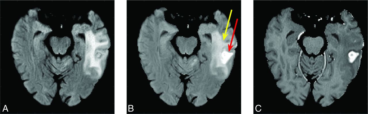
  • FIG 2.
    • Download figure
    • Open in new tab
    • Download powerpoint
    FIG 2.

    Unenhanced T2 FLAIR (A), T2FLAIRc (B), and T1c (C) are shown for a treated brain metastasis in the left temporal lobe (a case of radiation necrosis). T2FLAIRc demonstrates hybrid contrast between T2 FLAIR and T1c with edema (yellow arrow) and contrast enhancement (red arrow).
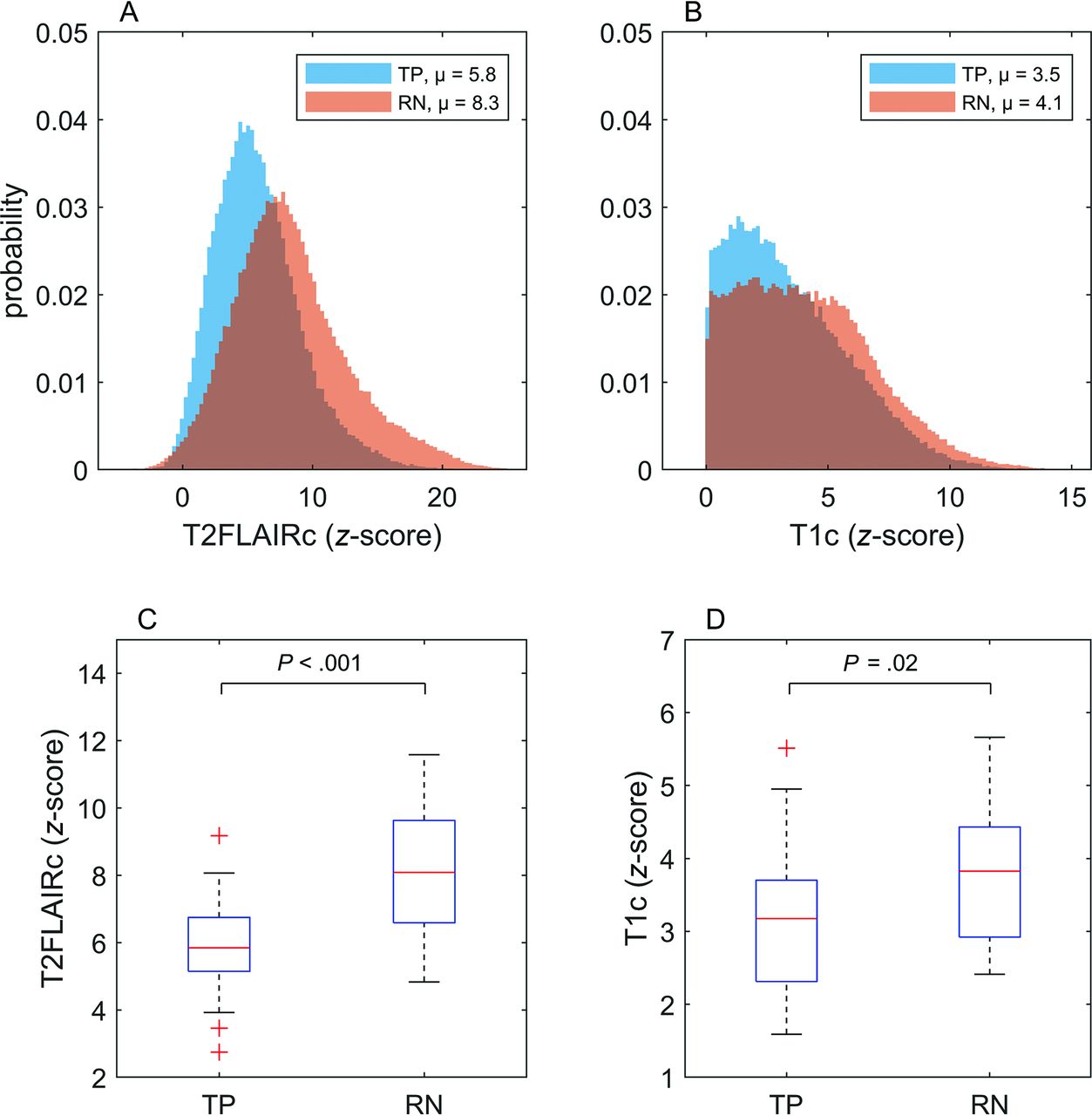
  • FIG 3.
    • Download figure
    • Open in new tab
    • Download powerpoint
    FIG 3.

    Histograms and boxplots illustrating the complete set of all enhancing voxels across all patients with TP and RN for T2FLAIRc and T1c. Mean signal intensities for each group are indicated in the figure legends for the histograms. The boxplots depict the mean and IQR. P values for unpaired t tests are shown and considered significant at P < .05.
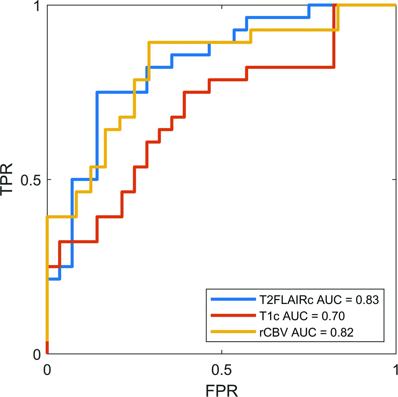
  • FIG 4.
    • Download figure
    • Open in new tab
    • Download powerpoint
    FIG 4.

    ROC curves for T2FLAIRc, T1c, and rCBV from DSC perfusion. The AUC for T2FLAIRc was significantly higher compared with T1c (P = .04) but not for rCBV (P = .9). FPR indicates false-positive rate; TPR, true-positive rate.
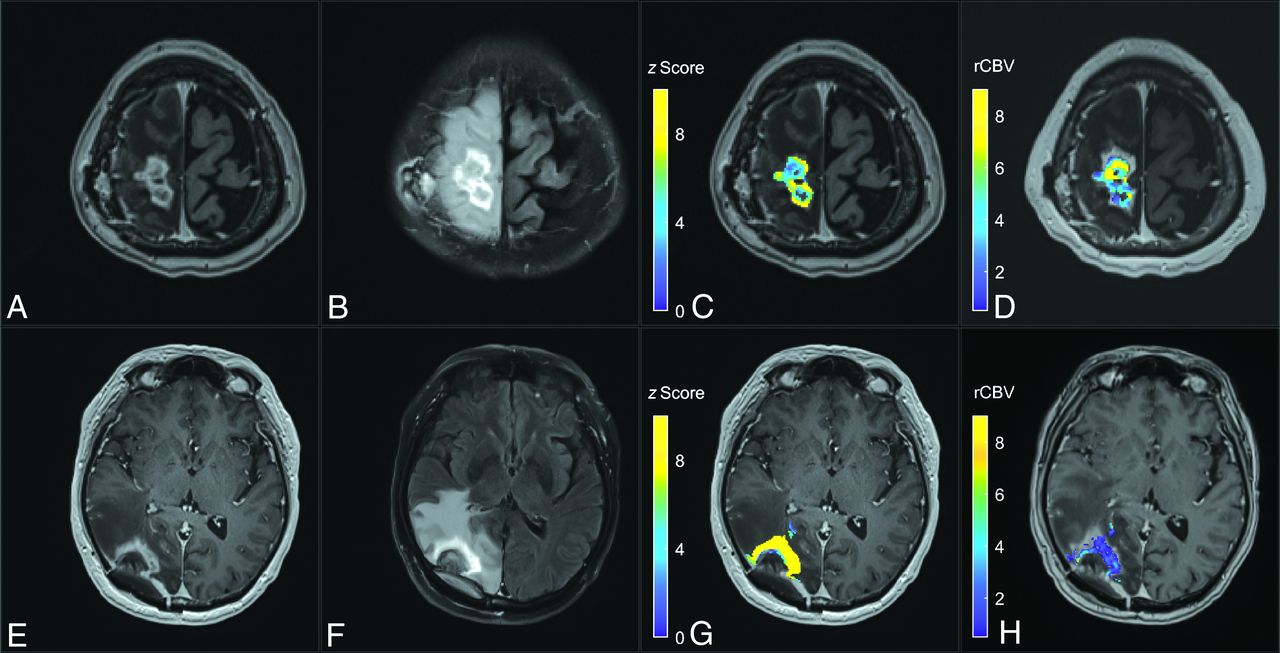
  • FIG 5.
    • Download figure
    • Open in new tab
    • Download powerpoint
    FIG 5.

    Comparison of a T2FLAIRc z score parameter map with the rCBV map for cases of TP (upper row) and RN (lower row). A and E, Axial T1c MPRAGE. B and F, Corresponding T2FLAIRc. C and G, T2FLAIRc z score for enhancing tumor voxels overlayed on T1c images shows lower values (mean, 5.8 [SD 2.0]) for TP and higher values (mean, 12.6 [SD 5.7]) for RN. D and H, The rCBV map from follow-up dynamic susceptibility perfusion MRI shows higher values for TP (mean, 4.8 [SD 2.3]) and lower values for RN (mean, 1.3 [SD 1.2]).

Tables

  • Figures
  • Patient and tumor characteristicsa

    CharacteristicTotal (n = 56)Tumor Progression (n = 28)Radiation Necrosis (n = 28)P Value
    Age (yr)61.9 ± 12.760.2 ± 12.563.6 ± 12.8.32
    Sex1.00
     Male17 (30.4%)8 (28.5%)9 (32%)
     Female39 (69.6%)20 (71.4%)19 (68%)
    Primary cancer type.50
     NSCLC25 (44.6%)14 (50%)11 (39.2%)
     Breast18 (32.1%)9 (32.1%)9 (32.1%)
     Melanoma4 (7.1%)1 (3.6%)3 (10.7%)
     RCC4 (7.1%)1 (3.6%)3 (10.7%)
     Other5 (8.9%)3 (10.7%)2 (7.1%)
    Total dose (Gy)25 (IQR,20–30)23.3 (IQR,19.5–27.5)27.3 (IQR,20–30).25
    No. of fractions3 (IQR, 1–5)3 (IQR, 1–5)5 (IQR, 1–5).72
    Time from SRS to DSC (days) 358 (IQR, 262− 86)394 (IQR, 347−586)296 (IQR, 222−588).06
    Time from index MR to DSC (days)73 [SD, 34]68 [SD, 32]78 [SD, 35].30
    Systemic therapy25 (44.6%)12 (42.9%)13 (46.4%)1.00
    Lesion outcome.76
     Pathology14 (25%)8 (29%)6 (21%)
     Follow-up42 (75%)20 (71%)22 (79%)
    Scanner field strength.42
     1.5T28 (50%)16 (57%)12 (43%)
     3T28 (50%)12 (43%)16 (57%)
    • Note:—NSCLC indicates non-small cell lung cancer; RCC, renal cell carcinoma.

    • a Categoric variables are presented as a proportion or percentage of patients. Continuous variables are presented as a mean (SD) for normally distributed variables and median (IQR) for non-normally distributed variables. Categoric variables were compared using the χ2 or Fisher exact test, when appropriate. Continuous variables were compared using the Wilcoxon rank-sum test for nonparametric group comparisons and the t test for parametric group comparisons. Results were considered significant with P < .05.

PreviousNext
Back to top

In this issue

American Journal of Neuroradiology: 46 (1)
American Journal of Neuroradiology
Vol. 46, Issue 1
1 Jan 2025
  • Table of Contents
  • Index by author
  • Complete Issue (PDF)
Advertisement
Print
Download PDF
Email Article

Thank you for your interest in spreading the word on American Journal of Neuroradiology.

NOTE: We only request your email address so that the person you are recommending the page to knows that you wanted them to see it, and that it is not junk mail. We do not capture any email address.

Enter multiple addresses on separate lines or separate them with commas.
Gadolinium-Enhanced T2 FLAIR Is an Imaging Biomarker of Radiation Necrosis and Tumor Progression in Patients with Brain Metastases
(Your Name) has sent you a message from American Journal of Neuroradiology
(Your Name) thought you would like to see the American Journal of Neuroradiology web site.
CAPTCHA
This question is for testing whether or not you are a human visitor and to prevent automated spam submissions.
Cite this article
Chris Heyn, Jonathan Bishop, Alan R. Moody, Tony Kang, Erin Wong, Peter Howard, Pejman Maralani, Sean Symons, Bradley J. MacIntosh, Julia Keith, Mary Jane Lim-Fat, James Perry, Sten Myrehaug, Jay Detsky, Chia-Lin Tseng, Hanbo Chen, Arjun Sahgal, Hany Soliman
Gadolinium-Enhanced T2 FLAIR Is an Imaging Biomarker of Radiation Necrosis and Tumor Progression in Patients with Brain Metastases
American Journal of Neuroradiology Jan 2025, 46 (1) 129-135; DOI: 10.3174/ajnr.A8431

Citation Manager Formats

  • BibTeX
  • Bookends
  • EasyBib
  • EndNote (tagged)
  • EndNote 8 (xml)
  • Medlars
  • Mendeley
  • Papers
  • RefWorks Tagged
  • Ref Manager
  • RIS
  • Zotero
0 Responses
Respond to this article
Share
Bookmark this article
Gad FLAIR T2 for Radiation Necrosis Brain Mets
Chris Heyn, Jonathan Bishop, Alan R. Moody, Tony Kang, Erin Wong, Peter Howard, Pejman Maralani, Sean Symons, Bradley J. MacIntosh, Julia Keith, Mary Jane Lim-Fat, James Perry, Sten Myrehaug, Jay Detsky, Chia-Lin Tseng, Hanbo Chen, Arjun Sahgal, Hany Soliman
American Journal of Neuroradiology Jan 2025, 46 (1) 129-135; DOI: 10.3174/ajnr.A8431
del.icio.us logo Twitter logo Facebook logo Mendeley logo
  • Tweet Widget
  • Facebook Like
  • Google Plus One
Purchase

Jump to section

  • Article
    • Graphical Abstract
    • Abstract
    • ABBREVIATIONS:
    • MATERIALS AND METHODS
    • RESULTS
    • DISCUSSION
    • CONCLUSIONS
    • Footnotes
    • References
  • Figures & Data
  • Supplemental
  • Info & Metrics
  • Responses
  • References
  • PDF

Related Articles

  • PubMed
  • Google Scholar

Cited By...

  • No citing articles found.
  • Crossref
  • Google Scholar

This article has not yet been cited by articles in journals that are participating in Crossref Cited-by Linking.

More in this TOC Section

  • Brain Tumor Subtyping Image-Based search via MRI
  • Neuroimaging of Erdheim-Chester Disease
  • CE MRI for Brain Metastasis Detection
Show more Brain Tumor Imaging

Similar Articles

Advertisement

Indexed Content

  • Current Issue
  • Accepted Manuscripts
  • Article Preview
  • Past Issues
  • Editorials
  • Editor's Choice
  • Fellows' Journal Club
  • Letters to the Editor
  • Video Articles

Cases

  • Case Collection
  • Archive - Case of the Week
  • Archive - Case of the Month
  • Archive - Classic Case

More from AJNR

  • Trainee Corner
  • Imaging Protocols
  • MRI Safety Corner
  • Book Reviews

Multimedia

  • AJNR Podcasts
  • AJNR Scantastics

Resources

  • Turnaround Time
  • Submit a Manuscript
  • Submit a Video Article
  • Submit an eLetter to the Editor/Response
  • Manuscript Submission Guidelines
  • Statistical Tips
  • Fast Publishing of Accepted Manuscripts
  • Graphical Abstract Preparation
  • Imaging Protocol Submission
  • Evidence-Based Medicine Level Guide
  • Publishing Checklists
  • Author Policies
  • Become a Reviewer/Academy of Reviewers
  • News and Updates

About Us

  • About AJNR
  • Editorial Board
  • Editorial Board Alumni
  • Alerts
  • Permissions
  • Not an AJNR Subscriber? Join Now
  • Advertise with Us
  • Librarian Resources
  • Feedback
  • Terms and Conditions
  • AJNR Editorial Board Alumni

American Society of Neuroradiology

  • Not an ASNR Member? Join Now

© 2025 by the American Society of Neuroradiology All rights, including for text and data mining, AI training, and similar technologies, are reserved.
Print ISSN: 0195-6108 Online ISSN: 1936-959X

Powered by HighWire