Advertisement

Main menu

  • Home
  • Content
    • Current Issue
    • Accepted Manuscripts
    • Article Preview
    • Past Issue Archive
    • Video Articles
    • AJNR Case Collection
    • Case of the Week Archive
    • Case of the Month Archive
    • Classic Case Archive
  • Special Collections
    • AJNR Awards
    • Low-Field MRI
    • Alzheimer Disease
    • ASNR Foundation Special Collection
    • Photon-Counting CT
    • View All
  • Multimedia
    • AJNR Podcasts
    • AJNR SCANtastic
    • Trainee Corner
    • MRI Safety Corner
    • Imaging Protocols
  • For Authors
    • Submit a Manuscript
    • Submit a Video Article
    • Submit an eLetter to the Editor/Response
    • Manuscript Submission Guidelines
    • Statistical Tips
    • Fast Publishing of Accepted Manuscripts
    • Graphical Abstract Preparation
    • Imaging Protocol Submission
    • Author Policies
  • About Us
    • About AJNR
    • Editorial Board
    • Editorial Board Alumni
  • More
    • Become a Reviewer/Academy of Reviewers
    • Subscribers
    • Permissions
    • Alerts
    • Feedback
    • Advertisers
    • ASNR Home

User menu

  • Alerts
  • Log in

Search

  • Advanced search
American Journal of Neuroradiology

American Journal of Neuroradiology

ASHNR American Society of Functional Neuroradiology ASHNR American Society of Pediatric Neuroradiology ASSR
  • Alerts
  • Log in

Advanced Search

  • Home
  • Content
    • Current Issue
    • Accepted Manuscripts
    • Article Preview
    • Past Issue Archive
    • Video Articles
    • AJNR Case Collection
    • Case of the Week Archive
    • Case of the Month Archive
    • Classic Case Archive
  • Special Collections
    • AJNR Awards
    • Low-Field MRI
    • Alzheimer Disease
    • ASNR Foundation Special Collection
    • Photon-Counting CT
    • View All
  • Multimedia
    • AJNR Podcasts
    • AJNR SCANtastic
    • Trainee Corner
    • MRI Safety Corner
    • Imaging Protocols
  • For Authors
    • Submit a Manuscript
    • Submit a Video Article
    • Submit an eLetter to the Editor/Response
    • Manuscript Submission Guidelines
    • Statistical Tips
    • Fast Publishing of Accepted Manuscripts
    • Graphical Abstract Preparation
    • Imaging Protocol Submission
    • Author Policies
  • About Us
    • About AJNR
    • Editorial Board
    • Editorial Board Alumni
  • More
    • Become a Reviewer/Academy of Reviewers
    • Subscribers
    • Permissions
    • Alerts
    • Feedback
    • Advertisers
    • ASNR Home
  • Follow AJNR on Twitter
  • Visit AJNR on Facebook
  • Follow AJNR on Instagram
  • Join AJNR on LinkedIn
  • RSS Feeds

AJNR Awards, New Junior Editors, and more. Read the latest AJNR updates

Research ArticleNeurointervention

Impact of Patient Head Posture on Lens Radiation Exposure During Cerebral Angiography

Su Jeong Yang, Jong-Tae Yoon, Jae-Chan Ryu, Byung Jun Kim, Mi Hyeon Kim, Boseong Kwon, Deok Hee Lee and Yunsun Song
American Journal of Neuroradiology January 2025, 46 (1) 102-106; DOI: https://doi.org/10.3174/ajnr.A8447
Su Jeong Yang
aFrom the Department of Radiology (S.J.Y.), Ajou University Hospital, Ajou University School of Medicine, Suwon, Republic of Korea
  • Find this author on Google Scholar
  • Find this author on PubMed
  • Search for this author on this site
  • ORCID record for Su Jeong Yang
Jong-Tae Yoon
bBiomedical Engineering Research Center (J.-T.Y., M.H.K.), Asan Institute for Life Sciences, Asan Medical Center, Seoul, Korea
  • Find this author on Google Scholar
  • Find this author on PubMed
  • Search for this author on this site
Jae-Chan Ryu
cDepartment of Neurology (J.-C.R), Korea University Ansan Hospital, Korea University College of Medicine, Ansan, Korea
  • Find this author on Google Scholar
  • Find this author on PubMed
  • Search for this author on this site
  • ORCID record for Jae-Chan Ryu
Byung Jun Kim
dAdvanced Therapies (B.J.K.), Siemens Healthineers Ltd, Seoul, Korea
  • Find this author on Google Scholar
  • Find this author on PubMed
  • Search for this author on this site
Mi Hyeon Kim
bBiomedical Engineering Research Center (J.-T.Y., M.H.K.), Asan Institute for Life Sciences, Asan Medical Center, Seoul, Korea
  • Find this author on Google Scholar
  • Find this author on PubMed
  • Search for this author on this site
  • ORCID record for Mi Hyeon Kim
Boseong Kwon
eDepartment of Radiology (B.K., D.H.L., Y.S.), Research Institute of Radiology, Asan Medical Center, University of Ulsan College of Medicine, Seoul, Korea
  • Find this author on Google Scholar
  • Find this author on PubMed
  • Search for this author on this site
  • ORCID record for Boseong Kwon
Deok Hee Lee
eDepartment of Radiology (B.K., D.H.L., Y.S.), Research Institute of Radiology, Asan Medical Center, University of Ulsan College of Medicine, Seoul, Korea
  • Find this author on Google Scholar
  • Find this author on PubMed
  • Search for this author on this site
  • ORCID record for Deok Hee Lee
Yunsun Song
eDepartment of Radiology (B.K., D.H.L., Y.S.), Research Institute of Radiology, Asan Medical Center, University of Ulsan College of Medicine, Seoul, Korea
  • Find this author on Google Scholar
  • Find this author on PubMed
  • Search for this author on this site
  • ORCID record for Yunsun Song
  • Article
  • Figures & Data
  • Supplemental
  • Info & Metrics
  • Responses
  • References
  • PDF
Loading

Article Figures & Data

Figures

  • Tables
  • Figure
    • Download figure
    • Open in new tab
    • Download powerpoint
  • FIG 1.
    • Download figure
    • Open in new tab
    • Download powerpoint
    FIG 1.

    Illustration of FOV change according to the head flexion angle: (A) neutral position, (B) flexion position, and (C) extension position. The dotted line represents the line from the petrous apex to the lens, while the curved arrow indicates the flexion angle, defined as the angle between the line from the petrous apex to the lens and the horizontal line of the FOV, presented as a white box. In the neutral position (A), the lens was located at the border of the FOV. In the flexion position (B), the lens shifted into the FOV. However, the lens was outside of the FOV and significantly further from the FOV in the extended position (C).

  • FIG 2.
    • Download figure
    • Open in new tab
    • Download powerpoint
    FIG 2.

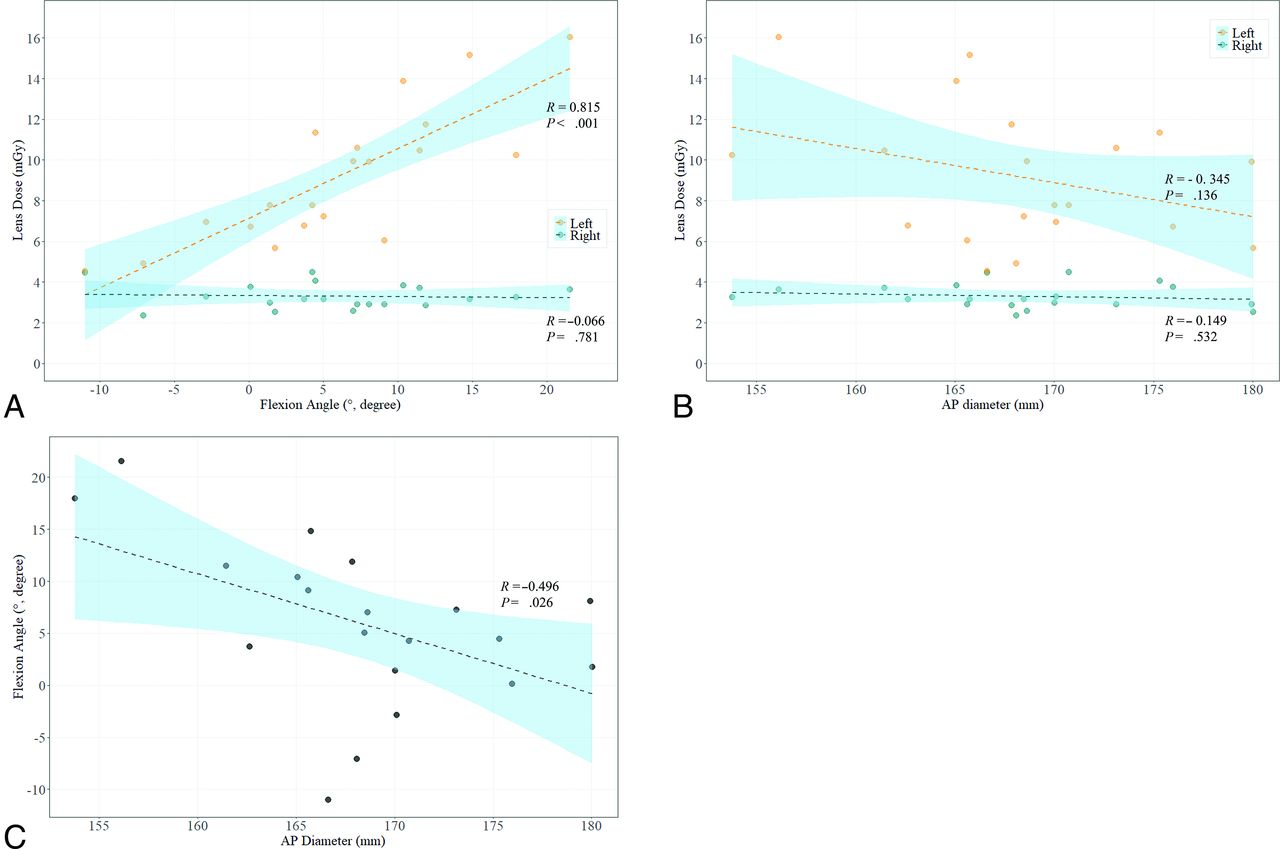
    Scatterplots illustrating the relationships between (A) head flexion angle and lens dose, (B) AP diameter of the head and lens dose, and (C) head flexion angle and AP diameter of the head.

  • FIG 3.
    • Download figure
    • Open in new tab
    • Download powerpoint
    FIG 3.

    Illustration of head position changes relative to the AP diameter of the head and the corresponding body shape: (A) a flexion position for an individual with a small head and body size, (B) a neutral position for an individual with an average-sized head and body, and (C) an extension position for an individual with a large head and body.

Tables

  • Figures
  • Baseline characteristics of the patients (n = 20)

    CharacteristicsValues
    Age, years62.3 ± 9.9
    Female sex12 (60.0)
    AP diameter of the head, mm168.3 ± 6.8
    Flexion angle of the head, degrees6.0 ± 7.8
    Radiation dose to the lens, mGya,b
    Right eye3.30 ± 0.60
    Left eye9.18 ± 3.31
    Kerma area product, Gy·cm²21.6 ± 6.0
    AK, mGy134.0 ± 32.1
    • ↵a The radiation dose was measured during DSA and fluoroscopy.

    • ↵b P value was < .001. Paired t test was used to compare the lens doses between the eyes.

    • Data are presented as mean ± standard deviation or number (%).

PreviousNext
Back to top

In this issue

American Journal of Neuroradiology: 46 (1)
American Journal of Neuroradiology
Vol. 46, Issue 1
1 Jan 2025
  • Table of Contents
  • Index by author
  • Complete Issue (PDF)
Advertisement
Print
Download PDF
Email Article

Thank you for your interest in spreading the word on American Journal of Neuroradiology.

NOTE: We only request your email address so that the person you are recommending the page to knows that you wanted them to see it, and that it is not junk mail. We do not capture any email address.

Enter multiple addresses on separate lines or separate them with commas.
Impact of Patient Head Posture on Lens Radiation Exposure During Cerebral Angiography
(Your Name) has sent you a message from American Journal of Neuroradiology
(Your Name) thought you would like to see the American Journal of Neuroradiology web site.
CAPTCHA
This question is for testing whether or not you are a human visitor and to prevent automated spam submissions.
Cite this article
Su Jeong Yang, Jong-Tae Yoon, Jae-Chan Ryu, Byung Jun Kim, Mi Hyeon Kim, Boseong Kwon, Deok Hee Lee, Yunsun Song
Impact of Patient Head Posture on Lens Radiation Exposure During Cerebral Angiography
American Journal of Neuroradiology Jan 2025, 46 (1) 102-106; DOI: 10.3174/ajnr.A8447

Citation Manager Formats

  • BibTeX
  • Bookends
  • EasyBib
  • EndNote (tagged)
  • EndNote 8 (xml)
  • Medlars
  • Mendeley
  • Papers
  • RefWorks Tagged
  • Ref Manager
  • RIS
  • Zotero
0 Responses
Respond to this article
Share
Bookmark this article
Impact of Head Posture on Angiography Exposure
Su Jeong Yang, Jong-Tae Yoon, Jae-Chan Ryu, Byung Jun Kim, Mi Hyeon Kim, Boseong Kwon, Deok Hee Lee, Yunsun Song
American Journal of Neuroradiology Jan 2025, 46 (1) 102-106; DOI: 10.3174/ajnr.A8447
del.icio.us logo Twitter logo Facebook logo Mendeley logo
  • Tweet Widget
  • Facebook Like
  • Google Plus One
Purchase

Jump to section

  • Article
    • Graphical Abstract
    • Abstract
    • ABBREVIATIONS:
    • MATERIALS AND METHODS
    • RESULTS
    • DISCUSSION
    • CONCLUSIONS
    • Footnotes
    • References
  • Figures & Data
  • Supplemental
  • Info & Metrics
  • Responses
  • References
  • PDF

Related Articles

  • PubMed
  • Google Scholar

Cited By...

  • No citing articles found.
  • Crossref
  • Google Scholar

This article has not yet been cited by articles in journals that are participating in Crossref Cited-by Linking.

More in this TOC Section

  • Rescue Reentry in Carotid Near-Occlusion
  • Contour Neurovascular System: Five Year Follow Up
  • Effect of SARS-CoV2 on Endovascular Thrombectomy
Show more Neurointervention

Similar Articles

Advertisement

Indexed Content

  • Current Issue
  • Accepted Manuscripts
  • Article Preview
  • Past Issues
  • Editorials
  • Editor's Choice
  • Fellows' Journal Club
  • Letters to the Editor
  • Video Articles

Cases

  • Case Collection
  • Archive - Case of the Week
  • Archive - Case of the Month
  • Archive - Classic Case

More from AJNR

  • Trainee Corner
  • Imaging Protocols
  • MRI Safety Corner
  • Book Reviews

Multimedia

  • AJNR Podcasts
  • AJNR Scantastics

Resources

  • Turnaround Time
  • Submit a Manuscript
  • Submit a Video Article
  • Submit an eLetter to the Editor/Response
  • Manuscript Submission Guidelines
  • Statistical Tips
  • Fast Publishing of Accepted Manuscripts
  • Graphical Abstract Preparation
  • Imaging Protocol Submission
  • Evidence-Based Medicine Level Guide
  • Publishing Checklists
  • Author Policies
  • Become a Reviewer/Academy of Reviewers
  • News and Updates

About Us

  • About AJNR
  • Editorial Board
  • Editorial Board Alumni
  • Alerts
  • Permissions
  • Not an AJNR Subscriber? Join Now
  • Advertise with Us
  • Librarian Resources
  • Feedback
  • Terms and Conditions
  • AJNR Editorial Board Alumni

American Society of Neuroradiology

  • Not an ASNR Member? Join Now

© 2025 by the American Society of Neuroradiology All rights, including for text and data mining, AI training, and similar technologies, are reserved.
Print ISSN: 0195-6108 Online ISSN: 1936-959X

Powered by HighWire