Advertisement

Main menu

  • Home
  • Content
    • Current Issue
    • Accepted Manuscripts
    • Article Preview
    • Past Issue Archive
    • Video Articles
    • AJNR Case Collection
    • Case of the Week Archive
    • Case of the Month Archive
    • Classic Case Archive
  • Special Collections
    • AJNR Awards
    • Low-Field MRI
    • Alzheimer Disease
    • ASNR Foundation Special Collection
    • Photon-Counting CT
    • View All
  • Multimedia
    • AJNR Podcasts
    • AJNR SCANtastic
    • Trainee Corner
    • MRI Safety Corner
    • Imaging Protocols
  • For Authors
    • Submit a Manuscript
    • Submit a Video Article
    • Submit an eLetter to the Editor/Response
    • Manuscript Submission Guidelines
    • Statistical Tips
    • Fast Publishing of Accepted Manuscripts
    • Graphical Abstract Preparation
    • Imaging Protocol Submission
    • Author Policies
  • About Us
    • About AJNR
    • Editorial Board
    • Editorial Board Alumni
  • More
    • Become a Reviewer/Academy of Reviewers
    • Subscribers
    • Permissions
    • Alerts
    • Feedback
    • Advertisers
    • ASNR Home

User menu

  • Alerts
  • Log in

Search

  • Advanced search
American Journal of Neuroradiology

American Journal of Neuroradiology

ASHNR American Society of Functional Neuroradiology ASHNR American Society of Pediatric Neuroradiology ASSR
  • Alerts
  • Log in

Advanced Search

  • Home
  • Content
    • Current Issue
    • Accepted Manuscripts
    • Article Preview
    • Past Issue Archive
    • Video Articles
    • AJNR Case Collection
    • Case of the Week Archive
    • Case of the Month Archive
    • Classic Case Archive
  • Special Collections
    • AJNR Awards
    • Low-Field MRI
    • Alzheimer Disease
    • ASNR Foundation Special Collection
    • Photon-Counting CT
    • View All
  • Multimedia
    • AJNR Podcasts
    • AJNR SCANtastic
    • Trainee Corner
    • MRI Safety Corner
    • Imaging Protocols
  • For Authors
    • Submit a Manuscript
    • Submit a Video Article
    • Submit an eLetter to the Editor/Response
    • Manuscript Submission Guidelines
    • Statistical Tips
    • Fast Publishing of Accepted Manuscripts
    • Graphical Abstract Preparation
    • Imaging Protocol Submission
    • Author Policies
  • About Us
    • About AJNR
    • Editorial Board
    • Editorial Board Alumni
  • More
    • Become a Reviewer/Academy of Reviewers
    • Subscribers
    • Permissions
    • Alerts
    • Feedback
    • Advertisers
    • ASNR Home
  • Follow AJNR on Twitter
  • Visit AJNR on Facebook
  • Follow AJNR on Instagram
  • Join AJNR on LinkedIn
  • RSS Feeds

AJNR Awards, New Junior Editors, and more. Read the latest AJNR updates

Research ArticleBrain Tumor Imaging

Interrater Agreement of BT-RADS for Evaluation of Follow-up MRI in Patients with Treated Primary Brain Tumor

Michael Essien, Maxwell E. Cooper, Ashwani Gore, Taejin L. Min, Benjamin B. Risk, Gelareh Sadigh, Ranliang Hu, Michael J. Hoch and Brent D. Weinberg
American Journal of Neuroradiology September 2024, 45 (9) 1308-1315; DOI: https://doi.org/10.3174/ajnr.A8322
Michael Essien
aFrom the Department of Radiology and Imaging Sciences (M.E., M.E.C., A.G., T.L.M., R.H., B.D.W.),
  • Find this author on Google Scholar
  • Find this author on PubMed
  • Search for this author on this site
  • ORCID record for Michael Essien
Maxwell E. Cooper
aFrom the Department of Radiology and Imaging Sciences (M.E., M.E.C., A.G., T.L.M., R.H., B.D.W.),
  • Find this author on Google Scholar
  • Find this author on PubMed
  • Search for this author on this site
  • ORCID record for Maxwell E. Cooper
Ashwani Gore
aFrom the Department of Radiology and Imaging Sciences (M.E., M.E.C., A.G., T.L.M., R.H., B.D.W.),
  • Find this author on Google Scholar
  • Find this author on PubMed
  • Search for this author on this site
  • ORCID record for Ashwani Gore
Taejin L. Min
aFrom the Department of Radiology and Imaging Sciences (M.E., M.E.C., A.G., T.L.M., R.H., B.D.W.),
  • Find this author on Google Scholar
  • Find this author on PubMed
  • Search for this author on this site
  • ORCID record for Taejin L. Min
Benjamin B. Risk
bRollins School of Public Health (B.B.R.), Emory University, Atlanta, Georgia
  • Find this author on Google Scholar
  • Find this author on PubMed
  • Search for this author on this site
  • ORCID record for Benjamin B. Risk
Gelareh Sadigh
bRollins School of Public Health (B.B.R.), Emory University, Atlanta, Georgia
  • Find this author on Google Scholar
  • Find this author on PubMed
  • Search for this author on this site
  • ORCID record for Gelareh Sadigh
Ranliang Hu
aFrom the Department of Radiology and Imaging Sciences (M.E., M.E.C., A.G., T.L.M., R.H., B.D.W.),
  • Find this author on Google Scholar
  • Find this author on PubMed
  • Search for this author on this site
  • ORCID record for Ranliang Hu
Michael J. Hoch
cDepartment of Radiological Sciences (G.S.), University of California Irvine, Orange, California
  • Find this author on Google Scholar
  • Find this author on PubMed
  • Search for this author on this site
  • ORCID record for Michael J. Hoch
Brent D. Weinberg
aFrom the Department of Radiology and Imaging Sciences (M.E., M.E.C., A.G., T.L.M., R.H., B.D.W.),
  • Find this author on Google Scholar
  • Find this author on PubMed
  • Search for this author on this site
  • ORCID record for Brent D. Weinberg
  • Article
  • Figures & Data
  • Supplemental
  • Info & Metrics
  • Responses
  • References
  • PDF
Loading

Article Figures & Data

Figures

  • Tables
  • FIG 1.
    • Download figure
    • Open in new tab
    • Download powerpoint
    FIG 1.

    Bar graph showing relative frequency of BT-RADS scores (from original reports) among selected MR imaging examinations (n = 103).
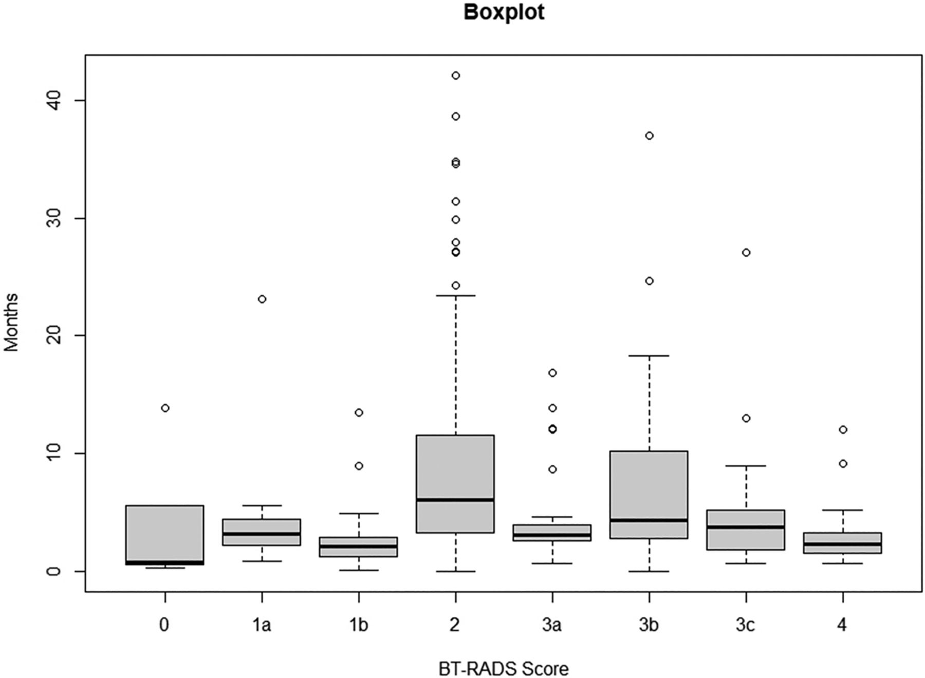
  • FIG 2.
    • Download figure
    • Open in new tab
    • Download powerpoint
    FIG 2.

    Boxplot graph showing time (in months) from the oldest MRI to the prior MRI used by readers to assign a BT-RADS score. The maximum and minimum values are represented at either end of the whiskers. The box represents the interquartile range (25th percentile to the 75th percentile), with the median value represented by the thick horizontal black line within the box. Outliers are shown away from the whiskers and box by the symbol (○).

  • FIG 3.
    • Download figure
    • Open in new tab
    • Download powerpoint
    FIG 3.

    Violin graph showing boxplot inside an attenuation plot. The width of the attenuation plot at any region corresponds to the frequency of the data points at that region. For the boxplot, the maximum and minimum values are represented at either end of the whiskers. The box represents the interquartile range (25th percentile to the 75th percentile), with the median value represented by a thick horizontal black line either within the box or at either end of the box. The mean value is represented by the black triangle (▴). Outliers are shown away from the whiskers and box by small black dots (●). On a scale of 1–5 (with 1 = very little, and 5 = great deal), all 6 readers rated how much perfusion contributed to their interpretation of scan and assignment of BT-RADS score.
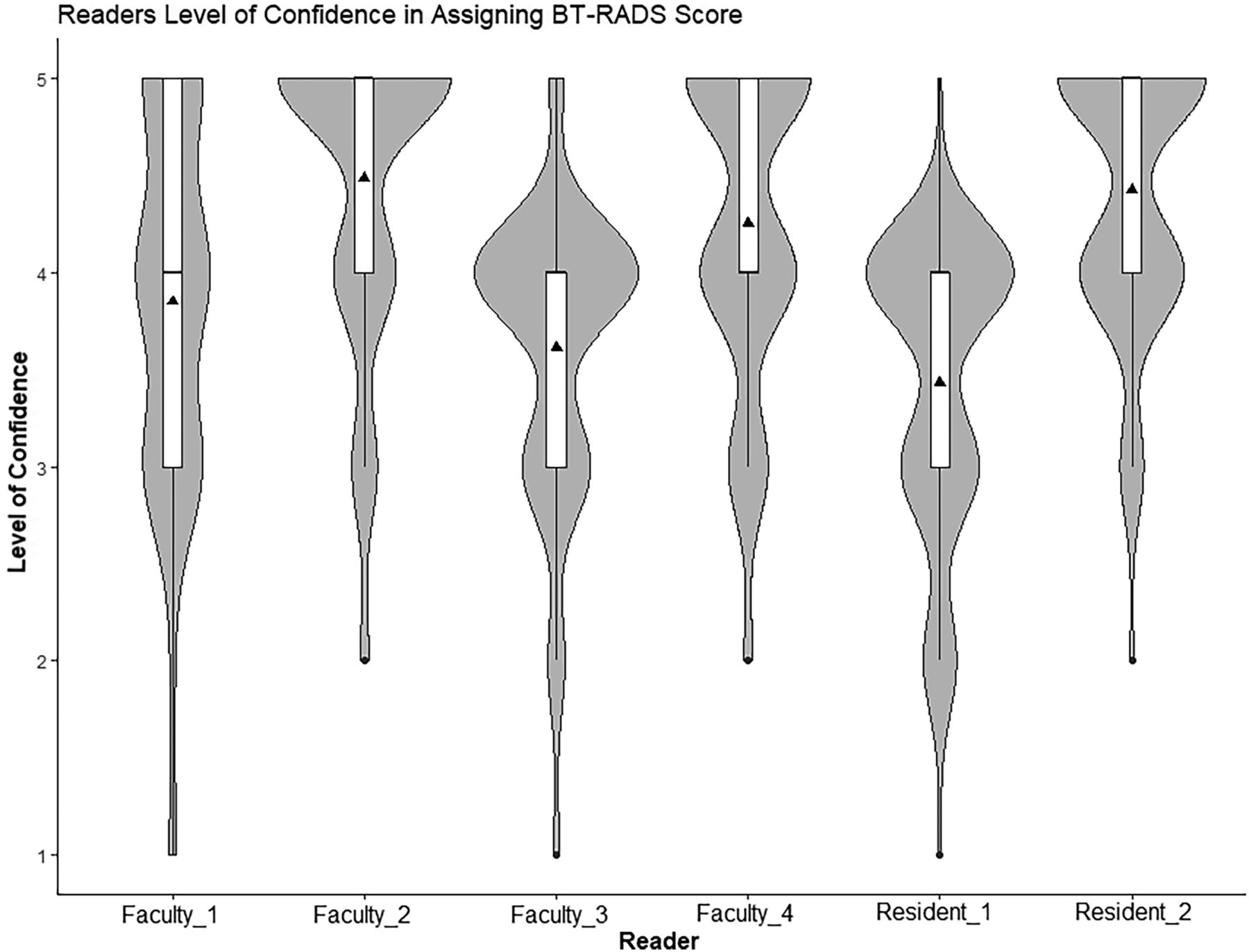
  • FIG 4.
    • Download figure
    • Open in new tab
    • Download powerpoint
    FIG 4.

    Violin graph showing boxplot inside an attenuation plot. The width of the attenuation plot at any region corresponds to the frequency of the data points at that region. For the boxplot, the maximum and minimum values are represented at either end of the whiskers. The box represents the interquartile range (25th percentile to the 75th percentile), with the median value represented by a thick horizontal black line either within the box or at either end of the box. The mean value is represented by the black triangle (▴). Outliers are shown away from the whiskers and box by small black dots (●). On a scale of 1–5 (with 1 = not sure at all, and 5 = absolutely sure), all 6 readers rated their level of confidence for the BT-RADS score they assigned to each case. Neuroradiologists (Faculty_1, Faculty _2, Faculty _3, and Faculty _4); Radiology residents (Resident_1 and Resident_2).

  • FIG 5.
    • Download figure
    • Open in new tab
    • Download powerpoint
    FIG 5.

    Imaging from a patient with agreement among all readers. A 48-year-old man with IDH–wild-type glioblastoma. FLAIR (A) and T1 postcontrast (B) imaging 18 months after surgery showing abnormal FLAIR and enhancing treated tumor in the left temporal lobe. FLAIR (C) and T1 postcontrast (D) 2 months later showing marked increase in the size of abnormal masslike FLAIR and enhancement (white arrows) with >25% increase in cross-sectional area. All readers gave the study a score of BT-RADS 4.

  • FIG 6.
    • Download figure
    • Open in new tab
    • Download powerpoint
    FIG 6.

    Imaging from a patient with disagreement among readers. A 45-year-old woman with IDH–wild-type glioblastoma. FLAIR (A) and T1 postcontrast (B) imaging 3 months after surgery and 1 month after completing radiation showing multifocal abnormal FLAIR in the bilateral frontal lobes with minimal enhancement in the left frontal lobe (white arrow). FLAIR (C) and T1 postcontrast (D) 2 months later (3 months after completing radiation) showing marked increase in left frontal edema (white arrows) and enhancement (white arrow). One-half of the readers (2 neuroradiologists and 1 resident) gave the study a score of BT-RADS 3a (pseudoprogression) and one-half of the readers gave the study a score of BT-RADS 4 (progression). The post hoc reference score was BT-RADS 4.

Tables

  • Figures
    • View popup
    Table 1:

    Patient and tumor characteristics

    Characteristic
    Number of patients98
    Number of men52 (53%)
    Number of MRI examinations evaluated103
    Median age [IQR] (yr)53 [41–66]
    Number of patients with previous surgery97 (99%)
    Number of patients with completed radiation therapy89 (91%)
    Number of patients taking steroids18 (18%)
    Number of patients taking bevacizumab20 (20%)
    Tumor classification
     Astrocytoma79 (77%)
     Grade 214 (14%)
     Grade 320 (19%)
     Grade 444 (43%)
     Anaplastic pilocytic astrocytoma1 (1%)
     Oligodendroglioma24 (23%)
      Grade 216 (15%)
      Grade 38 (8%)
    Tumor type by IDH-mutational status
    Astrocytoma
     IDH–wild-type40 (51%)
     IDH-mutant36 (45%)
     Unknown3 (4%)
    Oligodendroglioma
     IDH-mutant19 (79%)
     Unknown5 (21%)
    • View popup
    Table 2:

    Percent agreement between neuroradiologist blinded score and consensus score; blinded score and post hoc reference score (n = 46)

    ReaderAgreement (%) with Consensus ScoreAgreement (%) with Post Hoc Reference Score
    Faculty_16552
    Faculty_24646
    Faculty_35241
    Faculty_46348
    • Note:—Faculty_1, Faculty_3, and Faculty_4 had 3 years of practice experience. Faculty_2 had 1 year of practice experience.

    • View popup
    Table 3:

    Under- and overestimation of disagreement of neuroradiologist blinded score compared with post hoc reference score

    ReaderUnderestimation/Overestimation
    Faculty_114/8
    Faculty_212/13
    Faculty_320/7
    Faculty_417/7
    • Note:—Faculty_1, Faculty_3, and Faculty_4 had 3 years of practice experience. Faculty_2 had 1 year of practice experience.

PreviousNext
Back to top

In this issue

American Journal of Neuroradiology: 45 (9)
American Journal of Neuroradiology
Vol. 45, Issue 9
1 Sep 2024
  • Table of Contents
  • Index by author
  • Complete Issue (PDF)
Advertisement
Print
Download PDF
Email Article

Thank you for your interest in spreading the word on American Journal of Neuroradiology.

NOTE: We only request your email address so that the person you are recommending the page to knows that you wanted them to see it, and that it is not junk mail. We do not capture any email address.

Enter multiple addresses on separate lines or separate them with commas.
Interrater Agreement of BT-RADS for Evaluation of Follow-up MRI in Patients with Treated Primary Brain Tumor
(Your Name) has sent you a message from American Journal of Neuroradiology
(Your Name) thought you would like to see the American Journal of Neuroradiology web site.
CAPTCHA
This question is for testing whether or not you are a human visitor and to prevent automated spam submissions.
Cite this article
Michael Essien, Maxwell E. Cooper, Ashwani Gore, Taejin L. Min, Benjamin B. Risk, Gelareh Sadigh, Ranliang Hu, Michael J. Hoch, Brent D. Weinberg
Interrater Agreement of BT-RADS for Evaluation of Follow-up MRI in Patients with Treated Primary Brain Tumor
American Journal of Neuroradiology Sep 2024, 45 (9) 1308-1315; DOI: 10.3174/ajnr.A8322

Citation Manager Formats

  • BibTeX
  • Bookends
  • EasyBib
  • EndNote (tagged)
  • EndNote 8 (xml)
  • Medlars
  • Mendeley
  • Papers
  • RefWorks Tagged
  • Ref Manager
  • RIS
  • Zotero
0 Responses
Respond to this article
Share
Bookmark this article
BTRADS Agreement in Follow-Up MRI for Brain Tumors
Michael Essien, Maxwell E. Cooper, Ashwani Gore, Taejin L. Min, Benjamin B. Risk, Gelareh Sadigh, Ranliang Hu, Michael J. Hoch, Brent D. Weinberg
American Journal of Neuroradiology Sep 2024, 45 (9) 1308-1315; DOI: 10.3174/ajnr.A8322
del.icio.us logo Twitter logo Facebook logo Mendeley logo
  • Tweet Widget
  • Facebook Like
  • Google Plus One
Purchase

Jump to section

  • Article
    • Abstract
    • ABBREVIATIONS:
    • MATERIALS AND METHODS
    • RESULTS
    • DISCUSSION
    • CONCLUSIONS
    • Footnotes
    • References
  • Figures & Data
  • Supplemental
  • Info & Metrics
  • Responses
  • References
  • PDF

Related Articles

  • PubMed
  • Google Scholar

Cited By...

  • No citing articles found.
  • Crossref
  • Google Scholar

This article has not yet been cited by articles in journals that are participating in Crossref Cited-by Linking.

More in this TOC Section

  • Brain Tumor Subtyping Image-Based search via MRI
  • Neuroimaging of Erdheim-Chester Disease
  • CE MRI for Brain Metastasis Detection
Show more Brain Tumor Imaging

Similar Articles

Advertisement

Indexed Content

  • Current Issue
  • Accepted Manuscripts
  • Article Preview
  • Past Issues
  • Editorials
  • Editor's Choice
  • Fellows' Journal Club
  • Letters to the Editor
  • Video Articles

Cases

  • Case Collection
  • Archive - Case of the Week
  • Archive - Case of the Month
  • Archive - Classic Case

More from AJNR

  • Trainee Corner
  • Imaging Protocols
  • MRI Safety Corner
  • Book Reviews

Multimedia

  • AJNR Podcasts
  • AJNR Scantastics

Resources

  • Turnaround Time
  • Submit a Manuscript
  • Submit a Video Article
  • Submit an eLetter to the Editor/Response
  • Manuscript Submission Guidelines
  • Statistical Tips
  • Fast Publishing of Accepted Manuscripts
  • Graphical Abstract Preparation
  • Imaging Protocol Submission
  • Evidence-Based Medicine Level Guide
  • Publishing Checklists
  • Author Policies
  • Become a Reviewer/Academy of Reviewers
  • News and Updates

About Us

  • About AJNR
  • Editorial Board
  • Editorial Board Alumni
  • Alerts
  • Permissions
  • Not an AJNR Subscriber? Join Now
  • Advertise with Us
  • Librarian Resources
  • Feedback
  • Terms and Conditions
  • AJNR Editorial Board Alumni

American Society of Neuroradiology

  • Not an ASNR Member? Join Now

© 2025 by the American Society of Neuroradiology All rights, including for text and data mining, AI training, and similar technologies, are reserved.
Print ISSN: 0195-6108 Online ISSN: 1936-959X

Powered by HighWire