Advertisement

Main menu

  • Home
  • Content
    • Current Issue
    • Accepted Manuscripts
    • Article Preview
    • Past Issue Archive
    • Video Articles
    • AJNR Case Collection
    • Case of the Week Archive
    • Case of the Month Archive
    • Classic Case Archive
  • Special Collections
    • AJNR Awards
    • Low-Field MRI
    • Alzheimer Disease
    • ASNR Foundation Special Collection
    • Photon-Counting CT
    • View All
  • Multimedia
    • AJNR Podcasts
    • AJNR SCANtastic
    • Trainee Corner
    • MRI Safety Corner
    • Imaging Protocols
  • For Authors
    • Submit a Manuscript
    • Submit a Video Article
    • Submit an eLetter to the Editor/Response
    • Manuscript Submission Guidelines
    • Statistical Tips
    • Fast Publishing of Accepted Manuscripts
    • Graphical Abstract Preparation
    • Imaging Protocol Submission
    • Author Policies
  • About Us
    • About AJNR
    • Editorial Board
    • Editorial Board Alumni
  • More
    • Become a Reviewer/Academy of Reviewers
    • Subscribers
    • Permissions
    • Alerts
    • Feedback
    • Advertisers
    • ASNR Home

User menu

  • Alerts
  • Log in

Search

  • Advanced search
American Journal of Neuroradiology

American Journal of Neuroradiology

ASHNR American Society of Functional Neuroradiology ASHNR American Society of Pediatric Neuroradiology ASSR
  • Alerts
  • Log in

Advanced Search

  • Home
  • Content
    • Current Issue
    • Accepted Manuscripts
    • Article Preview
    • Past Issue Archive
    • Video Articles
    • AJNR Case Collection
    • Case of the Week Archive
    • Case of the Month Archive
    • Classic Case Archive
  • Special Collections
    • AJNR Awards
    • Low-Field MRI
    • Alzheimer Disease
    • ASNR Foundation Special Collection
    • Photon-Counting CT
    • View All
  • Multimedia
    • AJNR Podcasts
    • AJNR SCANtastic
    • Trainee Corner
    • MRI Safety Corner
    • Imaging Protocols
  • For Authors
    • Submit a Manuscript
    • Submit a Video Article
    • Submit an eLetter to the Editor/Response
    • Manuscript Submission Guidelines
    • Statistical Tips
    • Fast Publishing of Accepted Manuscripts
    • Graphical Abstract Preparation
    • Imaging Protocol Submission
    • Author Policies
  • About Us
    • About AJNR
    • Editorial Board
    • Editorial Board Alumni
  • More
    • Become a Reviewer/Academy of Reviewers
    • Subscribers
    • Permissions
    • Alerts
    • Feedback
    • Advertisers
    • ASNR Home
  • Follow AJNR on Twitter
  • Visit AJNR on Facebook
  • Follow AJNR on Instagram
  • Join AJNR on LinkedIn
  • RSS Feeds

AJNR Awards, New Junior Editors, and more. Read the latest AJNR updates

Research ArticleNeurointervention

Tumor Embolization via the Meningohypophyseal and Inferolateral Trunk in Patients with Skull Base Tumors Using the Distal Balloon Protection Technique

Kei Yamashiro, Motoharu Hayakawa, Kazuhide Adachi, Mitsuhiro Hasegawa and Yuichi Hirose
American Journal of Neuroradiology May 2024, 45 (5) 618-625; DOI: https://doi.org/10.3174/ajnr.A8169
Kei Yamashiro
aFrom the Department of Neurosurgery (K.Y., M. Hayakawa), Fujita Health University Okazaki Medical Center, Okazaki, Aichi, Japan
bDepartment of Neurosurgery (K.Y., M. Hayakawa, K.A., Y.H.), Fujita Health University, Toyoake, Aichi, Japan
  • Find this author on Google Scholar
  • Find this author on PubMed
  • Search for this author on this site
  • ORCID record for Kei Yamashiro
Motoharu Hayakawa
aFrom the Department of Neurosurgery (K.Y., M. Hayakawa), Fujita Health University Okazaki Medical Center, Okazaki, Aichi, Japan
bDepartment of Neurosurgery (K.Y., M. Hayakawa, K.A., Y.H.), Fujita Health University, Toyoake, Aichi, Japan
  • Find this author on Google Scholar
  • Find this author on PubMed
  • Search for this author on this site
  • ORCID record for Motoharu Hayakawa
Kazuhide Adachi
bDepartment of Neurosurgery (K.Y., M. Hayakawa, K.A., Y.H.), Fujita Health University, Toyoake, Aichi, Japan
  • Find this author on Google Scholar
  • Find this author on PubMed
  • Search for this author on this site
Mitsuhiro Hasegawa
cTokyo D-tower Hospital (M. Hasegawa), Tokyo, Japan
  • Find this author on Google Scholar
  • Find this author on PubMed
  • Search for this author on this site
Yuichi Hirose
bDepartment of Neurosurgery (K.Y., M. Hayakawa, K.A., Y.H.), Fujita Health University, Toyoake, Aichi, Japan
  • Find this author on Google Scholar
  • Find this author on PubMed
  • Search for this author on this site
  • ORCID record for Yuichi Hirose
  • Article
  • Figures & Data
  • Supplemental
  • Info & Metrics
  • Responses
  • References
  • PDF
Loading

Article Figures & Data

Figures

  • FIG 1.
    • Download figure
    • Open in new tab
    • Download powerpoint
    FIG 1.

    Schematic representation of tumor embolization using the distal balloon protection technique. First, a GC is placed in the ICA (A). Then, the balloon catheter is guided to the proximity of the bifurcation of the ophthalmic artery (B). Next, a microcatheter is inserted into the MHT or ILT (C). If cannulation is difficult, the microcatheter is guided near the orifice of the MHT or ILT. The balloon is then inflated to occlude the internal carotid and ophthalmic arteries (D). The embolic particles are injected from the microcatheter into the MHT or ILT (E). BC indicates balloon catheter; MC, microcatheter.

  • FIG 2.
    • Download figure
    • Open in new tab
    • Download powerpoint
    FIG 2.

    Schematic representation of aspiration and removal of embolic particle reflux into the internal carotid artery. In the GC method, embolic particles are aspirated and removed using the CG, while the ICA is occluded with a balloon (A). In the AC method, a GC different from the one used for embolization is placed in the common carotid artery (for convenience, the GC is depicted in the ICA), and the AC is guided to the vicinity of the meningohypophyseal or inferolateral trunk to aspirate and remove embolic particles (B). In the AC and FR method, after aspiration and removal of embolic particles by the aspiration method, the balloon GC is inflated to reverse blood flow in the ICA to remove embolic particles more reliably (C). BC indicates balloon catheter; BGC, balloon guide catheter.
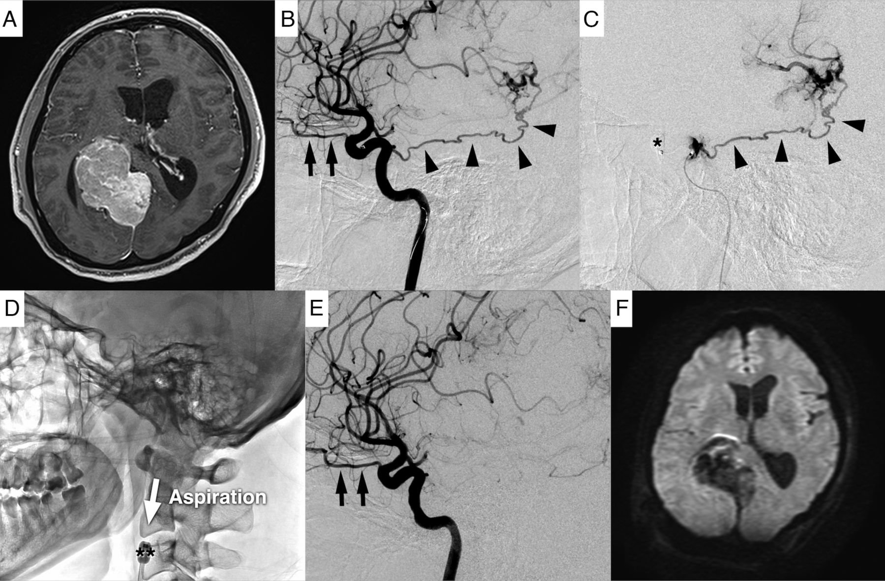
  • FIG 3.
    • Download figure
    • Open in new tab
    • Download powerpoint
    FIG 3.

    A case of tumor embolization via the MHT using the distal balloon protection technique. Preoperative tumor embolization was performed for a right falcotentorial meningioma through the right MHT (A and B). In this case, the microcatheter was guided to the vicinity of the orifice of the MHT because cannulation of the microcatheter into the MHT was not possible (C). Next, the ICA and ophthalmic artery were occluded with a balloon (C), and the MHT was embolized with embolic particles. After embolization, the AC was guided to the vicinity of the MHT to aspirate and remove the embolic particles. This step was followed by inflating the balloon GC and deflating the balloon catheter to reverse blood flow in the ICA to remove the embolic particles (D). Finally, complete embolization of the MHT was confirmed (E). Postoperative MR imaging showed no embolic cerebral infarction (F). The black arrow indicates the ophthalmic artery; black arrowhead, MHT; single asterisk, balloon catheter; double asterisk, balloon GC.

PreviousNext
Back to top

In this issue

American Journal of Neuroradiology: 45 (5)
American Journal of Neuroradiology
Vol. 45, Issue 5
1 May 2024
  • Table of Contents
  • Index by author
  • Complete Issue (PDF)
Advertisement
Print
Download PDF
Email Article

Thank you for your interest in spreading the word on American Journal of Neuroradiology.

NOTE: We only request your email address so that the person you are recommending the page to knows that you wanted them to see it, and that it is not junk mail. We do not capture any email address.

Enter multiple addresses on separate lines or separate them with commas.
Tumor Embolization via the Meningohypophyseal and Inferolateral Trunk in Patients with Skull Base Tumors Using the Distal Balloon Protection Technique
(Your Name) has sent you a message from American Journal of Neuroradiology
(Your Name) thought you would like to see the American Journal of Neuroradiology web site.
CAPTCHA
This question is for testing whether or not you are a human visitor and to prevent automated spam submissions.
Cite this article
Kei Yamashiro, Motoharu Hayakawa, Kazuhide Adachi, Mitsuhiro Hasegawa, Yuichi Hirose
Tumor Embolization via the Meningohypophyseal and Inferolateral Trunk in Patients with Skull Base Tumors Using the Distal Balloon Protection Technique
American Journal of Neuroradiology May 2024, 45 (5) 618-625; DOI: 10.3174/ajnr.A8169

Citation Manager Formats

  • BibTeX
  • Bookends
  • EasyBib
  • EndNote (tagged)
  • EndNote 8 (xml)
  • Medlars
  • Mendeley
  • Papers
  • RefWorks Tagged
  • Ref Manager
  • RIS
  • Zotero
0 Responses
Respond to this article
Share
Bookmark this article
Skull Base Tumor Embolization with Protection
Kei Yamashiro, Motoharu Hayakawa, Kazuhide Adachi, Mitsuhiro Hasegawa, Yuichi Hirose
American Journal of Neuroradiology May 2024, 45 (5) 618-625; DOI: 10.3174/ajnr.A8169
del.icio.us logo Twitter logo Facebook logo Mendeley logo
  • Tweet Widget
  • Facebook Like
  • Google Plus One
Purchase

Jump to section

  • Article
    • Abstract
    • ABBREVIATIONS:
    • MATERIALS AND METHODS
    • RESULTS
    • DISCUSSION
    • CONCLUSIONS
    • Acknowledgments
    • Footnotes
    • References
  • Figures & Data
  • Supplemental
  • Info & Metrics
  • Responses
  • References
  • PDF

Related Articles

  • PubMed
  • Google Scholar

Cited By...

  • Embolization of Posterior Fossa Meningiomas Supplied with Meningohypophyseal Trunk by Using n-BCA and Dual Balloon Protection
  • Crossref
  • Google Scholar

This article has not yet been cited by articles in journals that are participating in Crossref Cited-by Linking.

More in this TOC Section

  • Rescue Reentry in Carotid Near-Occlusion
  • Contour Neurovascular System: Five Year Follow Up
  • Effect of SARS-CoV2 on Endovascular Thrombectomy
Show more Neurointervention

Similar Articles

Advertisement

Indexed Content

  • Current Issue
  • Accepted Manuscripts
  • Article Preview
  • Past Issues
  • Editorials
  • Editor's Choice
  • Fellows' Journal Club
  • Letters to the Editor
  • Video Articles

Cases

  • Case Collection
  • Archive - Case of the Week
  • Archive - Case of the Month
  • Archive - Classic Case

More from AJNR

  • Trainee Corner
  • Imaging Protocols
  • MRI Safety Corner
  • Book Reviews

Multimedia

  • AJNR Podcasts
  • AJNR Scantastics

Resources

  • Turnaround Time
  • Submit a Manuscript
  • Submit a Video Article
  • Submit an eLetter to the Editor/Response
  • Manuscript Submission Guidelines
  • Statistical Tips
  • Fast Publishing of Accepted Manuscripts
  • Graphical Abstract Preparation
  • Imaging Protocol Submission
  • Evidence-Based Medicine Level Guide
  • Publishing Checklists
  • Author Policies
  • Become a Reviewer/Academy of Reviewers
  • News and Updates

About Us

  • About AJNR
  • Editorial Board
  • Editorial Board Alumni
  • Alerts
  • Permissions
  • Not an AJNR Subscriber? Join Now
  • Advertise with Us
  • Librarian Resources
  • Feedback
  • Terms and Conditions
  • AJNR Editorial Board Alumni

American Society of Neuroradiology

  • Not an ASNR Member? Join Now

© 2025 by the American Society of Neuroradiology All rights, including for text and data mining, AI training, and similar technologies, are reserved.
Print ISSN: 0195-6108 Online ISSN: 1936-959X

Powered by HighWire