
Article Figures & Data
Figures
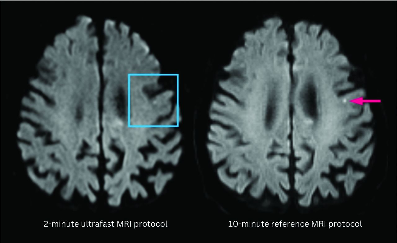
- FIG 1.
A punctate focus of restricted diffusion was less conspicuous on the ultrafast DWI image (left) compared with the reference DWI image (right). The decreased conspicuity may be due to a combination of differences in image quality between the 2 protocols and section positioning leading to partial volume averaging.
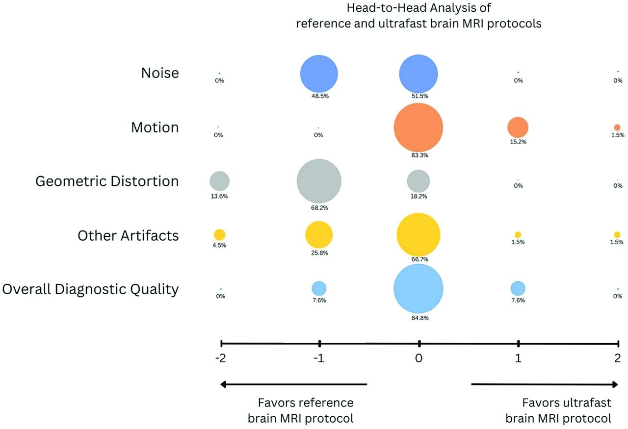
- FIG 2.
Bubble plot shows head-to-head comparison between the reference brain MR protocol and the ultrafast ms-EPI protocol. Negative scores indicate preference of the clinical reference protocol; 0 indicates equivalence between the 2 protocols; and positive scores indicate preference of the ultrafast ms-EPI protocol. The reference protocol was preferred by the evaluating neuroradiologists in terms of image noise and geometric distortion (P < .05 for both). The ultrafast protocol was preferred over the reference protocol in terms of motion artifacts (P < .05). There was no significant difference between the 2 protocols in terms of overall diagnostic quality.
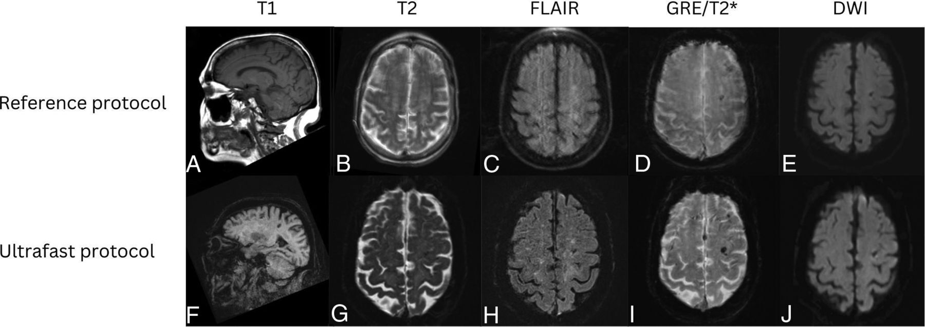
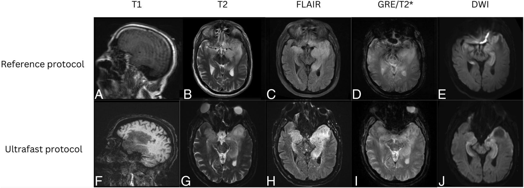
- FIG 3.
Patient with a diagnosis of amyloid angiopathy. Reference sagittal T1-weighted (A), axial T2-weighted (B), FLAIR (C), SWI (D), and DWI (E) show scattered foci of susceptibility signal in the left greater than right frontal lobes consistent with chronic microhemorrhages. The findings were more conspicuous on the ultrafast sagittal T1-weighted (F), axial T2-weighted (G), FLAIR (H), SWI (I), and DWI (J) because of motion artifact on the reference MR images.
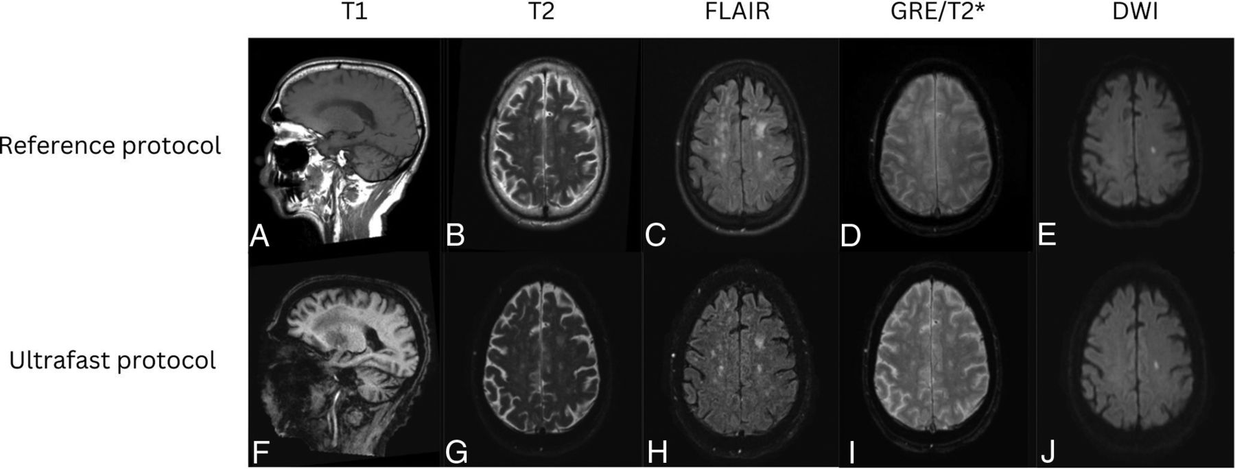
- FIG 4.
Patient with a diagnosis of herpes simplex virus encephalitis. Reference sagittal T1-weighted (A), axial T2-weighted (B), FLAIR (C), SWI (D), and DWI (E) show prominent T1 hypointensity and T2/FLAIR hyperintensity in the left greater than right mesial temporal lobes with scatter foci of susceptibility signal in the left temporal lobe consistent with microhemorrhages. There was associated restricted diffusion in the left greater than right mesial temporal lobes. These signal abnormalities were all seen with similar conspicuity on the ultrafast sagittal T1-weighted (F), and axial T2-weighted (G), FLAIR (H), SWI (I), and DWI (J).
- FIG 5.
Patient with punctate subacute infarct in the left corona radiata. Reference sagittal T1-weighted (A), axial T2-weighted (B), FLAIR (C), SWI (D), and DWI (E) show a punctate focus of restricted diffusion with associated FLAIR hyperintensity in the left centrum semiovale on a background of white matter T2/FLAIR hyperintensities that likely represent chronic small vessel ischemic disease. The same findings were seen on the ultrafast sagittal T1-weighted (F), axial T2-weighted (G), FLAIR (H), SWI (I), and DWI (J).
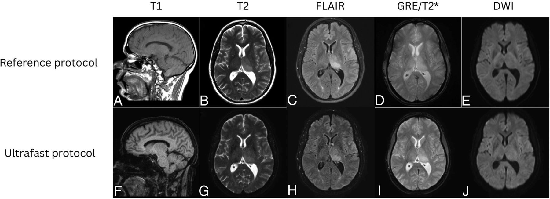
- FIG 6.
Patient with a low-grade left thalamic glioma. Reference sagittal T1-weighted (A), and axial T2-weighted (B), FLAIR (C), SWI (D), and DWI (E) show an ill-defined, mildly expansile, T1 hypointense, and T2/FLAIR hyperintense lesion centered in the left thalamus, compatible with diagnosis of low-grade glioma. Similar findings were appreciated on the ultrafast sagittal T1-weighted (F), axial T2-weighted (G), FLAIR (H), SWI (I), and DWI (J).


