Advertisement

Main menu

  • Home
  • Content
    • Current Issue
    • Accepted Manuscripts
    • Article Preview
    • Past Issue Archive
    • Video Articles
    • AJNR Case Collection
    • Case of the Week Archive
    • Case of the Month Archive
    • Classic Case Archive
  • Special Collections
    • AJNR Awards
    • Low-Field MRI
    • Alzheimer Disease
    • ASNR Foundation Special Collection
    • Photon-Counting CT
    • View All
  • Multimedia
    • AJNR Podcasts
    • AJNR SCANtastic
    • Trainee Corner
    • MRI Safety Corner
    • Imaging Protocols
  • For Authors
    • Submit a Manuscript
    • Submit a Video Article
    • Submit an eLetter to the Editor/Response
    • Manuscript Submission Guidelines
    • Statistical Tips
    • Fast Publishing of Accepted Manuscripts
    • Graphical Abstract Preparation
    • Imaging Protocol Submission
    • Author Policies
  • About Us
    • About AJNR
    • Editorial Board
    • Editorial Board Alumni
  • More
    • Become a Reviewer/Academy of Reviewers
    • Subscribers
    • Permissions
    • Alerts
    • Feedback
    • Advertisers
    • ASNR Home

User menu

  • Alerts
  • Log in

Search

  • Advanced search
American Journal of Neuroradiology

American Journal of Neuroradiology

ASHNR American Society of Functional Neuroradiology ASHNR American Society of Pediatric Neuroradiology ASSR
  • Alerts
  • Log in

Advanced Search

  • Home
  • Content
    • Current Issue
    • Accepted Manuscripts
    • Article Preview
    • Past Issue Archive
    • Video Articles
    • AJNR Case Collection
    • Case of the Week Archive
    • Case of the Month Archive
    • Classic Case Archive
  • Special Collections
    • AJNR Awards
    • Low-Field MRI
    • Alzheimer Disease
    • ASNR Foundation Special Collection
    • Photon-Counting CT
    • View All
  • Multimedia
    • AJNR Podcasts
    • AJNR SCANtastic
    • Trainee Corner
    • MRI Safety Corner
    • Imaging Protocols
  • For Authors
    • Submit a Manuscript
    • Submit a Video Article
    • Submit an eLetter to the Editor/Response
    • Manuscript Submission Guidelines
    • Statistical Tips
    • Fast Publishing of Accepted Manuscripts
    • Graphical Abstract Preparation
    • Imaging Protocol Submission
    • Author Policies
  • About Us
    • About AJNR
    • Editorial Board
    • Editorial Board Alumni
  • More
    • Become a Reviewer/Academy of Reviewers
    • Subscribers
    • Permissions
    • Alerts
    • Feedback
    • Advertisers
    • ASNR Home
  • Follow AJNR on Twitter
  • Visit AJNR on Facebook
  • Follow AJNR on Instagram
  • Join AJNR on LinkedIn
  • RSS Feeds

AJNR Awards, New Junior Editors, and more. Read the latest AJNR updates

Research ArticleBrain Tumor Imaging

Identification of a Single-Dose, Low-Flip-Angle–Based CBV Threshold for Fractional Tumor Burden Mapping in Recurrent Glioblastoma

Aliya Anil, Ashley M. Stokes, John P. Karis, Laura C. Bell, Jennifer Eschbacher, Kristofer Jennings, Melissa A. Prah, Leland S. Hu, Jerrold L. Boxerman, Kathleen M. Schmainda and C. Chad Quarles
American Journal of Neuroradiology October 2024, 45 (10) 1545-1551; DOI: https://doi.org/10.3174/ajnr.A8357
Aliya Anil
aFrom the Cancer System Imaging (A.A., C.C.Q.), The University of Texas MD Anderson Cancer Center, Houston, Texas
  • Find this author on Google Scholar
  • Find this author on PubMed
  • Search for this author on this site
  • ORCID record for Aliya Anil
Ashley M. Stokes
bDivision of Neuroimaging Research and Barrow Neuroimaging Innovation Center (A.M.S.), Barrow Neurological Institute, Phoenix, Arizona
  • Find this author on Google Scholar
  • Find this author on PubMed
  • Search for this author on this site
  • ORCID record for Ashley M. Stokes
John P. Karis
cDepartment of Neuroradiology (J.P.K.), Barrow Neurological Institute, Phoenix, Arizona
  • Find this author on Google Scholar
  • Find this author on PubMed
  • Search for this author on this site
  • ORCID record for John P. Karis
Laura C. Bell
dClinical Imaging Group (L.C.B.), Genentech Inc., San Francisco, California
  • Find this author on Google Scholar
  • Find this author on PubMed
  • Search for this author on this site
  • ORCID record for Laura C. Bell
Jennifer Eschbacher
eDepartment of Neuropathology (J.E.), Barrow Neurological Institute, Phoenix, Arizona
  • Find this author on Google Scholar
  • Find this author on PubMed
  • Search for this author on this site
  • ORCID record for Jennifer Eschbacher
Kristofer Jennings
fDepartment of Biostatistics (K.J.), The University of Texas MD Anderson Cancer Center, Houston, Texas
  • Find this author on Google Scholar
  • Find this author on PubMed
  • Search for this author on this site
  • ORCID record for Kristofer Jennings
Melissa A. Prah
gDepartment of Biophysics (M.A.P., K.M.S.), Medical College of Wisconsin, Milwaukee, Wisconsin
  • Find this author on Google Scholar
  • Find this author on PubMed
  • Search for this author on this site
  • ORCID record for Melissa A. Prah
Leland S. Hu
hDepartment of Radiology (L.S.H.), Division of Neuroradiology, Mayo Clinic Arizona, Phoenix, Arizona
  • Find this author on Google Scholar
  • Find this author on PubMed
  • Search for this author on this site
  • ORCID record for Leland S. Hu
Jerrold L. Boxerman
iDepartment of Diagnostic Imaging (J.L.B.), Rhode Island Hospital, Providence, Rhode Island
  • Find this author on Google Scholar
  • Find this author on PubMed
  • Search for this author on this site
  • ORCID record for Jerrold L. Boxerman
Kathleen M. Schmainda
gDepartment of Biophysics (M.A.P., K.M.S.), Medical College of Wisconsin, Milwaukee, Wisconsin
  • Find this author on Google Scholar
  • Find this author on PubMed
  • Search for this author on this site
  • ORCID record for Kathleen M. Schmainda
C. Chad Quarles
aFrom the Cancer System Imaging (A.A., C.C.Q.), The University of Texas MD Anderson Cancer Center, Houston, Texas
  • Find this author on Google Scholar
  • Find this author on PubMed
  • Search for this author on this site
  • ORCID record for C. Chad Quarles
  • Article
  • Figures & Data
  • Supplemental
  • Info & Metrics
  • Responses
  • References
  • PDF
Loading

Article Figures & Data

Figures

  • FIG 1.
    • Download figure
    • Open in new tab
    • Download powerpoint
    FIG 1.

    A sRCBV comparison between the single-dose, LFA protocol and the double-dose, MFA protocol on the mean tumor ROI across all the patients (n = 52) included in the study. This result shows a strong agreement between the 2 protocols with a concordance correlation coefficient value of 0.99.
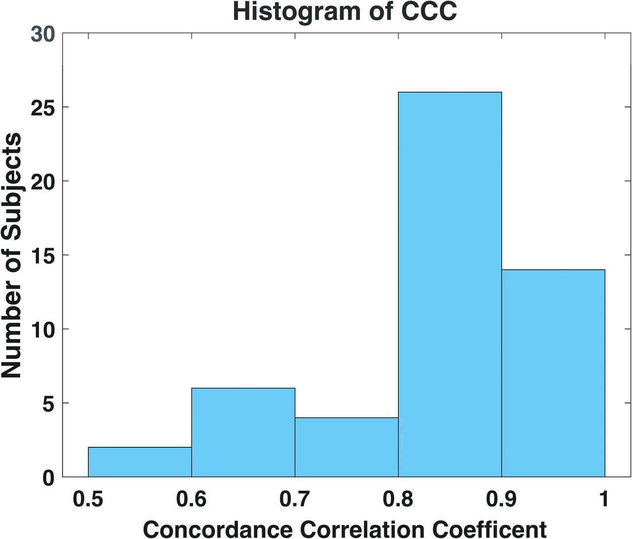
  • FIG 2.
    • Download figure
    • Open in new tab
    • Download powerpoint
    FIG 2.

    Histogram of CCC across each patient, ranging from 0.52 to 0.96. Seventy-seven percent of patients exhibit a CCC greater than 0.8.

  • FIG 3.
    • Download figure
    • Open in new tab
    • Download powerpoint
    FIG 3.

    The area under the ROC curve (AUC) for the optimal LFA-based thresholds is found to be 0.95 and 0.96 for sRCBV <1.0 and >1.56, respectively.

  • FIG 4.
    • Download figure
    • Open in new tab
    • Download powerpoint
    FIG 4.

    Boxplots (with individual datapoints) showing the consistency between percentage of tumor voxels (sRCBV >1.0) in the enhancing tumor for the double-dose, MFA and single-dose, LFA protocols.

  • FIG 5.
    • Download figure
    • Open in new tab
    • Download powerpoint
    FIG 5.

    A–C, The patient shown is a 73-year-old woman with grade-IV glioblastoma presenting 15 months after surgical resection. D–F, The patient shown is a 34-year-old man with grade-IV glioblastoma presenting 20 months after surgical resection. Images include anatomic postcontrast T1-weighted images (A and D), FTB maps for the single-dose, LFA protocol (B and E) by using 1.0 and 1.37, and the reference double-dose, MFA protocol (C and F) by using 1.0 and 1.56 superimposed on the contrast-enhanced T1-weighted images. Blue, yellow, and red voxels represent PTRE (FTBlow, sRCBV <1.0), tumor/treatment effect admixture (FTBmid, 1.0 > sRCBV <1.37 [LFA], 1.56 [MFA]), and high tumor cell probability (FTBhigh, sRCBV >1.37 [LFA], 1.56 [MFA]), respectively. The Dice similarity coefficients comparing the LFA and MFA sRCBV for PTRE and tumor recurrence for the patient in (A–C) are 0.85 and 0.87, respectively. For the patient in (D–F), the coefficients are 0.77 for PTRE and 0.96 for tumor recurrence.
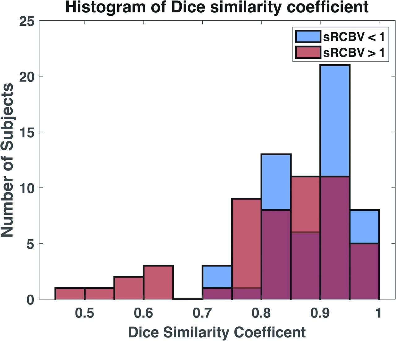
  • FIG 6.
    • Download figure
    • Open in new tab
    • Download powerpoint
    FIG 6.

    Histogram of Dice similarity coefficient between the regions of PTRE (sRCBV <1.0) and tumor recurrence (sRCBV >1.0) for each patient. Ninety-two percent and 67% of the subjects show a Dice similarity coefficient greater than 0.8 for sRCBV <1.0 and sRCBV >1.0, respectively.

PreviousNext
Back to top

In this issue

American Journal of Neuroradiology: 45 (10)
American Journal of Neuroradiology
Vol. 45, Issue 10
1 Oct 2024
  • Table of Contents
  • Index by author
  • Complete Issue (PDF)
Advertisement
Print
Download PDF
Email Article

Thank you for your interest in spreading the word on American Journal of Neuroradiology.

NOTE: We only request your email address so that the person you are recommending the page to knows that you wanted them to see it, and that it is not junk mail. We do not capture any email address.

Enter multiple addresses on separate lines or separate them with commas.
Identification of a Single-Dose, Low-Flip-Angle–Based CBV Threshold for Fractional Tumor Burden Mapping in Recurrent Glioblastoma
(Your Name) has sent you a message from American Journal of Neuroradiology
(Your Name) thought you would like to see the American Journal of Neuroradiology web site.
CAPTCHA
This question is for testing whether or not you are a human visitor and to prevent automated spam submissions.
Cite this article
Aliya Anil, Ashley M. Stokes, John P. Karis, Laura C. Bell, Jennifer Eschbacher, Kristofer Jennings, Melissa A. Prah, Leland S. Hu, Jerrold L. Boxerman, Kathleen M. Schmainda, C. Chad Quarles
Identification of a Single-Dose, Low-Flip-Angle–Based CBV Threshold for Fractional Tumor Burden Mapping in Recurrent Glioblastoma
American Journal of Neuroradiology Oct 2024, 45 (10) 1545-1551; DOI: 10.3174/ajnr.A8357

Citation Manager Formats

  • BibTeX
  • Bookends
  • EasyBib
  • EndNote (tagged)
  • EndNote 8 (xml)
  • Medlars
  • Mendeley
  • Papers
  • RefWorks Tagged
  • Ref Manager
  • RIS
  • Zotero
0 Responses
Respond to this article
Share
Bookmark this article
Low-Flip-Angle CBV Threshold in Glioblastoma
Aliya Anil, Ashley M. Stokes, John P. Karis, Laura C. Bell, Jennifer Eschbacher, Kristofer Jennings, Melissa A. Prah, Leland S. Hu, Jerrold L. Boxerman, Kathleen M. Schmainda, C. Chad Quarles
American Journal of Neuroradiology Oct 2024, 45 (10) 1545-1551; DOI: 10.3174/ajnr.A8357
del.icio.us logo Twitter logo Facebook logo Mendeley logo
  • Tweet Widget
  • Facebook Like
  • Google Plus One
Purchase

Jump to section

  • Article
    • Abstract
    • ABBREVIATIONS:
    • MATERIALS AND METHODS
    • RESULTS
    • DISCUSSION
    • CONCLUSIONS
    • Acknowledgments
    • Footnotes
    • References
  • Figures & Data
  • Supplemental
  • Info & Metrics
  • Responses
  • References
  • PDF

Related Articles

  • PubMed
  • Google Scholar

Cited By...

  • No citing articles found.
  • Crossref
  • Google Scholar

This article has not yet been cited by articles in journals that are participating in Crossref Cited-by Linking.

More in this TOC Section

  • Brain Tumor Subtyping Image-Based search via MRI
  • Neuroimaging of Erdheim-Chester Disease
  • CE MRI for Brain Metastasis Detection
Show more Brain Tumor Imaging

Similar Articles

Advertisement

Indexed Content

  • Current Issue
  • Accepted Manuscripts
  • Article Preview
  • Past Issues
  • Editorials
  • Editor's Choice
  • Fellows' Journal Club
  • Letters to the Editor
  • Video Articles

Cases

  • Case Collection
  • Archive - Case of the Week
  • Archive - Case of the Month
  • Archive - Classic Case

More from AJNR

  • Trainee Corner
  • Imaging Protocols
  • MRI Safety Corner
  • Book Reviews

Multimedia

  • AJNR Podcasts
  • AJNR Scantastics

Resources

  • Turnaround Time
  • Submit a Manuscript
  • Submit a Video Article
  • Submit an eLetter to the Editor/Response
  • Manuscript Submission Guidelines
  • Statistical Tips
  • Fast Publishing of Accepted Manuscripts
  • Graphical Abstract Preparation
  • Imaging Protocol Submission
  • Evidence-Based Medicine Level Guide
  • Publishing Checklists
  • Author Policies
  • Become a Reviewer/Academy of Reviewers
  • News and Updates

About Us

  • About AJNR
  • Editorial Board
  • Editorial Board Alumni
  • Alerts
  • Permissions
  • Not an AJNR Subscriber? Join Now
  • Advertise with Us
  • Librarian Resources
  • Feedback
  • Terms and Conditions
  • AJNR Editorial Board Alumni

American Society of Neuroradiology

  • Not an ASNR Member? Join Now

© 2025 by the American Society of Neuroradiology All rights, including for text and data mining, AI training, and similar technologies, are reserved.
Print ISSN: 0195-6108 Online ISSN: 1936-959X

Powered by HighWire